Synaptic plasticity of Inhibitory synapse promote synchrony in inhibitory network in presence of heterogeneity and noise
Abstract
Recently spike timing dependent plasticity was observed in inhibitory synapse in the layer II of entorhinal cortex. The rule provides an interesting zero in the region of and in addition the dynamic range of the rule lie in gamma frequency band. We propose a robust mechanism based on this observed synaptic plasticity rule for inhibitory synapses for two mutually coupled interneurons to phase lock in synchrony in the presence of intrisic heterogeneity in firing. We study the stability of the phase locked solution by defining a map for spike times dependent on the phase response curve for the coupled neurons. Finally we present results on robustness of synchronization in the presence of noise.
pacs:
Valid PACS appear hereIt is generally accepted that inhibitory interneurons are important for synchrony in neocortex. Several studies have reported the role for inhibitory interneurons in generating stable synchronous rhythms in neocortex Benardo ; Jefferys ; Michelson ; Whittington ; Bragin . Cortical oscillations in the gamma frequency band (20-80 Hz), are thought to be involved in binding of object properties, a process of great significance for the functioning of the brain. These experimental findings have led to numerous theoretical studies of synchrony among inhibitory interneurons van ; Ernst ; Traub ; Wang . The principle result of these studies showed that depending on the decay time of the inhibitory synaptic coupling, the mutually coupled inhibitory neurons oscillate in synchrony ( in phase locking) or in antisynchrony (out of phase locking). However much of the above investigations did not explore the effects of heterogeneity in the intrinsic firing rates nor did they take into account noise, which is invariably present in neuronal systems.
In another set of theoretical investigations, White explored the implications of small heterogeneity for the degradation of synchrony of fast spiking inhibitory neurons and the mechanism by which the degradation occur. They found that introduction of even small amounts of heterogeneity in the external drive, resulted in significant reduction in coherence of neuronal spiking. It is important then to understand what mediates observed in vivo synchrony of neuronal networks under biological realistic conditions of noise induced unreliability and intrinsic heterogeneity in spiking rates of the neuronal ensemble.
In this letter we propose a robust mechanism based on spike timing dependent synaptic plasticity of inhibitory synapses Haas by which two coupled interneurons can phase lock in synchrony even under conditions of mild heterogeneity in the firing rates of the coupled neurons and in the presence of noise. Earlier work Valentin has explored the function of synaptic plasticity in the excitatory synapse in improving synchronization in unidirectionally coupled neuronal network. We consider a network of two coupled interneurons with self inhibition as shown in Figure 1b. The self-inhibition is introduced because biological neural networks often have local inhibitory interneurons which deliver feedback inhibition to the cells activating those interneurons Sheperd . The importance of self inhibition that simulates the network effect, is explored in details in Talathi_Neurosci .
Each neuron in the coupled network is modelled as
where (i=A,B) is the membrane potential , is the gaussian synaptic noise of amplitude satisfying and . , is the external drive, , is the synaptic current due to self inhibition and is the synaptic current from mutual inhibition. is the dynamic synapse, whose strength is determined by the inhibitory synaptic plasticity rule, and is the synaptic strength of self inhibition. (r=Na, K, L) are reversal potentials of the sodium and potassium ion channels and the leak channel respectively. is the reversal potential of the inhibitory synapse. give the fraction of bound receptors and satisfy the first order kinetic equation, where is the presynaptic voltage. It involves two time constants, , the docking time for the neurotransmitter and , the undocking time constant for the neurotransmitter binding. is the sigmoidal function given by, . The gating variables X(t),(X=m,h,n), satisfy first order kinetic equations, . We have used standard functions and and parameters for the model Talathi , such that the dynamics of the neurons to spiking is through saddle node bifurcation and the model represents a type I neuron Ermentrout . The model parameters are within physiological range and give high spike rates typical of interneurons.
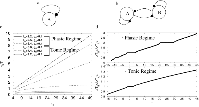
We consider two regimes of operation of the network shown in Figure 1b. The two regimes called the phasic regime and the tonic regime White are determined from the firing characteristics of a single self inhibited neuron (Figure 1a). In the phasic regime the network period depends on the synaptic decay constant White and in the tonic regime the synaptic dynamics weakly affect the network period. These two regimes of operation are clearly illustrated in the plot of , which is the ratio of synaptic decay constant to the firing frequency of the self inhibited neuron versus in Figure 1b for various values of and . These two regimes of network oscillations were observed earlier White , with the synaptic model for the inhibitory synapse, obeying the first order kinetic equation, . Changing in this situation not only changes the decay time of the synapse but also the rise time given by and the saturation level of the synapse . As a result a narrow region of higher harmonic phase locking was observed between the coupled interneurons in the phasic regime. We have therefore considered the synaptic model presented above where we have control over the decay time for the synapse independent of the rise time and the saturation level of the synapse. Earlier work Chow has shown that in presence of mild heterogeneity, coherence in neuronal firing is observed much more for phasic regime than in tonic regime. In the results presented here we therefore consider the phasic regime of the network operation and study the effect of STDP in inhibitory synapses, in maintaining coherence in neuronal firing in presence of heterogeneity and noise. Details on the calculations for the tonic regime will appear elsewhere Talathi_Neurosci
In all the subsequent calculations, we fix the parameters of the model in the phasic regime, (, , ms, ms) and study the effect of the dynamic synapse in maintaining synchrony in presence of heterogeneity and intrinsic noise. In figure 1d, we plot the ratio of mean firing periods for the two coupled neurons A and B as function of heterogeneity in the external drive, defined as in absence of noise i.e., . As can be seen from Figure 1d, (top panel), the region of 1:1 locking, as function of heterogeneity is much broader in the phasic regime of network operation as compared to the tonic regime, where we set . In addition, higher order synchronization are also present in the phasic regime, as a result coherence between the two neurons is preserved more often in the phasic regime of the network operation.
A spike timing dependent plasticity (STDP) rule for inhibitory synapses has been recently reported in Haas and it has the form , where . is the time of presynaptic spike stimulation and is the time of a spike generated by the postsynaptic neuron. is the scaling factor accounting for the amount of change in inhibitory conductance induced by the synaptic plasticity rule and is the normalizing constant. An empirical fit of the above function to the data gives, and , giving a window of ms over which the efficacy of synaptic plasticity is non zero. This implies that in the physiologically important regime of gamma oscillations (25-80 Hz), STDP rule of inhibitory synapses can play a significant role in modulating the firing dynamics of the neuronal network.
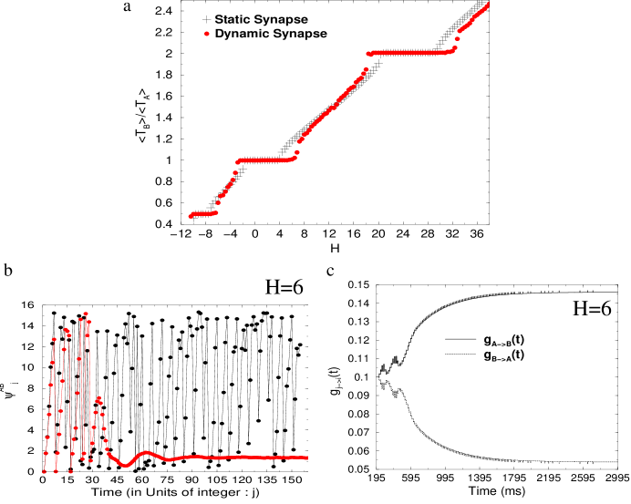
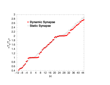
We now consider two situations in the phasic regime: when the strength of mutual inhibition is static: and when the mutual inhibition strength is dynamic and governed by observed synaptic plasticity rule, i.e., and . In order to take into account the effect of multiple spike pairs, we follow Froemke and define , where is the difference in spike times of neurons A and B respectively. gives the efficacy of spike in A and B and is defined as . We take ms, an average of the efficacy values given in Froemke as experimental results on contribution of multiple spike pairing to inhibitory synaptic plasticity are as yet unknown. In Figure 2a we plot the ratio as function of heterogeneity H in the static and dynamic case.
As shown in Figure 2a there is considerable increase in 1:1, 1:2 and 2:1 synchronization windows mediated by the dynamic synapse in the phasic regime. This implies an increased probability of observing coherence in the firing pattern of the mutually coupled interneurons even in presence of mild heterogeneity as has been reported in many in vivo experimental data. In Figure 2b ,we show the evolution of phase difference in the static and dynamic case, with heterogeneity of 6 %. We see that in the static case the phase difference grows linearly modulo , representing situation of asynchronous firing. However in the dynamic case, after the initial transient is over, the phase difference saturates to a fixed value, (1.4 in this example), representing stable 1:1 locking between the two mutually coupled neurons. In Figure 2c we plot the evolution of the synaptic strength as function of time. We see that the STDP rule results in modulating the synaptic strength so as to phase lock the two mutually coupled neurons.

In order to understand the dynamics of phase locking under heterogeneity in the presence of dynamic synapse, we consider the situation initially in the static case. Let the two coupled neurons fire heterogeneously with intrinsic firing period and when they are uncoupled. In the case of 6 % heterogeneity, we have so that . Let be the phase response curve of the neuron model. It is known that for type I neuron models, the phase response curve is positive Ermentrout , and as a result every time a spike arrives, the phase of subsequent spike is delayed. In the situation considered, with initial phase difference between the two neurons set to zero, the spike times of individual firing neurons can be written as, , (K=A,B) with , where and . is the time of spike of neuron K. The map evolving the phase difference is then,
| (2) | |||||
where . The fixed point of the map is then given by
Numerical solution to above equation gives the fixed points, at as can be seen in Figure 3, where we plot versus . Stability of the fixed point would require
For the set of parameters considered however, the numerical value obtained for for the fixed point.
Thus we see that when the synapse is static the phase difference is unstable and the two coupled interneurons fire asynchronously in the situation of 6% heterogeneity. Now consider the situation in the dynamic synapse case. Again setting the initial phase difference zero, in the presence of dynamic synapse with STDP, we have , where phase shift given by the phase response curve also depends on the dynamics of the synaptic coupling strength governed by the STDP rule. The map function for evolution of the phase shift is then obtained as,
| (3) |
where . The fixed point of the map is then given by . As can be seen from Figure 3b, where we plot , in the steady state (), when and , we obtain, and . Stability of the fixed point given by , implies is stable as can be also seen from the time evolution of phase in Figure 2b. Thus we see that STDP of inhibitory synapse, modulates the phase response curve such that the network locks into synchrony even under mild heterogeneity.
In Figure 4 we present results on synchrony in presence of noise. We set the noise amplitude in equation 1. For mild noise, STDP of inhibitory synapse, is able to maintain synchrony between the two coupled interneurons under conditions of mild heterogeneity in the drive.
We have also tested the dynamics of the network in the tonic regime. STDP of inhibitory synapse, also significantly increases the window of synchronous oscillations by the same mechanism Talathi_Neurosci .
It has been suggested in Haas that plasticity of inhibitory synapses may play an important role in balancing the effect of excitatory synapse preventing run away behavior typically observed in epileptogenesis. In this work we present an important function for STDP in inhibitory synapse in maintaining synchrony in networks of coupled interneurons, under biologically realistic situation of mild heterogeneity and noise.
Acknowledgements
This work was partially funded by a grant from the National
Science Foundation, NSF PHY0097134. SST was partially supported by
the NSF sponsored Center for Theoretical Biological Physics at
UCSD. We would like to thank Henry Abarbanel, Julie Haas, Thomas
Nowotny and Johnathan Driscoll for instructive feedbacks that
improved this work significantly.
References
- (1) Benardo L.S., J Neurophysiol, 77 3134-3144, (1997).
- (2) Jefferys J.G., Traub R.D., Whittington M.A., Trends Neurosci, 19 202-208, (1996).
- (3) Michelson H.B., Wong R.K., J Physiol (Lond), 477 35-45, (1994).
- (4) Whittington M.A., Traub R.D., Jefferys J.G., Nature, 373 612-615, (1995).
- (5) Bragin A., Jando G., Nadasdy Z., Hetke J., Wise K., Buzsaki G.,J Neurosci, 15 47-60, (1995).
- (6) van Vreeswijk C., Abbott L.F., J Comp Neurosci, 1 313-321, (1994).
- (7) Ernst U., Pawelzik K., Geisel T., Phys Rev Lett, 74 1570-1573, (1995).
- (8) Traub R.D., Jefferys J.G., Whittington M.A., J Comp Neurosci, 416 433-438, (2002).
- (9) Wang X.J., Rinzel J., Neural Compute, 4 84-97, (1992).
- (10) Froemke R.C., Dan Y., Nature, 416 433-438, (2002).
- (11) Kempter R., Gerstner W., Hemmen van J.L., Physical Review E, 8 979-1001, (1996).
- (12) Roberts P.D., J Comp Neurosci, 7 235-246, (1999).
- (13) White A.J., Chow C.C., Ritt J., Trevino C.S., Kopell N., J Comp Neurosci, 5 5-16, (1998).
- (14) Chow C.c., White J.A., Ritt J., Kopell N., J Comp Neurosci, 5, 407-420, (1998).
- (15) Haas J., Nowotny T., Abarbanel H.D.I.A., J Neurophysiol, Submitted.
- (16) Zhigulin V.P., Rabinovich M.I., Huerta R., Abarbanel H.D.I.,Phys Rev E, 67 021901, (2003).
- (17) Shepherd G.M., The Synaptic Organization of the Brain, 3rd ed, Oxford University Press, New York, (1990).
- (18) Abarbanel, H.D. I. and Talathi S.S., Phys Rev Lett, 96, 148104 (2006).
- (19) Talathi S.S. and Abarbanel H.D.I., Unpublished Results.
- (20) Ermentrout B., Neural Compute 8 979-1001, (1996).