Budded membrane microdomains as regulators for cellular tension
Abstract
We propose a mechanism for the control of the mechanical properties of the membrane of living cells that is based on the exchange of membrane area between the cell membrane and a membrane reservoir. The reservoir is composed of invaginated membrane microdomains which are liable to flatten upon increase of membrane strain, effectively controlling membrane tension. We show that the domain shape transition is first order, allowing for coexistence between flat and invaginated domains. During coexistence, the membrane tension is controlled by the domains elasticity and by the kinetics of the shape transition. We show that the tension of the plasma membrane of living cells is inherently transient and dynamical, and that valuable insights into the organization of the cell membrane can be obtained by studying the variation of the cell membrane tension upon mechanical perturbation.
Endocytosis, exocytosis, cell motility and many other crucial cellular processes are known to be influenced by the tension of the cell membranesheetz . The force needed to pull a tubular membrane tether from the cell with an optical trap is often used as a probe of the mechanical state of the membranesheetz_cytoskel . The mechanical response of living cells to such perturbation involves cytoskeleton deformation, the breaking of membrane-cytoskeleton bondssheetz_pip2 and changes in membrane morphology. The level of membrane tension in cells is thought to primarily reflects cytoskeleton anchoring to the membranesheetz_cytoskel . Nevertheless, there is experimental evidence that Surface Area Regulation (SAR) occurs morris_SAR and is able to buffer the increase of tension upon mechanical perturbation. SAR could be achieved by the transfer of membrane area from a reservoir to the plasma membrane, during which the level of membrane tension is at least partly controlled by the dynamical response of the reservoir. One possible manifestation of the exchange of membrane with a reservoir is the presence of a plateau in the force-extension curve of tether extraction experiments, as observed in sheetz_buffer . The plateau ends when the putative membrane reservoir is emptied. In this particular experiment the reservoir is able to react quite fast to the perturbation (s), which could indicate i) that the reservoir is permanently connected to the plasma membrane and ii) that the regulation is purely physical and does not involve biological signaling.
Our goal in this paper is to study the influence of membrane morphology, and in particular the flattening of invaginated ”bud-like” domains, on the mechanical response of the membrane. Such domains include caveolae that form shaped invaginationscav_review reactive to membrane stresscav_stress ; woodman . Striking experiments, showing Caveolae flattening under tensionflat_cav , support the idea that the delivery of invaginated membrane area to the plasma membrane is controlled by tensioncav_reservoir . The shape of membrane domains in artificial membrane systems, such as giant vesicles, is also known to be dependent on the membrane tensionmembr_domains1 . Indeed, domain budding has been observed upon the decrease of membrane stressmembr_domains2 .
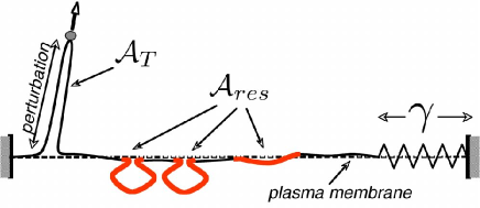
Our model is summarised in Fig.1. Here the cytoskeleton is included as linear elastic element (spring) mechanically coupled to the cell membrane. Although crude, this approximation is consistent with the experimental results of sheetz_buffer (see SUPP ). Furthermore, it allows us isolate and study the response of the reservoir, which can be thought of as one element in a chain of response that transmits stress to the cytoskeleton. Cytoskeleton elasticity and membrane cytoskeleton anchoring are reflected by an effective stretching coefficient of order SUPP . Within this framework, the variation of the cell tension with the tether area reads:
| (1) |
where and are the cell tension and reservoir area at rest (without tether, . If membrane area is delivered to the plasma membrane, as during exocytosis, the “tether” area is negative. One can see that the membrane tension can be maintained constant upon tether pulling only if the decrease of reservoir area matches the increase of tether area.
The membrane domains constituting the reservoir are described within a very general theoretical framework. It is based on the domain bending rigidity, that disfavors the budded state, and its composition difference with the rest of the membrane, that promotes domain invagination in order to reduce the length of the domain periphery (Fig.2). Domains are treated as spherical caps of fixed area and adjustable curvature . Their shape is uniquely characterized by the parameter (Fig.2 and Eq.(2)), equal to unity for a fully budded domain (a sphere), and that vanishes for a flat domain. The energy of a domain contains a surface tension-independent part . This itself involves a term arising from the line tension membr_domains1 , proportional to the length of the cap edge (neck), and a term giving the bending energy of the cap, proportional to the bending rigidity and the squared curvaturesafran . Simple geometry then gives
| (2) |
If the domain size exceeds a critical value , an invaginated sphere () has a lower energy than a flat domain () and budding is expectedlipowsky_budding . We assume that in what follows. Typical values of the parameters and correspond to a critical size , similar to the size of caveolae. In practice invaginated domains remain attached to the mother membrane by a small neck. Here, this is controlled phenomenologically by assigning the value () to the ratio of invagination radius to neck size, giving an upper bound to the shape parameter.
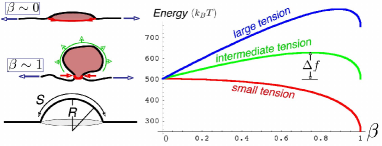
Including the membrane tension , the domain energy reads: . Increasing membrane tension increases the energy of curved states and promotes the flat state (see Fig.2). The flat states eventually becomes stable for a critical tension :
| (3) |
As can be seen in Fig.2, the budded and flat domain shapes are separated by an energy barrier for intermediate tension. The existence of this barrier is crucial to their function as tension regulators, as it allows the coexistence of flat and invaginated domains. Variation of the membrane strain (the tether length) may occur without change of tension by adjusting the fraction of budded domains.
The energy scales in this system (with ) are
| (4) |
The energy of surface tension competes with the line and bending energies for , or (corresponding to tether forces of order ). This is precisely in the range of mechanical tension recorded for cellular membranessheetz_cytoskel , which is very encouraging for the biological relevance of our model. One notes that the energy scale is very large compared to the thermal energy , or to the energy of any “active temperature” present in biological systemsmembrane_activity . This has two important physical consequences: (i) the shape transition of a domain is very discontinuous, a domain snaps open rather than continuously flattening upon tension increase, and (ii) the budding and flattening transitions should actually occur at different tensions, for which the respective energy barriers are of order . In biological systems, the “temperature” might be seen as a parameter reflecting cellular activity, such as the polymerization of the actin cortex near the membrane, and the activity of membrane pumps. For simple cells such as Red Blood Cells, it is typically a few times the thermodynamic temperaturemembrane_activity .
The bottleneck for the shape transition is the maximum of energy, which corresponds to a shape parameter . The budding and flattening tensions ( and respectively) at which the corresponding energy barrier vanishes are:
| (5) |
where characterize the size of the invagination neck (see above).
We investigate the tension regulation performed by a collection of domains, of total area (where , is the largest value of shape parameter consistent with the existence of a finite size neck).
When flat and budded domains coexist, a fraction of domains are invaginated, and the reservoir area is . The total membrane energy, including the contribution of each of the membrane domains (Eq.(2)) and the total work done against membrane tension can be written:
| (6) |
Optimizing the energy for the fraction of invaginated domains leads directly to the regulation of membrane tension, when flat and budded domains coexist (). Substituting in Eq.(6) the expression for the surface tension Eq.(1) (with ), we find that the tension is set to the value of Eq.(3), which depends on the characteristics of the membrane reservoir (, , and ), but not on the tether area. Regulation is achieved by adjusting the fraction of budded domains to:
| (7) |
where is the fraction of budded domains corresponding to the tension at rest , and where a normalized stretching coefficient , with dimension of energy, is introduced for convenience.
If the reservoir is given time to equilibrate, regulation starts for a level of perturbation corresponding to a tether area (at which all domains are budded, ), and ends at a tether area (at which all domains are flat, ). The tension of the cell membrane is then set to the values , for any perturbation within the range . If the perturbation is very fast, one expect a large difference between the regulated tension upon tether pulling and tether retraction, in agreement with Eq.(5).
To obtain the full kinetic response of the membrane to strain, we describe the transition as a classical Kramers’ processvankampen , were the transition time between two states is exponential with the energy barrier that has to be overcome in the process: . Here, is the characteristic fluctuation time of the domain shape, and may be an effective temperature resulting from the large biochemical activity near the cell membranemembrane_activity . The transition time is very much dependent upon the membrane tension. Assuming that the transition of a single domain occurs with negligible change of tension (this implies ), the transition is fully described by the energy , with given by Eq.(1). Assuming for simplicity that both domain flattening and budding involve the same fluctuation time , the kinetic evolution of the fraction is given by
| (8) |
where the maximum of energy corresponds to the least favorable domain shape
In order to mimic the tether pulling experiment, where the tether is typically extracted at constant speed () in sheetz_buffer , we consider the reservoir response to a perturbation applied with a given rate : . If the perturbation is applied slowly (), the reservoir has time to equilibrate (), and the fraction of budded domain is found from Eq.(8) to be given by , with. The fraction is the equivalent of the equilibrium fraction (Eq.(7)), that takes thermal fluctuations into account ( if ). Thermal fluctuations smoothen the transition between budded and flat domains by allowing states of non-minimal energy to be populated. As a consequence, the tension is not perfectly constant during the transition, and the slope at mid plateau is of order . If, on the other hand, the perturbation is applied very fast, the shape transition requires small energy barriers, which means high tension for bud flattening, and low tension for domain budding, respectively and give by Eq.(5).
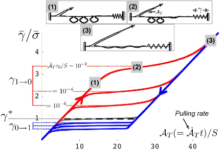
The physical mechanism at the origin of tension regulation and the membrane hysteretic response to tether extraction and retraction are shown in Fig.3. To obtain an analytical expression of the plateau height with the perturbation rate, we approximate that the tension is almost constant during regulation () so that the energy barrier is of order . The plateau tension upon increase and decrease of the perturbation are then respectively given by and , with . As expected for activated processes, the dependence of the tension at transition with the rate of perturbation is logarithmic. The same is true for the slope of the plateau, which can be estimated by identifying the plateau inflexion point. The condition imposes , corresponding to a plateau slope . As the effective membrane temperature may be inferred independently from measurement of membrane fluctuationsmembrane_activity , the study of both the height and slope of the tension plateau gives valuable information of the kinetics of area transfer between the reservoir and the plasma membrane.
The difference between the quasi-static and dynamic plateau can be of order and correspond to a difference in force of order . This is precisely the scale of the forces measured upon tether extraction in sheetz_buffer . This possibility thus exists that the initial increase of tension observed prior to the plateau in sheetz_buffer is of purely kinetic origin and originate from the slow response of an already partially unfolded reservoir at rest. In this case, the membrane tension of the cell at rest would be of Eq.(5), fully controlled by the mechanical properties of the membrane domains forming the reservoirSUPP .
In summary, we have derived the mechanical reactivity of a cell membrane, in contact with a reservoir composed of invaginated membrane domains liable to flatten under strain. The flattening transition is first order, which means that the invaginations snap open above a critical strain rather than continuously flattening, an observation consistent with experimental evidences from the membrane invagination caveolaecav_reservoir . As a consequence, the cell mechanical response shows a plateau during the transition, corresponding to the coexistence of flat and invaginated domains. This study provides the basis for a mechanical regulation of the tension of the cell membrane. The mechanism at the origin of this regulation is of purely physical origin, consistent with the fast timescale () of the observed cellular response. In practice, a ring of specialized membrane proteins such as dynamincav-dynamin is often present at the neck of membrane invagination. These proteins most probably influence the domain line energy, and might even dominate the energy required to flatten the domain. The regulation of membrane tension by membrane invaginations rely on the existence of two well defined domain shapes, separated by an energy barrier. If anything, neck proteins can only increase the energy barrier to flattening, thereby reinforcing tension regulation.
This work also opens the possibility of a new quantitative “force spectroscopy” of the cell membrane. One could thereby obtain structural information on the membrane organization, in much the same way information on a protein structure can be gathered from force measurement upon protein unfoldingprotein_unfolding . As a first step, one may identify the fairly regular oscillation of the force during regulation in sheetz_buffer , to the flattening of single domains. Preliminary analysisSUPP hints at domains of area .
References
- (1) D. Raucher, & M.P. Sheetz (1999) J. Cell Biol. 144, 497-506 D. Raucher, & M.P. Sheetz (2000) J. Cell Biol. 148, 127-136
- (2) M.P. Sheetz (2001) Nature Rev. Mol. Cell Bio. 2, 392-396
- (3) D. Raucher, T. Stauffer, W. Chen, K. Shen, S. Guo, JD. York, MP. Sheetz, and T. Meyer. (2000) Cell 100, 221 228
- (4) C.E. Morris & U. Homann (2000) J. Membrane Biol. 179, 79-102
- (5) D. Raucher, & M.P. Sheetz (1999) Biophys. J. 77, 1992-2002
- (6) K. Simons & E. Ikonen (1997) Nature 387, 569-572 Schlegel, A., D. Volonté, J.A. Engelman, F. Galbiati, P. Mehta, X.L. Zhang, P.E. Scherer, and M.P. Lisanti. (1998) Cell Signal 10:457-463
- (7) H. Park, Y.-M. Go, P.L. St.John, M.C. Maland, M.P. Lisanti, D.R. Abrahamson, & H. Jo (1998) J. Biological Chemistry 273 32304-32311
- (8) S.E. Woodman, F. Sotgia, F. Galbiati, C. Minetti & Lisanti, M. P. (2004)Neurology 62 p.538-543.
- (9) L. Prescott & M.W. Brightman. (1976) Tissue Cell. 8 pp.248-58
- (10) A.F. Dulhunty and C. Franzini-Armstrong (1975) J Physiol. 250, 513-39.
- (11) T. Baumgart, S. T. Hess, & W. W. Webb (2003) Nature 425 p. 821-824 G. Staneva, M. I. Angelova, & K. Koumanov (2004) Chemistry and Physics of Lipids 129 p. 53 62
- (12) A. Roux & P. Bassereau private communication.
- (13) P. Sens & M.S. Turner, in preparation
- (14) S.A. Safran 1994. Statistical Thermodynamics of Surfaces, Interfaces and Membranes. Perseus, Cambridge, MA.
- (15) R. Lipowsky (1992) J. Phys. II France 2 1825-1840 R. Lipowsky (1993) Biophys. J. 64 1133-1138
- (16) S. Tuvia, A. Almagor, A. Bitler, S. Levin, R. Korenstein & S. Yedgar (1997) Proc. Natl. Acad. Sci. USA 94, pp. 5045 5049 N.Gov, A. Zilman, & S.A. Safran (2005) Cond-Mat 0207514
- (17) N.G. Van Kampen (1992) Stochastic processes in Physics and Chemistry, Elsevier, Amsterdam
- (18) E. Evans (2001) Annu. Rev. Biophys. Biomol. Struct.30 pp105-28
- (19) Q. Yao, J. Chen, H. Cao, J.D. Orth, J.M. McCaffery, R.V. Stan & M.A. McNiven MA. (2005) J Mol Biol. 29, 491-501.
- (20) M. Rief, M. Gautel, F. Oesterhelt, J.M. Fernandez & H.E. Gaub (1997) Science 276, 1109 1112