Mathematical modeling of filamentous microorganisms
† Dept. Biology, Virginia Polytechnic Institute,
Blacksburg, VA, 24061, USA.)
Abstract
Growth patterns generated by filamentous organisms (e.g. actinomycetes and fungi) involve spatial and temporal dynamics at different length scales. Several mathematical models have been proposed in the last thirty years to address these specific dynamics. Phenomenological macroscopic models are able to reproduce the temporal dynamics of colony-related quantities (e.g. colony growth rate) but do not explain the development of mycelial morphologies nor the single hyphal growth. Reaction-diffusion models are a bridge between macroscopic and microscopic worlds as they produce mean-field approximations of single-cell behaviors. Microscopic models describe intracellular events, such as branching, septation and translocation. Finally, completely discrete models, cellular automata, simulate the microscopic interaction among cells to reproduce emergent cooperative behaviors of large colonies. In this comment, we review a selection of models for each of these length scales, stressing their advantages and shortcomings.
1 Introduction
The panorama of mathematical models of filamentous microorganisms is quite large, for several reasons. To begin with, filamentous organisms grown on solid media, give rise to morphologies (circular colonies with branched mycelia) that are easily observable but need to be put in the rigorous frame of mathematics to be properly understood (see [6, 5, 36] for examples of mathematical models of microorganisms growth patterns). Qualitative arguments that have proved very useful in other areas of biology simply could not match the dynamical and spatially extended problems introduced by mycelial growth. A second reason of interest concerns industrial applications of filamentous organisms, since they are among the main producers of enzymes and antibiotics (for a recent review on fungal morphology and industrial application see [42]). Not surprisingly, several mathematical models have been produced to improve growth and catabolyte production of fungi and actinomycetes. Finally, multicellular life-style of bacteria has recently attracted much attention, and particularly developmental processes related with multicellularity in prokaryotes [53]. Among bacteria, Streptomocyes is one of the most striking examples of a multicellular bacteria, and differentiation is a well established and documented process in this genus.
In this review, we will not cover extensively the published models of growth of filamentous organisms, rather we will discuss a few examples of models at different organization level, ranging from the single cell to the whole colony level. Modeling mycelial growth can be undertaken at different levels of complexity and length scale. Microscopic models are focused on single cell scale, so they deal with tip growth, branching and septation (see for example [14, 33]), while macroscopic models study the evolution of quantities on the scale of the whole colony, such as biomass or total hyphal length (see for example [56, 15, 16]). 111Notice that “microscopic” and “macroscopic” in this context do not always correspond to the same terms used in other biological or physical modeling contexts, for instance in our case microscopic means the level of a single cell or a single hypha, which is in fact a macroscopic physical object itself, with its own complex dynamics.
In principle, microscopic, single-cell description can be applied individually to an huge number of cells, to simulate the macroscopic behavior of the whole colony. Unfortunately, this approach presents many shortcomings: first the many non-linearities present in these models require large scale simulations which can be computationally inefficient and unfeasible even using modern computers. In addition, a big number of equations implies a big number of parameters, which are often difficult to estimate from experiments. The uncertainty of their values results in many different possible outcomes in simulations. Finally the results are often difficult to understand and analyze, so that even relatively simple phenomena can be obscured by the complexity of the model.
A possible link between microscopic and macroscopic approaches consists in models whose variables are local densities of hyphae and concentrations of growth-related substances. In these models, which can be viewed as a local mean-field approximation of microscopic models, single-cell individuality and local fluctuations in the number of cells or molecules are lost, nevertheless some spatial structure is retained so that they are able to reproduce some of the macroscopic growth patterns of the colony. Mathematically, these are reaction-diffusion models [19, 20, 21, 60, 9], composed of a set of coupled partial differential equations with reaction and diffusion terms. Reaction-diffusion models have been widely used, especially to describe pattern-formation.
In the following sections, we first will give a short summary of the main biological features of growth of filamentous microorganisms (Section 2). Then, we will present examples of macroscopic, microscopic, reaction-diffusion and completely discrete (cellular automata) models for mycelial growth in fungal and bacterial colonies (Section 3), stressing the advantages and limitations of the different approaches. Finally, in Section 4, we will discuss pattern formation for aerial mycelium. General conclusions are drawn in the last section.
2 Growth of filamentous microorganisms
In bacteria that divide by binary cell division, mother and daughter cells separate at the end of the cell replication process, with a minimal spatial displacement: they basically pile up on each other. Cell colony increases in size, and exploits the nearest resources, until they have been depleted. The limits of this strategy are particularly evident for growth on solid (typically agar) media, where colonies are quite small and stop growth after a few days. Particularly, these cells are neither able to escape from a region of low nutrient concentration, nor able to avoid depletion of resources by restricting growth to a few specialized cells. In the contrary, a filamentous prokaryote such as Streptomyces is able to colonize a whole agar plate, even in minimal medium. How is that possible?
In filamentous organisms, cells are organized in hyphae, filaments of cells divided by septa (for reviews on filamentous organisms growth and life cycle see Refs. [48, 57]). At cell division, mother and daughter cells are not physically separated, but are kept together by the septum, Fig. 1. This physical constraint has wide effects on the mechanisms of growth and division: are mother and daughter cells identical concerning growth and division? The answer to this question lies at the core of the strategies implemented by filamentous organisms to exploit environmental resources, and consequently to the growth kinetics and patterns observed on solid media.
In this section, we set the stage to answer this key question, describing the basic mechanisms of cell division and hyphal growth, and we explore the most common morphologies and growth kinetics of filamentous organisms on solid media.
2.1 Hyphal growth
A typical colony of fungi or actinomycetes grown on solid media is formed by two different classes of mycelia: vegetative and aerial mycelium. The former penetrates the medium, and is responsible for absorbing nutrients from the environment, while the latter, developed from the vegetative mycelium, releases spores in the air.
We shall describe hyphal growth and kinetics following the development of a colony, from germination to the formation of a fully developed colony. A rigorous general description of filamentous organisms is impossible since growth mechanisms differ between fungi and actinomycetes, and even among one actinomycete (or fungus) species to another. However, it is possible to describe some general properties which apply to a hypothetical filamentous organism, keeping in mind that every specific case will somehow differ from this scheme.
A colony originates from a single spore, which germinates in favorable conditions. Cell elongation is localized to the tips, where new cell wall is continuously assembled [44, 25]. In a typical cell division cycle, Fig. 1, the cell grows, replicates its DNA, and the genetic material is segregated to mother and daughter cells. Finally, once the mother cell has reached a critical size (roughly, it has doubled its original size), septum formation separates mother and daughter cells [30, 23].
This cell division process gives rise to an apical and a subapical cell. They differ greatly, since the apical cell is actively growing, growth being localized again at the hyphal tip, while the subapical cell does not grow, but supports tip growth by producing material that is delivered to the apical cell. Successive rounds of coordinated elongation and septa formation produce a hypha formed by several different cells separated by septa [30]. Notice the difference of this process from cell division occuring in cells that replicate by binary division: in a colony of E. coli every cell of the population can replicate. In filamentous organisms the very mechanism of cell division implies differentiation for cells located in different positions along an hypha.
The hypothetical organism we have described so far is formed by a hypha growing at the tips. Extension rate increases exponentially during the first hours, synchronous with periodical rounds of DNA synthesis, and with the lengthening of the supporting hypha [31]. Tip growth rate reaches a maximum when growth rate equals the rate of transport to the tips of material needed for cell wall construction. After this phase, tips growth cannot match the production of cell wall material and elongation rate is constant. However, the excess material is not wasted, but used by a second growing point branched from the hypha, Fig. 1. The very same growth properties described for the original hypha apply to the new branch: cell growth is localized at the tips, new tips can be formed by branching.
Generally, in rich media branching is highly favored, while it is inhibited in poor media. However, during early growth, both the total number of branches and the total mycelial length increase exponentially with the same specific growth rate (), so that the colony expands exponentially. Their ratio is called the hyphal growth unit, and it reaches a constant value after an oscillatory transient ( a more quantitative analysis of this phase of growth is given in Section 3).
The relationship between branching, growth and septa formation varies with the species and with the strains, both in actinomycetes and fungi. For example, Streptomyces granaticolor shows a pattern similar to fungi where branches are localized near to septa [30], while in Streptomyces coelicolor branching occurs far from them [48]. Moreover, in Streptomyces coelicolor apparently branching and septation can be completely separated, as mutants unable to septate are still capable of branching, and develop a vital mycelium [41].
2.2 Colony morphology and kinetics
After a few hours, a colony usually develops a circular shape, and the colony radius elongates with a linear extension rate, driven by the growing tips at the colony margins (see Fig. 2). More precisely, it is possible to define a “peripheral growth zone” with width [55], actually a ring of hyphae, that is actively involved in colony elongation. Both hyphal tips, where the growth process occurs, and supporting cells, that produce material needed for growth to be delivered to the tips, belong to the peripheral growth zone.
An empirical equation describes the relationship between colony radial rate (, at this stage, colony radius is an obvious choice to measure colony growth), and the specific growth rate [55]
| (1) |
This empirical relationship is useful to understand the strategy adopted by filamentous organisms to exploit resources [48]. Both in a rich and poor environment, a colony grows to completely cover an agar dish, but the total biomass produced is different in the two media. When the environment is poor in nutrients, branching is kept low, the number of tips is low and therefore can be kept high. This way, even though is small, the colony moves with an high extension rate through a region that cannot provide enough nutrients to maintain a large number of cells. That is why, for example, Streptomyces can fill a whole petri dish even growing in minimal media. On the other hand, if the area is rich in nutrients, branching is very high, to exploit completely the available resources. The resulting is small, because many growing tips need to be supported, but the extension rate is kept high by a large . A very similar strategy is adopted by fungi, but the dimensions are different: in Streptomyces are typically one order of magnitude smaller than in fungi.
Even though only cells located at the margins are responsible for radial growth, growth occurs in the center of a colony as well. In Streptomyces granaticolor, Kretschmer [30] reports various cycles of DNA replication, branching and septation in subapical cells, as certified by the smaller average size of subapical not branched cells compared to apical cells. However, eventually, branching and septation cease completely in the center of a colony. Cells located in this area are the first to detect a lack of nutrients and the presence of a high concentration of catabolites [52]. Not surprisingly some of them differentiate into aerial mycelium, that will give rise to spores. These specialized cells will be transported by physical or biological vectors to new regions where they will develop into new colonies.
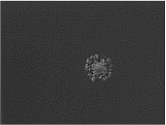
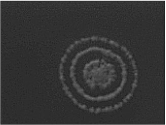
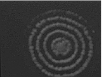
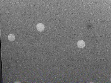
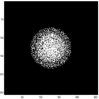
A detailed description of aerial mycelium formation is beyond the scope of this comment (see [12]); however, it is important to stress that differentiation relies upon intercellular signaling. For example, in S. griseus a substance termed A-factor is involved in triggering aerial mycelium production [26]. Extracellular diffusion of the inducers of aerial mycelium guarantees a global coupling of the cell population. Aerial mycelium growth is supported by other cells that undergo lysis, thus providing nutrients for the aerial mycelium [40]. At this stage, Streptomyces produce antibiotics to prevent other bacteria to have access to this source of nutrients [11]. Usually aerial mycelium grows in a compact circular pattern but different patterns can develop as well. S. rutgersensis grown on minimal media forms concentric rings of aerial mycelium (see Fig. 7) [7]. Similar but order of magnitude larger rings are also observed in Neurospora crassa [17]. Mathematical models have been produced to explain the generation of these patterns: they will be presented in Section 4.
3 Modeling fungal growth
3.1 Macroscopic models
One of the first empirical macroscopic model was proposed by Trinci [56] to explain mycelial growth in its early stage. Let us call the number of branches and the total mycelial length at time . During the early stages of growth, the temporal evolution of these quantities is well reproduced by the following equations:
| (2) | |||||
| (3) |
with and constants and the growth rate. From Eqs. (2-3), we immediately get:
that is, the ratio (called hyphal growth unit) is constant over time.
These phenomenological relations are in agreement with growth patterns of several filamentous fungi (see for example [10]) and bacteria (see for example, Figure 2 in [2]).
During the initial phase of growth, after a short lag period, also the radius of the colony grows exponentially. (see Ref. [29] for a model of this phase of growth). Then, as described in Section 2, linear radial growth sets up while hyphae within the interior of the colony do not grow further [56]. During this phase, there is a quadratic increase in biomass. In fact,
(with the time from the beginning of linear growth phase) and assuming biomass uniformly distributed throughout the colony, (i.e. the total biomass proportional to the colony surface).
That is, total biomass grows quadratically in time.
This simple model found many experimental confirmations, but it does not supply any information on the local distribution of the biomass, and in particular on the different behaviors between center and border of the colony. Using a reaction-diffusion model Ferret et al. [22] have predicted the evolution of the biomass and tips density under different growing conditions. In presence of unlimited nutrients and no spatial constraints, Eqs. (3-2) are recovered, and the biomass increases exponentially with time. However, as mentioned above, growth stops in the center of the colony: in the model the same happens when the biomass density reaches a maximum value, , while at the colony border the growth continues freely. These differential growing velocities are modeled by introducing a collision probability as a function of local biomass density . In the the central part of the colony, the local density quickly reaches its maximum value, , there is no free space available for growth, collisions are unavoidable and the free space (proportional to ) is zero: the mycelium cannot grow further. On the other hand, at the colony border, no collisions happen, , and the hyphae can grow freely outward. Assuming a linear model for (see Fig. 2 of the cited paper), that fulfills these two limiting cases, the model is able to predict the temporal behavior of total biomass and the colony border evolution (i.e. internal and external radius, defined as the radius of the not-growing core, and the radius of the whole colony, respectively). The model has been validated by comparing its predictions to the behaviors of two different fungi: Gibberella fujikuroi and Aspergillus oryzae.
These macroscopic models are suitable for modeling the growth of the whole colony, but they are not able to grab all the complex fine structure of mycelial growth. In fact, in addiction to tip extension and branching, filamentous growth is characterized by septation and internal transport mechanisms (translocation): these microscopic features will be taken into account in the next class of models.
3.2 Microscopic models
Septation and branching
Once the nuclear material is replicated, mycelial microorganisms divide a hyphal compartment in two parts by septation, Fig. 1. Branching usually occurs after septation, with a fixed time delay (see for example the case of Geotrichum candidum in [23]). A model that includes branching and septation has been introduced by Yang et al. [60] The model includes a deterministic part, that mirrors tip growth and septation, and a stochastic part to keep track of new branch initiations. Septation is triggered when the amount of nuclear material has been doubled and segregated. Due to the random behavior of branching, new branches are supposed to be generated at random around their respective septa according to a normally distributed probability with a fixed variance . In the limiting case, , new branches grow just behind septa. Tip growth and new branching directions are calculated according to a random model described in [61]. In short, experimental observations in 2-D colonies reveal that tip growth angles and branching outgrowth angles are normally distributed around zero. In the model, that represents growth in a 3-D environment. The orientation of the primary hyphal segment with respect to the straight direction is obtained sampling the experimental distribution. In the plane perpendicular to the growth axis, the direction is fixed by an additional random angle assumed to be uniformly distributed between . A simile procedure is used for determining the branching angle in the plane perpendicular to the hyphal growth axis (see Fig. 4).
Simulation results are in good agreement with experimental data for the fungus Geotrichum candidum (taken from Ref. [23]) and for the filamentous bacterium Streptomyces tendae. Both germ tube growth and temporal behavior of the number of tips and septa can be reproduced. In addition, the morphology of the growing mycelium appears similar to that observed in experiments (see Fig. 10 in [23]).
Translocation
Another peculiar aspect of the internal kinetics of a growing mycelium is the capability to distribute nutrients through the mycelial network (translocation), both passively (due to diffusion through the hyphae) and actively (due to active and hence energy-dependent metabolism) [45, 46]. This way, fungi can absorbe nutrients in some parts of the mycelium and transport them through the network, particularly where food is scarce. This mechanism can play a crucial role for colonies growing in highly heterogeneous environments, such as most of the natural ones.
Among those that have been recently proposed [15, 16], we will describe here a recent model introduced by Boswell et al. [9] to study the fungus Rhizocotonia soloni. The model includes five components (densities): active and inactive hyphal densities, tip density, internal and external substrate concentrations. Inactive mycelia, usually the center of the colony, do not contribute to translocation, while translocation occurs in active hyphae. Internal passive translocation is modeled as a diffusion-like term for internal substrate, , that depends on mycelial concentration , i.e. in presence of a dense mycelial network the diffusion process is much faster than in a sparse one. Active translocation is modeled by assuming that active mechanisms are due to the continuous substrate demand by growing tips, i.e. the flux follows the gradient of tips density . So, active translocation moves substrate from low-density tip areas to high density ones (so called tip-driven diffusion) through the mycelial network (i.e. it also depends on ). In mathematical terms:
| (4) |
The parameters are obtained from experiments. Using this model the authors explore the interplay between active and passive translocation in a growing colony in two kinds of growth media, homogeneous (nutrients are homogeneously spread on the dish) 222The effects of different, but homogeneous, growth media have also been studied in Aspergillus Oryzae colonies [35, 38, 39], and will be discussed further in the case of Streptomyces colonies in Section 4. and heterogeneous (substrates are patchy distributed). Simulations suggest that diffusive translocation is used for random exploration while tip-driven translocation is used for optimizing the distribution of available resources. This view is coherent with the idea that optimal search combines a random process with some more complex, but usually more demanding, strategies of resource exploitation.
Single hypha models
Going down in the length scale, we find single hypha models. The average kinetics implemented in the previous models do not describe properly the evolution of individual hyphae. Indeed, due to the complexity of the hyphal structure and the different growth rates of individuals branches within an hypha (see [56]), stochasticity has to be taken into account.
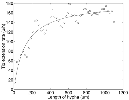
Christiansen et al. [14], studied the growth of a single hyphal element of Aspergillus oryzae, combining image analysis with a model that makes use of Monte Carlo simulations. They consider the growth starting from a single spore; at each time step ( ), the probability of branching is: . The branching rate is computed from a phenomenological model of tip kinetics 333 A detailed model of the elongation process of the growing tip can be found in Ref. [51]. , and from observed distribution of the number of new branches as function of hyphal length . From experimental observations, tip velocity as function of hyphal length can be fitted by the following function (see Fig. 3):
| (5) |
where is the maximum tip extension rate, is an empirical saturation constant, that depends on environmental conditions (glucose concentrations), and varies among different types of branches ( is usually larger for the germtube than for secondary hyphae).
In the model is calculated from:
with a normally distributed random number, and the parameters and fitted from experimental values of . Eq. (5) is a good approximation of the average behavior of tip growth rate ; but detailed experimental measurements show that a decrease in tip extension rate occurs in correspondence to the formation of new apical branches (see Fig. 3 adapted from Fig. 2. in Ref. [14], but also the discussion in Ref. [28] where no correlations between tip extension rates and branching are found). Following Ref. [14], we will neglect these fluctuations in the current analysis. After the hyphal length has reached a certain threshold (that depends on the individual hypha, and differs greatly between primary, germtube, and secondary hyphae), the number of new branches formed on a hyphae grows linearly as function of length:
| (6) |
where is a branching constant (actually, it assumes two different values for the germtube and for secondary hyphae). From Eqs. (5) and (6), the branching intensity (i.e. the number of branches per time unity) can be derived:
| (7) |
Using these values of we can simulate the growth of a single hyphal element using a standard Monte Carlo technique. In a standard simulation, a single spore has been considered, the tip grows with a rate and at each time step a new branch is formed with a probability . Although hyphae grow only at the tips, with a saturating velocity, the total length of mycelium increases exponentially due to the exponential increase in the number of branches (after the minimal length has been reached). The simulated growth kinetics agree with the observed experimental patterns for the total length and the number of tips for a single hypha.
A detailed study of single hypha growth has been presented as well by Hutchinson et al. [28]. They record the temporal evolution of primary hyphae and first and second order branches in six colonies a few hours after germination. Using the experimentally observed distributions, they proposed a model of mycelial growth: hyphae perform a straight-line growth for a length sampled from an experimental fitted distribution. At this point, branching can occur, and the direction for the new branch is obtained by sampling from experimental distributions. This model is capable of reproducing the morphology of the early stages of Mucor hiemalis growth, and represents one of the first model where a single hypha dynamics (microscopic) is used for generating macroscopic growth patterns (morphology of the mycelia).
From the single hypha level, we could go down one level in the analysis to the genetic and molecular basis of filamentous growth (i.e. to the genetic networks underlying these processes). However, although progresses have been recently made in elucidating the molecular basis of mycelial growth and development (for example, see [13] for a review on Streptomyces), no mathematical link between physiology of cell behavior and molecular interactions have been successfully produced so far.
3.3 Cellular Automata
Cellular Automata (CA) are fully discrete dynamical systems, i.e. their dynamical variables are defined at the nodes of a lattice (spatial discretization), with values taken from a finite set (state discretization) and evolve in discrete time steps (temporal discretization). They were introduced more than half century ago by John Von Neumann’s [59]. As the choice of the name suggests, the original idea is that the complex behavior characterizing many biological systems should originate as a collective effect from simple local interaction rules (cell-interactions). Since then, CA have been applied to a vast range of biological and physical problems (see for example the reviews [18, 8, 3] and the “classical” works [59, 24, 62]) 444Another class of fully discrete models that emphasize local interactions as opposed to global ones are Lindenmayer Systems (L-systems) [34, 50]. L-systems are basically a set of string re-writing rules applied recursively. They are closely related to cellular automata, but usually they lack an explicit spatial extension. These rules can mirror growth process, and they are generally used for generation of fractals and modeling of plants. An applications to fungal growth, Mucor hiemalis, was proposed by Soddel et al. [54]. We will not discuss these models here, but we refer the readers to the cited paper and references therein.
A CA model for fungal growth has been proposed by Lazlo & Silman [33]. They consider a two dimensional square grid (that mirrors the dish) where each site has neighbor sites (Moore neighborhood). Each site can be either occupied by a mycelium or empty. A probabilistic rule is set for updating the status of each site of the lattice simultaneously. In its simplest version the rule is the following: each empty site will become occupied with a probability dependent on the number of neighbor occupied sites :
-
•
(one neighbor site occupied)
-
•
-
•
-
•
(overcrowded)
Even using only this simple rule, interesting patters are generated, and the temporal behaviors of biomass and radius of the colony are well reproduced. This basic model has been extended considering three different states of differentiation: vegetative mycelium, differentiating mycelia (an intermediary state) and spore formation, and setting the corresponding probability of transition according to local occupancy rules. Still keeping the model simple (one single byte is enough to represent the state of a site) the automaton is able to produce patterns for spore and mycelia, reminiscent of the experimental growth patterns.
One of the advantages of CA, is that they can be used in testing hypotheses about the interactions among cells: some schematization (rules) of microscopic dynamics can be easily implemented as cellular automata; then we can observe the macroscopic patterns generated and compare it with experimental patterns. This class of models is particularly useful when the macroscopic patterns observed do not depend on the fine details of microscopic interactions, but they emerge as a collective behavior of many cells (the site of the automaton). In this framework, cellular automata, despite their simplicity, turn out to be a valuable tools for the studies of emergent cooperative effects.
4 Aerial mycelium pattern formation
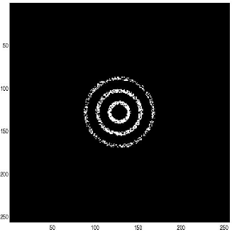
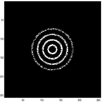
As described in Section 2.2, aerial mycelium can form concentric ring patterns in S. rutgersensis colonies (see Fig. 7). Ring patterns are found also in other bacterial and fungi colonies [1]. Many models can produce such patterns [37]; in the case of bacterial colonies, it was suggested that the interplay of front propagation and Turing instability leads to concentric rings and spot patterns [58]. In the more specific case of filamentous bacteria, a different approach based on competition for resources has been proposed by Bezzi et al. [7]. The model is based on the idea that growing hyphae have the biological objective to find nutrients to give rise to spores (aerial mycelia). As a result, we expect a strong competition to arise on minimal media for the energetic resources between neighbor substrate mycelia, whereas in maximal media, where there are sufficient nutrients, the competition is weaker.
If cells are connected mainly along the radial direction (hyphae start growing mainly radially from the initial spore), then competition will be stronger along this direction than along the tangential one. In other words, in the growing edge of the colony, the competition is not isotropic but, following the vegetative mycelium morphology, it will be stronger among cells belonging to neighboring circumferences (radial direction) than among cells belonging to the same (tangential direction).
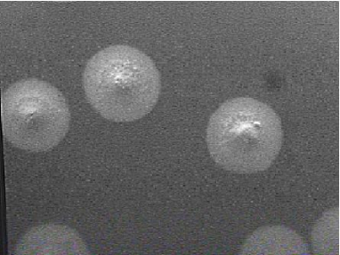
In presence of high concentrations of nutrients, the competition is rather weak and the final distribution of aerial mycelium has the shape of a gaussian-like distribution centered on the initial spore (similar to the one shown in Fig. 5, right). If the colony grows in a nutrient-limited environment, hyphae that are neighbor along the radial direction compete strongly for nutrients, aerial mycelia development is inhibited, and concentric ring patterns appear (see Fig. 5, left). Despite its simplicity (one differential equation) the model is able to capture many peculiar features of Streptomyces rutgersensis growth. However, since the model does not include explicitly the growth of vegetative mycelium, some important aspects, like the “interference” patterns among neighboring colonies (Fig. 6), are not reproduced. Clearly, a more detailed view is necessary; a coupled maps model including vegetative mycelia and tip growth is under development [4] whose results are shortly summarized in Fig. 8.
As in the case of vegetative mycelia growth, different approaches are required to describe the microscopic details, in which fluctuations and stochasticity play a major role. Monte Carlo simulations are a possible choice, an alternative strategy is using cellular automata, in which simple microscopic local rules (deterministic or probabilistic) can produce complex patterns.
Cellular automata have also been used for studying pattern formation in bacteria, in particular for Streptomyces rutgersensis [43]. In this two-dimensional model the building block is the individual cell. A hypha is composed of several cells, which do not move in space. Cells absorb food (energy) according to its local concentration, convert and store it internally and use the stored energy for metabolism, growth and reproduction. If the amount of stored energy lowers below a given threshold, the cells start to starve. The model assumes that this starvation phase triggers the formation of aerial mycelium. When a cell reaches a given dimension and has accumulated sufficient energy, it reproduces by binary division or gemmation. Despite many approximations introduced, the resulting patterns of vegetative mycelium are very reminiscent of experimental patterns: at the beginning of the life of the colony (i.e., for the first generations) the hyphae are distributed randomly in space, but then, when food becomes scarce, they are selected to align with food gradient.
Similar concentric rings patterns are also observed in fungi, among them: Neurospora crassa. In Ref. [17], Deutsch et al. present a cellular automaton for modeling Neurospora crassa growth patterns. The two-dimensional patterns are reproduced using a one-dimensional cellular automaton; but at each time step the one dimensional grid (called ”base space” for the automaton) represents a circle of increasing radius (so that the radial structure can be preserved). The model is capable of reproducing the morphologies of a single growing colony of Neurospora Crassa, but, due to the peculiar implementation of the radial symmetry, it is not applicable to the studies of interference patterns (similar to S. rutgersensis pattern shown in Fig. 6).
5 Conclusions
In filamentous organisms (e.g. fungi and actinomycetes), mother and daughter cells never detach: although they are separated by a septum, they remain physically connected. A filament of cells separated by septa is called an hypha, which is the building block of filamentous organisms. Hyphae are usually branched, and the complex of hyphae forming a fungal or actynomicete colony is called mycelium.
The mycelial organization has deep consequences on growth of filamentous organisms, because active growth is localized only at hyphal tips. Colony growth rate is not simply dictated by hyphal tips growth, but also by the frequency of formation of new tips – branching frequency. Spatial organization and temporal growth are intrinsically connected, and the morphology of a colony is a key element to be taken into account if we want to understand the growth of filamentous organisms.
Growth and mycelial pattern formation involve events occurring at very different length scales, ranging from a colony scale (macroscopic) to a single-cell scale (microscopic). For example, experimentally spatial organization is described by quantities like the angle between an hypha and a related branch, the distance between two consecutive branches, the distance between two consecutive septa, the time course of the total amount of branches, the frequency of branches along an hypha, and so on. Evidently, these quantities are measured at the microscopic scale, but the resulting mycelial morphology is observed at the colony scale. On the other hand, the temporal dynamics of colony growth (e.g. colony radius growth rate) is a macroscopic phenomenon, which is originated by tips growth, a single-cell event.
Mathematical models are needed to describe such complex phenomena that involve spatial and temporal dynamics, microscopic and macroscopic experimental observations. Indeed, several models have been developed in the last thirty years to reproduce growth kinetics and mycelial patterns in filamentous organisms. Different biological questions generate different type of modeling, that concur to draw a complete picture of filamentous organisms growth and pattern formation. Such models differ for the different organisms they apply to, as well as for the different scales of the experimental observations they simulate.
In this comment we review a selection of models, organized according to the scale of the experimental observation from which the model originates. We start with macroscopic models that reproduce colony growth rate experiments, then, moving down in length scale, to some reaction diffusion models, that include more detailed phenomena like septation and translocation, and finally we terminate with single hypha models, that concern single hypha growth and branching. We also review cellular automata models that implement microscopic interaction but at the same time they are able to reveal macroscopic patterns.
The last step of the length scale should be the molecular level, but, to our knowledge, there are no models that use the genetic data produced in the last twenty years to address growth kinetics and pattern formation in filamentous organisms. Although genetic analysis revealed some of the genes involved differentiation and the way they are connected (i.e. the underlying gene networks), models based on these data are still missing. Such models have been produced for several basic processes like the cell cycle, circadian rhythm and signal transduction pathways (for a review see [27]). By using similar mathematical analysis it should be possible to develop similar models to understand and modify in a predictable way filamentous organisms.
Acknowledgements
We thank F. Bagnoli, A. Mengoni and G. Mersi for many helpful discussions. We also thank M. Buiatti and O. Coenen for careful reading of the manuscript and suggesting many improvements. We thank P. Goatin for help in drawing the figures. MB was supported by IST-2001-35271 grant (MB) and AC by DARPA-Biocomputation Program (AFRL #F30602-02-0572). Correspondence should be addressed to Michele Bezzi, Sony Computer Science Laboratory, 6 rue Amyot - 75005 Paris France. E-mail: michele@csl.sony.fr
References
- [1] Adler, J. 1966. Chemotaxis in bacteria. Science 153:708-716.
- [2] Allan, E.J. and Prosser, J. I. 1983. Mycelial growth and branching of Streptomyces coelicolor A3(2) on solid medium, Journ. of General Microbiology, 129:2029-2036.
- [3] Bagnoli, F. 1998 .Cellular automata,Dynamical Modelling. In Biotechnologies, F. Bagnoli and S. Ruffo, editors, World Scientific, Singapore.
- [4] Bagnoli, F., Bezzi, M., Ciliberto, A and Mengoni, A., in preparation.
- [5] Ben-Jacob, E. , Cohen, I., Shochet, O., Aranson, I., Levine, H. and Tsimring L. 1995. Complex bacterial patterns. Nature, 373:566-7.
- [6] Ben-Jacob, E., Shochet, O., Tenenbaum, A., Cohen, I., Czirok, I. and Vicsek, T. 1994. Generic modelling of cooperative growth patterns in bacterial colonies. Nature, 368:46-9
- [7] Bezzi, M., Ciliberto, A. and Mengoni, A. 1999. Pattern formation by competition: a biological example. J. Biol. Phys., 25:279-288
- [8] Bezzi, M. 2001. Modeling Evolution and Immune System by cellular automata, Rivista del Nuovo Cimento 24:1-51.
- [9] Boswell G. P., Jacobs H., Davidson, F. A., Gadd, G. M and Ritz, K., 2002. Functional consequences of nutrient translocation in mycelial fungi, J. Theor. Biol., 217:459-77.
- [10] Bull, A. T., and Trinci A. J. P., 1977. The physiology and metabolic control of fungal growth, Adv. in Microbial Physiology, 50:1-84
- [11] Chater, K. F. 1992. Streptomyces coelicolor: a mycelial spore-bearing prokaryotes. In Molecular genetics of development, V.A. Russo, S. Brody, D. Cove and S. Ottolenghi, eds. Blackwell, Oxford.
- [12] Chater, K.F. and Losick, R. 1997. Mycelial life style of Streptomyces coelicolor A3(2) and its relatives. In Bacteria as multicellular organisms, J. A. Shapiro and M. Dworkin eds, Oxford University Press.
- [13] Chater, K. F. 1998. Taking a genetic scalpel to the Streptomyces colony, Microbiology, 144:1465-1478.
- [14] Christiansen, T., Spohr, A. and Nielsen, J., 1998. On-line Study of Growth Kinetics of Single Hyphae of Aspergillus oryzae in a Flow-Through Cell, Biotechnol. Bioengr., 63:147-153.
- [15] Davidson,F.A.1998. Modelling the qualitative response of fungal mycelia to heterogeneous environments. J. theor. Biol., 195:281-92.
- [16] Davidson,F.A. and Olsson,S.2000. Translocation induced outgrowth of fungi in nutrient-free environments.J. theor. Biol., 205:73-84.
- [17] Deutsch, A., Dress, A. and Rensing, L. 1993. Formation of morphological differentiation patterns in the ascomycete Neurospora crassa, Mech. Dev., 44:17-31.
- [18] Droz, M. and Chopard, B. 1998. Cellular Automata Modeling of Physical Systems, Cambridge University Press.
- [19] Edelstein, L. 1982. The propagation of fungal colonies: a model for tissue growth. J. theor. Biol., 98:679 701.
- [20] Edelstein, L. and Segel, L. A. 1983. Growth and metabolism in mycelial fungi. J. theor. Biol., 104:187 210.
- [21] Edelstein, L.-Keshet, L. and Ermentrout, B. 1989. Models for branching networks in two dimensions. SIAM J. Appl. Math., 49:1136 1157.
- [22] Ferret E., Simeon J. H., Molin, P. Jorquera, H., Acuna, G and Giral, R. 1999. Macroscopic growth of filamentous fungi on solid substrate explained by a microscopic approach, Biotechnol. Bioengr, 65:512-522.
- [23] Fiddy, C. and Trinci A.P.J. 1976. Nuclei septation, branching and growth of Geotrichum candidum J. Gen. Microbiol. 97:185-192.
- [24] Gardner, M. 1971. On cellular automata, self-reproduction, the Garden of Eden and the game ”life”. Sci. Am., 224, 112-117.
- [25] Gooday, G. W. 1971. An autoradiographic study of hyphal growth of some fungi, J. Gen. Microbiol., 67:125-131.
- [26] Hara, O. and Beppu, T. 1982. Mutants blocked in streptomycin production in Streptomyces griseus: the role of A-factor. J. antibiot., 35:349-358.
- [27] Hasty, J., J., McMillen, D., Farren, I. and Collins, J.J. 2001. Computational studies of gene regulatory networks: in numero molecular biology. Nat. Rev. Gen., 2:268-79.
- [28] Hutchinson, S. A., Sharma, P., Clarke, K.R. and Macdonald, I. 1980. Control of hyphal orientation in colonies of Mucor hiemalis, Trans. Br. Mycol. Soc., 75:177-191.
- [29] V. Kotov, and S. V. Reshetnikov A Stochastic model for early mycelial growth, Mycol. Res., 94:577-586, (1990).
- [30] Kretschmer, S. 1981. Dependence of the mycelial growth pattern on invidually regulated cell cycle in Streptomyces granaticolor, Zeit. Allg. Mikorbiol., 22:335-42.
- [31] Kretschmer, S. 1988. Stepwise increase of elongation rate in individual hypahe of Streptomyces granaticolor during outgrowth, J. Basic Microbiol., 1-2:35-43.
- [32] Kretschmer, S. 1991. Location of branches within the apical hyphal region of Streptomyces granaticolor mycelia, J. Basic Microbiol., 32:35-42.
- [33] Lazlo, A. J. and Silman, R. W. 1993. Cellular Automata simulations of fungal growth on solid substrates, Biotech. Adv. , 11:621-633.
- [34] Lindenmayer, A. 1968. Mathematical models for cellular interaction in development I. Filaments with one-sided inputs. Journal of Theoretical Biology, 18:280-289.
- [35] Lopez, J. M., and Jensen, H. J., Generic model of morphological changes in growing colonies of fungi, Phyisical Review E, 65, 021903.
- [36] Matsuyama, M. and Matsushita, M. 1993. Fractal morphogenesis by a bacterial cell population. Crit. Rev. Microb. 19:117-35
- [37] Matsushita, M., Wakita, J., Itoh, H.,R fols, I., Matsuyama, T., Sakaguchi, H. and Mimura M. 1998. Interface growth and pattern formation in bacterial colonies. Physica A, 249:517-524.
- [38] Matsuura S. and Miyazima S. 1992. Self-affine fractal growth front of Aspergillus oryzae. Physica A, 191:30-40.
- [39] Matsuura S. and Miyazima S. 1993, Colony of the fungus aspergillus oryzae and self-affine fractal geometry of growth fronts. Fractals, 1:11-19.
- [40] Mendez, C., Brana, A. F., Manzanal, M.B. and Hardisson, C. 1985. Role of substrate mycelium in colony development in Streptomyces, Can. J. Microbiol., 35:446-450.
- [41] McCormick, J., Su, E. P., Drisks, A. and Losick, R. 1997. Growth and viability of Streptomyces coelicolor mutant for the cell division gene fstZ. Mol. Microbiol., 14:243-254.
- [42] McIntyre, M., Muller, C., Dynesen, J. and Nielsen, J. 2001. Metabolic engineering of the morphology of aspergillus, Adv. Biochem. Engin. Biotech., 73:103-128.
- [43] Mersi, G. in preparation and also: Bagnoli, G., Bezzi, M., Ciliberto, A. and Mersi, G. A cellular automata model for pattern formation in bacterial colony: he case of vegetative mycelial growth in Streptomyces INFM meeting, book of abstracts III-66, 242, Bari 24-28/06/2002.
- [44] Miguelez, E.M., Martin, M.C., Manzanal M.B. and Hardisson C., 1992. Growth and morphogenesis in Streptomyces griseus, FEMS Microbiology Letters, 100:351-360.
- [45] Olsson, S. and Gray, S. N. 1998. Patterns and dynamics of 32 P-phosphate and labelled 2-aminoisobutyric acid (14C-AIB) translocation in intact basidiomycete mycelia. FEMS Microbiol. Ecol., 26:109 120.
- [46] Persson, C., Olsson, S. and Jansson, H.-B. 2000. Growth of Arthrobotrys superba from a birch wood food base into soil determined by radioactive tracing. FEMS Microbiol. Ecol., 31:47 51.
- [47] Prosser, J. I. and Trinci A. P. J. A model for hyphal growth and branching, J. Gen. Microbiol., 111:153-164.
- [48] Prosser, J. I., Tough, A.J. 1991. Growth mechanism and Growth Kinetics of filamentous Microorgisms. Biotechnology, 10:253-274.
- [49] Prosser, J. I. 1993. Growth kinetics of mycelial colonies and aggregates of ascomycetes, Mycol. Res., 97:516-28.
- [50] Prusinkiewicz, P. and Hanan, J. 1989. Lindenmayer systems, fractals, and plants. Lecture Notes in Biomathematics Springer-Verlag:Berlin
- [51] Regalado, C. M. and Sleeman, B. D., 1999. Aggregation and collapse in a mechanical model of fungal tip growth, J. Math. biol., 39:109-138.
- [52] Robson, G.D., Bell, S.D., Kuhn, P.J., Trinci, A.P. 1987. Glucose and penicillin concentrations in agar medium below fungal colonies, J. Gen. Microbiol., 133,:361-367.
- [53] Shapiro, J. A. and Dworkin, M. 1997. Bacteria as multicellular organisms, Oxford University Press.
- [54] Soddell, F., Seviour, R. and Soddell, J. 1994. Using Lindenmayer Systems to Investigate How Filamentous Fungi May Produce Round Colonies. In Complex Systems: Mechanism of Adaptation, 61-68. R. Stonier and X. Yu Eds. Amsterdam, IOS Press
- [55] Trinci A.P.J. 1971. Influence of the width of the peripheral growth zone on the radial growth rate of fungal colonies on solid media. J. Gen. Microbiol., 67:325-344.
- [56] Trinci A.P.J. 1974. A study of the kinetics of hyphal extension and branch initiation of fungal mycelia. J. Gen. Microbiol., 81:225-36.
- [57] Trinci, A. P. J., Wiebe, M. G. and Robson G. D. 1994, The mycelium as an integrated entity, In The Mycota I. Growth, Differentiation and sexuality, Wessels and Meinhardt Eds, Springer-Verlag, Berlin.
- [58] Tsimring L. et al. 1995. Aggregation Patterns in Stressed Bacteria. Phys. Rev. Lett., 75:1859-1862.
- [59] Von Neumann, J. 1966. Theory of Self-Reproducing Automata, University of Illinois Lectures on the Theory and Organization of Complicated Automata, Ed. A. W. Burkus, University of Illinois Press, Urbana.
- [60] Yang, H., R. King, U. Reichl, and E.D. Gilles, 1992. Mathematical model for apical growth, septation, and branching of mycelial organisms, Biotechnol. Bioengr., 39:49-58.
- [61] Yang, H., U. Reichl, R. King, and E.D. Gilles, 1992. Measurement and simulation of the morphological development of filamentous microorganisms, Biotechnol. Bioengr., 39:44-48.
- [62] Wolfram, S. 1986. Theory and Applications of Cellular Automata World Scientfic, Singapore.
Figures