Superstability of Surface Nanobubbles
Abstract
Shock wave induced cavitation experiments and atomic force microscopy measurements of flat polyamide and hydrophobized silicon surfaces immersed in water are performed. It is shown that surface nanobubbles, present on these surfaces, do not act as nucleation sites for cavitation bubbles, in contrast to the expectation. This implies that surface nanobubbles are not just stable under ambient conditions but also under enormous reduction of the liquid pressure down to MPa. We denote this feature as superstability.
pacs:
47.55.dp, 68.37.Ps, 68.08.-pIn recent years, numerous experiments revealed the existence of nanoscopic soft domains at the liquid-solid interface, see tyrrellPRL2001 ; simonsen ; holmberga ; agrawal ; zhangcraig ; lijuanzhang ; zhang06 ; jiong ; switkes ; steitz and references therein. Most experiments employ atomic force microscopy (AFM) tyrrellPRL2001 ; simonsen ; holmberga ; agrawal ; zhangcraig ; lijuanzhang ; zhang06 ; jiong , but other techniques switkes ; steitz have been used as well. The most consistent interpretation of these experiments is that the soft domains, which resemble spherical caps with heights of the order of and diameters of the order of , are so-called surface nanobubbles, i.e., nanoscale gas bubbles located at the liquid-solid interface. This claim is, for instance, supported by the fact that nanobubbles can be merged by the tip of an AFM to form a larger bubble simonsen , or by the fact that they disappear upon degassing of the liquid lijuanzhang ; zhang06 ; switkes , or by the gas concentration dependence of their density jiong .
Surface nanobubbles are puzzling objects. First, they should not exist: according to the experimental data these bubbles have a radius of curvature of the order of , and therefore (due to a large Laplace pressure inside of the bubbles) they should dissolve on timescales far below a second epstein ; ljunggren . In marked contrast the experiments show that nanobubbles are stable for hours. Second, they are potential candidates to explain various phenomena associated with the liquid-solid interface, such as liquid slippage at walls lb_pre ; lauga ; neto or the anomalous attraction of hydrophobic surfaces tyrrellPRL2001 in water. In addition, heterogeneous cavitation usually starts from gaseous nuclei at solid surfaces (see atchley and references therein), and surface nanobubbles are suggested as potential inception sites zhang06 ; bremond05 ; holmbergb . However, apart from convincing experimental evidence for the existence and stability of nanobubbles, still little is known. For instance, why are they apparently stable or how do they react to environmental changes?
In this Letter it is shown that surface nanobubbles, contrary to the expectation, do not act as nucleation sites for shock wave induced cavitation on surfaces, where a large tensile stress is created in the water. Hence, yet another puzzle is added to the nanobubble paradox: They are not only stable under ambient conditions but also under enormous reduction of the water pressure down to MPa. We denote this phenomenon as superstability.
To demonstrate the superstability of nanobubbles we combine cavitation experiments and AFM measurements. More precisely, cavitation experiments (similar to bremond05 ; bremond06 ; bremondPOF06 ) with different hydrophobic substrates submerged in water are performed: a shock wave generates a large tensile stress () in the water which leads to cavitation of bubbles at the substrates. The size of the cavitation bubbles is of the order of several hundred . Thus, though the bubbles originate from smaller nuclei, they can be visualized by optical means. In addition, AFM-measurements of the same substrates in water at ambient conditions are performed to proof and quantify the existence of stable nanobubbles on these substrates. Combining the cavitation and AFM experiments allows to study the relation between cavitation activity and nanobubbles. An analogous strategy has been used previously bremond06 to perfectly correlate the appearance of surface bubbles in cavitation experiments to the existence of gas-filled microcavities (i.e., microbubbles) of diameter of which had been etched into the surface. Is there a similar connection between cavitation on smooth unstructured surfaces and surface nanobubbles?
In other words: to what extent must the liquid pressure be reduced to grow a nanoscale bubble to a visible size (i.e., above microns)? A first estimate is obtained from the criterion that unstable growth of a bubble occurs when falls below the critical pressure , with the ambient static pressure and the Blake threshold brennen95 ; leighton94 . The hemispherical dynamics of a surface bubble under rapid decrease of the liquid pressure is close to that of a free bubble with the same radius of curvature bremondPOF06 . Therefore, though surface nanobubbles are spherical caps rather than free spherical bubbles, one may obtain a reasonable estimate by the assumption of a spherical bubble. Assuming a nanoscale bubble with radius and one arrives at which is exceeded in the experiments by more than an order of magnitude, see Fig. 1. Moreover, we solved the Rayleigh-Plesset equation brennen95 ; leighton94 (which describes the dynamics of a spherical bubble under variations of the liquid pressure) numerically for a gas bubble with the measured liquid pressure reduction as driving force. This calculation yields that bubbles down to a radius of curvature should grow to visible bubbles during the experiments. Hence, theoretically it should be no problem to nucleate a surface nanobubble to visible size, but is this reflected in the experiments?
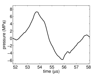
The setup for the cavitation experiments is similar to that used in bremond05 ; bremond06 ; bremondPOF06 . A shock wave generator generates a pressure signal in the water, consisting of a high pressure front followed by a large tensile stress, see Fig. 1. The substrate of interest is processed and handled inside a filtered flow bench and placed inside of a sterile flask filled with pure water (Milli-Q Synthesis A10, Millipore), ensuring cleanroom conditions throughout the experiment. The flask is placed inside the water tank such that the shock wave is focussed onto the substrate. The pressure signal at this position is recorded with a fibre optic probe hydrophone. The cavitation event is photographed by a CCD camera through a long-distance microscope. The major difference between the present setup and that of bremond05 ; bremond06 ; bremondPOF06 is the maintenance of cleanroom conditions by use of the protective flask. Compared to Fig. 2 of Ref. bremond05 less than 1 cavitation activity on the surface is observed when cleanroom conditions are preserved, indicating that contaminations play a dominant role for cavitation experiments under ambient lab conditions.
The AFM data are acquired on a VEECO/Digital Instruments (DI) multimode AFM equipped with a NanoScope IIIa controller (DI, Santa Barbara, CA) in tapping mode in water using a DI liquid cell and V-shaped Si3N4 cantilevers (Nanoprobes, DI). The data shown for case are obtained after mounting the sample into the AFM while keeping the sample surface covered by water at all times, as described previously morigaki .
Corresponding to different kinds of substrates and/or different procedures of substrate preparation, we present results associated with four different kinds of probes, labeled . Probes and use smooth polyamide surfaces as solid substrate. Polyamide is heated and molded between silicon and atomically smooth mica. The mica is removed when the polyamide is cooled down to room temperature, leaving a relatively smooth polyamide surface with a root mean square (rms) roughness of (measured by AFM on ) and a static contact angle of . Besides large smooth areas of many the production process also creates several microscopic cracks in the surface. In case these polyamide surfaces are used in the experiments without further treatment. In case the substrate is first covered by ethanol which is then flushed away by water. This ethanol-water exchange has been reported to induce the formation of surface nanobubbles, see zhangcraig ; jiong and references therein. Besides the explanation suggested in zhangcraig we note that also the exothermic mixing exo of ethanol and water might induce the formation of nanobubbles, since a temperature increase favors the formation of nanobubbles jiong . In the cavitation experiments a drop of ethanol is placed on the substrate such that it is completely covered by ethanol before it is submerged in water. Then the substrate is moved inside the protective flask for a minute to replace the miscible ethanol by water comment . In the AFM experiment for case a liquid cell is used.
Probes and use pieces of smooth hydrophobized silicon as the substrate. A Si(100) wafer is diced into chips () which are immersed for 15 minutes in a (5:1) Piranha cleaning mixture. Hereafter, the chips are hydrophobized by chemical vapor deposition of 1H,1H,2H,2H-perfluorodecyldimethylchlorosilane (PFDCS) bremond05 , yielding a rms value of (measured by AFM on ), a coating thickness (measured by ellipsometry), and an advancing contact angle . Note that the silane-film is not able to move; it is a stable self-assembled monolayer covalently bonded to the underlying substrate. Before immersion in water the probes are rinsed with ethanol and blown dry with argon gas bremond05 . Case additionally applies the ex situ ethanol-water exchange as described above.
In each of the cases substrates of the respective type are produced in an identical manner. One substrate is used in the cavitation experiments and one in the AFM measurements. Note that we checked that the observed cavitation activity and nanobubble density were reproducible among substrates of the same kind. Furthermore, in case the same substrate is used in both experiments. After the cavitation experiment (exposure to a single shock wave) the substrate is transported in water to the AFM, where it is mounted without exposure to air, whereafter the water-solid interface is imaged as it appears after the cavitation experiments.
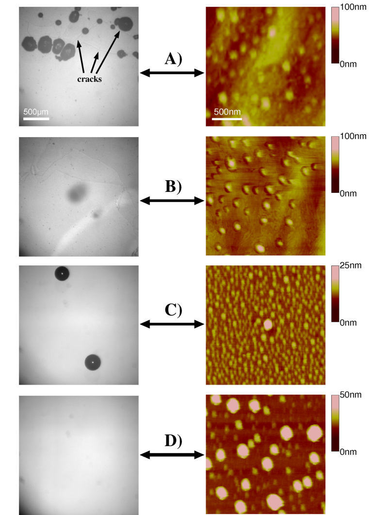
Do substrates with a high nanobubble density show a large cavitation activity? Fig. 2 illustrates the experimental results. The left panel shows typical recordings of the cavitation experiments for the cases . The right panel shows the corresponding AFM-measurements of the substrate surfaces immersed in water. Though the substrates have relatively large contact angles and the water pressure drops down to during the experiments there is hardly any cavitation on the smooth substrates . Note that the cavitation bubbles in originate exclusively from microscopic cracks in the surface, as can be seen in Fig. 2A). Applying the ethanol-water exchange, these microcracks do not lead to surface cavitation, see Fig. 2B). Contrary to the cavitation experiments, the AFM measurements show that all substrates are densely covered by surface nanobubbles, with number densities between 10 and 80 bubbles per . The sizes range from 3 to 40 nm in height and 60 to 300 nm in diameter. Several standard tests were performed (not shown) to ensure that the structures seen in the AFM images are indeed surface nanobubbles. Force-volume measurements tyrrellPRL2001 ; holmberga ; zhangcraig and tip manipulation of the bubbles simonsen are in accordance with previous studies. Furthermore, nanobubbles are not present when the substrates are immersed in ethanol, in agreement with jiong . Successive cycles of ethanol-water and water-ethanol exchange resulted in pictures without (in ethanol) and with nanobubbles (in water). Finally, when degassed ethanol is exchanged by degassed water, nanobubbles are not induced.
Thus the combination of the cavitation and the AFM experiments yields the remarkable result that the surface nanobubbles do not cavitate, in spite of the enormous tensile stress they are exposed to. This contradicts the expectation that the experimental pressure signal should be able to cavitate bubbles with an initial radius of curvature down to 8 nm. Case explicitly shows that nanobubbles are still present after the cavitation experiments, and that there is no cavitation activity at the surface induced by the shockwave. While it is already puzzling that surface nanobubbles are stable under ambient conditions, it is even more puzzling that they still exist after the passage of a shock wave with a large tensile stress down to . We denote this as superstability.
One may wonder what actually is happening with the surface nanobubbles when the shock wave is passing by. With the present technology it is impossible to AFM-image the nanobubbles (which takes order of minutes) during the shock wave passage (which is order of ). Therefore, evidence can only be indirect.
One may also question whether the nanobubbles survive the compression wave (with typical time scale so that the nanobubbles respond quasi-statically). During the compression phase, gas may diffuse into the neighboring liquid around the bubble. With a typical diffusion constant of we get as typical diffusion length scale . Hence the liquid close to the remaining void (100nm) will become supersaturated with gas. However, during the negative pressure phase, i.e., during the expansion of the bubble, all this gas will be recollected by the bubble, as has been shown in ref. hil96 (for micrometer bubbles).
In summary, it is demonstrated that in standard shock wave induced cavitation experiments surface nanobubbles do not act as nucleation sites. Cavitation bubbles originate from contaminations or from microscopic structures such as microcracks or microcrevices bremond06 ; bremondPOF06 , rather than from surface nanobubbles which densely populate the immersed substrates. This implies that surface nanobubbles are unexpectedly stable under large tensile stresses.
We thank Szczepan Zapotoczny for his contribution, K. A. Morch for discussions, and acknowledge financial support from STW (NanoNed Program), DFG (Grant No. DA969/1-1)), the MESA+ Institute, and CW-NWO (vernieuwingsimpuls program for H.S.).
References
- (1) J. W. G. Tyrrell and P. Attard, Phys. Rev. Lett. 87, 176104 (2001).
- (2) A. C. Simonsen, P. L. Hansen, and B. Klösgen, J. Colloid Interface Sci. 273, 291 (2004).
- (3) M. Holmberg et al., Langmuir 19, 10510 (2003).
- (4) A. Agrawal et al., Nano Lett. 5, 1751 (2005).
- (5) X. H. Zhang, N. Maeda, and V. S. J. Craig, Langmuir 22, 5025 (2006).
- (6) L. Zhang et al., Langmuir 22, 8109 (2006).
- (7) X. H. Zhang et al., Langmuir 22, 9238 (2006).
- (8) S. Yang et al., submitted.
- (9) M. Switkes and J. W. Ruberti, Appl. Phys. Lett. 84, 4759 (2004).
- (10) R. Steitz et al., Langmuir 19, 2409 (2003).
- (11) P. S. Epstein and M. S. Plesset, J. Chem. Phys. 18, 1505 (1950).
- (12) S. Ljunggren and J. C. Eriksson, Colloids Surf. A 129, 151 (1997).
- (13) E. Lauga and M. P. Brenner, Phys. Rev. E 70, 026311 (2004).
- (14) E. Lauga, M. P. Brenner, and H. A. Stone, in Handbook of Experimental Fluid Dynamics, edited by C. Tropea, J. Foss, and A. Yarin (Springer, New York 2005).
- (15) C. Neto et al., Rep. Prog. Phys. 68, 2859 (2005).
- (16) A. A. Atchley and A. Prosperetti, J. Acoust. Soc. Am. 86, 1065 (1989).
- (17) M. Holmberg et al., in: Fifth International Symposium on Cavitation (2003).
- (18) N. Bremond, M. Arora, C. D. Ohl and D. Lohse, J. Phys.: Condens. Matter 17, S3603 (2005).
- (19) N. Bremond, M. Arora, C. D. Ohl and D. Lohse, Phys. Fluids. 18, 121505 (2006).
- (20) N. Bremond, M. Arora, C. D. Ohl and D. Lohse, Phys. Rev. Lett. 96, 224501 (2006).
- (21) K. Morigaki et al., Langmuir 19, 6994 (2003).
- (22) C. E. Brennen, Cavitation and Bubble Dynamics (Oxford University Press, New York, 1995).
- (23) T. G. Leighton, The Acoustic Bubble (Cambridge University Press, Cambridge, 1994).
- (24) Rodd’s chemistry of carbon compounds, 2nd edition, vol 1, part B, edited by S. Coffey (Elsevier, Amsterdam, 1965).
- (25) We verified that our ex situ ethanol-water exchange generates surface nanobubbles, by placing the processed substrate in the AFM while keeping water on its surface. Then the substrate was AFM-scanned and many surface nanobubbles ( per ) were observed.
- (26) S. Hilgenfeldt, D. Lohse, and M. P. Brenner, Phys. Fluids 8, 2808 (1996).