Dynamical contribution into enzyme catalytic efficiency
Abstract
A realistic physical model for the so called rate promoting vibration (RPV) at enzyme action is constructed. The origin of the RPV is assumed to be an oscillating electric field produced by long-lived localized vibrational modes in protein dynamics, namely, by the so called discrete breather (DB) in secondary structure. The strength of interaction of the RPV with the reaction coordinate is evaluated and its effect on the reaction acceleration is assessed within the framework of modern theory for thermally activated escape rate at periodic driving. We reveal the phenomenon of resonant activation in our model elucidating why the frequency of the RPV in the range was chosen by the evolution of enzymes as an optimal one. The effect of the RPV on the reaction acceleration is shown to vary from moderate one (up to ) in the case of three-site DB to enormous (up to ) in the case of five-site DB and thus can significantly contribute into enzyme catalytic efficiency. Also the model is shown to be compatible with the known functional dependence of enzymatic reaction rates on solvent viscosity.
Keywords: enzyme catalysis; protein dynamics; discrete breather;
solvent viscosity
,
1 Introduction
Comprehension that specific protein dynamics contributes somehow into enormous reaction acceleration (up to ) by enzymes has a long history (see [1], [2], [3], [4], [5], [6], [7], [8] and refs. therein). It is supposed that an enzyme provides more efficient reaction activation compared to corresponding nonenzymatic reaction due to a dynamical mechanism and that the latter is significant enough to be a rival to the effect of lowering of the potential energy barrier (due to transition state stabilization) caused by specific interactions of the substrate molecule in an enzyme active site with catalytically active groups. The latter paradigm going back to Pauling is a cornerstone of traditional chemical enzymology [9] and is developed in a number of vigorously conquering approaches such as transition state stabilization by: i) electrostatic field of preorganized dipoles in an enzyme active site [10], [11], ii) low energy barrier hydrogen bonds [12], iii) entropic, strain and desolvation effects (leading mostly to ground state destabilization) [9], etc. Chemical enzymology is a vast field of activity and it will not be touched upon here. Instead we deal with physical aspects of enzyme action which are still poorly understood. The reason for that seems to be in the lack of adequate theoretical tools to evaluate the contribution of a dynamical mechanism in the reaction rate and in meager direct experimental evidence for its existence. The latter is altered to some extent by recent experiments [13], [14], [15], [16], [17], [18]. In these papers the importance of protein dynamics for hydrogen tunneling transfer enzymatic reactions was shown and the notion of the rate promoting vibration (RPV) was coined [13], [15] (some authors call it protein promoting vibration). To regret hydrogen tunneling transfer is involved only in minor and marginal part of enzymatic reactions and up to now there is no direct experimental evidence for the significance of the RPV in covalent bond cleavage between heavy atoms that is the most typical rate limiting step at enzyme action. For such enzymes the well known solvent viscosity effect on the reaction rate (see [5], [19] and refs. therein) still remains the most vivid testimony for manifesting itself of protein dynamics in enzyme catalysis. It is commonly accepted that this phenomenon is mediated by protein dynamics but the detailed mechanism is obscure.
An important question is what kind of motion in proteins is responsible for the dynamical contribution into enzyme catalytic efficiency? Some authors relate protein specific motion producing the RPV with conformational motion of peculiar amino-acid residues which may belong to enzyme active cite or be at a distance from it [17], [18]. In our opinion this scenario encounters difficulties at attempts to reconcile it with the experiments [20], [21], [22], [23]. The latter unequivocally testify that enzymes retain their catalytic capacity at temperatures below the so-called dynamical ”glass” transition where conformational motion is considerably suppressed (see, e.g., [24], [21] and refs. therein). Some guess on the origin of the RPV can be obtained by considering its frequency. The authors of [15] conclude that the estimated dominant peaks in the spectral densities of the RPV indicate motions on the frequency scale. There are another assessments of this value in literature: [6] and [25]. In the latter paper the RPV is attributed to the mode of Amide-VII of a peptide group vibrations. However this mode requires some torsion of the plane of a peptide group and is very energetically consumptive and unfavorable. In our paper [26] it was shown that there is another possible type of long-lived localized vibrational modes in protein dynamics with required frequency, namely, the so called discrete breather (DB) in protein secondary structures. The non-linear localized excitations named DBs or else intrinsic localized modes are time periodic spatially localized oscillations with significant amplitudes of several units in a chain of weakly coupled non-linear oscillators while others are at rest or oscillate with negligible amplitudes. They were discovered by Sievers and Takeno in 1988 [27] and proved by MacKay and Aubry [28] to be structurally stable. By now they are well understood and commonly appreciated as a generic phenomenon in nature (see [29] and refs. therein). DBs have become a new and very fruitful paradigm in nonlinear physics and in particular much work is being carried out at present to construct models for DBs in biomolecules. The paper [26] is a development along this line. It is shown there that the frequency of a DB in an -helix can be obtained equal to in accordance with the results of the experiments of [30] on far infra-red laser pulse spectroscopy of proteins. The DB is actually an oscillation of the planes of some peptide groups in an -helix or -sheet around their equilibrium positions with considerable amplitude (up to ) while neighboring peptide groups oscillate with much less amplitudes and more distant groups oscillate with negligible ones or stay at rest. Thus the DB is assumed to store and utilize the energy released at substrate binding by an enzyme. It is created by an external cause (binding energy) rather than by equilibrium thermal fluctuations though can be fed by the latter. As a peptide group is known to have a large dipole moment () parallel to its plane [31] the participation of the latter in the DB creates an oscillating electric field. Such fields (the so called electrostatic fluctuations) are for long supposed to be a key factor for enzyme catalysis [4]. The field can interact with the reaction coordinate because during the movement of the system along the latter a separation of charges takes place yielding the dipole moment of the reaction coordinate. Thus protein dynamics affects the potential surface for the reaction making the latter to be non-stationary. The shape (e.g., the height) of the reaction energy barrier becomes time dependent. As will be discussed below the oscillating electric field in accordance with the general theory of the effect of periodic driving force [32] ””heats up” the system by changing its effective temperature thus giving rise to lowering of the activation energy of escape which can be much bigger than the real temperature even for comparatively weak fields”.
The aim of the present paper is to construct a realistic physical model for the dynamical mechanism contributing into enzyme action and to evaluate its possible catalytic efficiency. We attain the latter within the framework of the archetypical model for the reaction rate, namely that of the overdamped limit of the Fokker-Planck equation (see [33] and refs therein) with non-stationary force field. This equation is a standard tool for studying phenomena with time dependent potentials mostly within the context of stochastic resonance [34], [35], thermally activated escape problem at periodic driving in the classical regime [36], [37], [38], [39], [40], [32], [41], [42], [43], [44], [45], [46] and resonant activation (see [47] and refs therein). For the estimate of the escape rate we apply the results of the papers [37], [39], [46].
The paper is organized as follows. In Sec.2 we consider the oscillating electric field created by a DB in protein secondary structure. In Sec.3 we evaluate the ability of this field to accelerate an enzymatic reaction. In Sec.4 we argue that our model is able to shed new light on the origin of solvent viscosity effect on enzymatic reaction rates. In Sec.5 the results are discussed and the conclusions are summarized.
2 Physical model for the rate promoting vibration
The possibility for existence of a stable zig-zag shape DB in protein secondary structures (-helix or -sheet) was shown in the paper [26]. The DB in our case is actually the out of phase oscillations of the planes of some adjacent peptide groups (due to rotations round their torsional angles) with considerable amplitudes up to from their equilibrium values while neighboring peptide groups oscillate with much less amplitudes and more distant groups oscillate with negligible ones or stay at rest. The dimensionless form of the equation of motion for the small deviation () of the angle of the plane for the i-th peptide group from its equilibrium value in a chain is
| (1) |
where is the coupling constant, is a nonlinear local potential and the dash denotes a derivative in coordinate. In [26] the following results were obtained:
-helix: .
anti-parallel -sheet: .
parallel -sheet: .
Since parallel -sheet is practically not represented in natural proteins [48] we omit it from further consideration. The linearizeation of (1) yields the dispersion relationship where is the wave number. The DB frequency and its harmonics must not coincide with linear modes spectrum [29], i.e., satisfy the requirement . There is no analytical solution of (1) for the general case of a multi-site DB [29]. However numerical simulations show that in our case of the DB in protein secondary structure the time dependence is qualitatively rather similar to harmonic oscillations [26]. The latter can be easily anticipated from (1) taking into account the smallness of the coupling constants and of the anharmonic corrections in the local potentials. That is why we restrict ourselves by a harmonic approximation as sufficient for further analysis
| (2) |
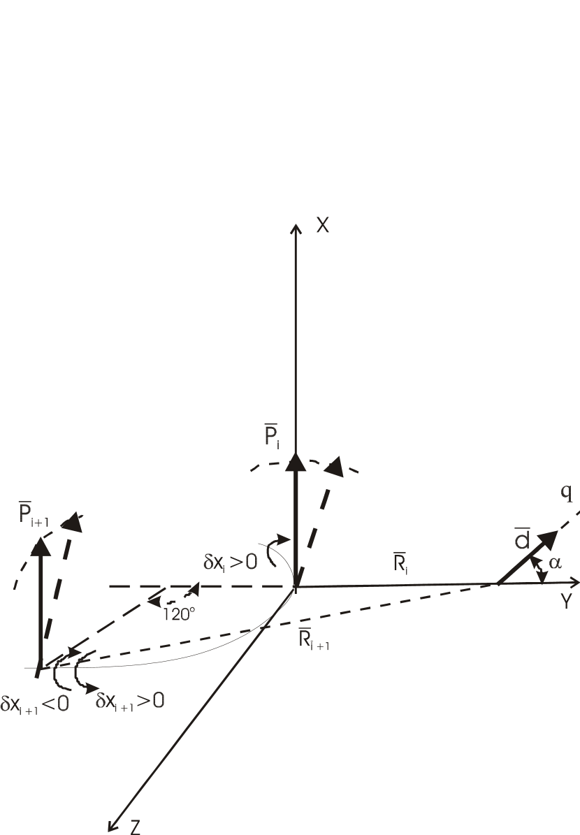
where is the amplitude and is the phase. At first we consider the simplest case of a three-site DB. In this case we can take into account only the motion of the central peptide group in the three-site DB undergoing maximal deviations and neglect others because in such DB the amplitude of the central peptide group by an order of magnitude exceeds those of the neighbors [26]. Returning to dimensional time we should replace by dimensional frequency () where is the effective local potential and is the moment of inertia of the peptide group. In [26] the following results for were obtained : 114.75 for -helix; 84.34 for anti-parallel -sheet and 85.49 for parallel -sheet, i.e., approximately for all of them. Now let us take into account that the peptide group is known to have a large dipole moment () parallel to its plane [31]. In an -helix the dipole moment of a peptide group is approximately parallel to its direction. If we place the origin of the Cartesian frame in the dipole moment and direct the axis X along the dipole moment at equilibrium position of the peptide group (and consequently along the axis of the -helix) then the dipole moment at time t has the projections: , , (see Fig.1). Taking into account that we have: .
We are going to examine the influence of the electric field produced by this oscillating dipole moment on the enzymatic reaction, i.e., on the covalent bond breaking between heavy atoms that is the most typical rate limiting step at enzyme action. We assume that the reaction coordinate is located at a distance from the dipole moment in the point and its direction constitutes the angle with the axis , i.e., (see Fig.1). The quasistationary electric field ( where is the speed of light) from the dipole moment in this point is
| (3) |
where is the dielectric constant for protein interior ( [31]). We attribute the dipole moment to the bond to be broken at the reaction where is the partial charge. The energy of the interaction of the electric field with the dipole moment is
| (4) |
The factor before in the right hand side is the strength of the interaction.
Now we can qualitatively consider the case of a multi-site DB where several peptide groups oscillate with non-negligible amplitudes. For instance in a five-site DB three of them have significant values compared with the neighbors (). This example is quite representative and we further restrict ourselves by it. On the one hand peptide groups in the zig-zag DB undergo out of phase oscillations, i.e., the deviations of the angular variables have opposite signs (). On the other hand the structure of an -helix is such that there are three peptide groups in its perpendicular cros-section, i.e., the radial angle between adjacent groups is (actually there are 3.6 peptide groups in the perpendicular cros-section, so that the angle is [31] but this subtliety does not modify our futher qualitative conclusions). Two circumstances compensate each other. That is why at opposite deviations of the angular variables the projections of linear deviations of the dipole moments of adjacent peptide groups on the axis have the same sign, i.e., are in phase (see Fig.1). If we neglect for simplicity the differences both in the distances from the dipoles to the location of the reaction coordinate and in angles between the dipoles and the direction of the reaction coordinate then we can assert that the oscillating electric field produced by three peptide groups of the five-site DB has the amplitude that is approximately thrice of that for one peptide group of the three-site DB and consequently the strength of the interaction in (4) should be multiplied by the factor . Taking into account the above mentioned differences makes it of course less than and we use the qualitative factor as sufficient for our further analysis.
In the next Sec. we consider the effect of the time dependent part of the interaction (4) on the enzymatic reaction.
3 Efficiency of the rate promoting vibration for reaction acceleration
We use the Kramers’ model for a chemical reaction as the escape of a Brownian particle with the mass from the well of a metastable potential (see for review [33] and refs. therein) in the presence of the additional time dependent term defined by (4). For the metastable potential we choose without any serious loss of generality the conventionally accepted simple form . This potential has a minimum at , a maximum at and the barrier height . In the overdamped limit (high friction case) the Langevin equation of motion along the reaction coordinate is
| (5) |
where is the friction coefficient, is the Boltzman constant, is the temperature and is the Gaussian white noise ( where is the Dirac -function) with zero mean (). We introduce the dimensionless variables and parameters
| (6) |
and denote
| (7) |
where is the dimensionless parameter characterizing the oscillating electric field strength and is the dimensionless parameter characterizing the intensity of thermal fluctuations (i.e., the temperature of the heat bath) relative the barrier height. Their ratio is the most important parameter of the model
| (8) |
Omitting the overlines we obtain
| (9) |
The corresponding overdamped limit of the Fokker-Planck equation for the probability distribution function [33] is
| (10) |
Before proceed further we should define the range of the parameters for our model. To be supported by evidence we take the numerals for a very typical and one of the most studied enzymatic reaction catalysed by Subtilisin. This enzyme belongs to serine proteases and brakes the bond between the atoms C and N in a peptide group of a substrate of protein nature. At physiological temperatures where enzymes normally work () the corresponding noncatalysed reaction typically has the rate constant while the enzymatic reaction has the rate constant (that of the rate limiting step) [49]. Thus the total catalytic effect is . Usually we do not know the friction coefficient . However assuming the single local-system relaxation time [33] to be typically we conclude that for the noncatalysed reaction we must have . We can evaluate from the Arrhenius factor of the Kramers’ rate
| (11) |
that in order to obtain such a reaction rate constant in the absence of the oscillating electric field () we should take . We also assume the distance from the minimum to the barrier to be typically . Taking the typical values ( of the electron charge () and assuming that the DB is arranged favorably respective the reaction coordinate () we obtain that for the ratio is for the case of the three-site DB and can attain the values for the five-site DB. It should be stressed that even in the latter case at our value of and satisfies the requirement , i.e., we are in the so called subthreshold driving regime. The latter provides that the potential surface always has a minimum and a maximum, i.e., the oscillating field is small enough not to distort the physical picture of the chemical reaction as the Brownian particle escape from the metastable state. We obtain from the dimensional frequency that the dimensionless one (see (6)) is . It should be stressed that we are very far from the stochastic resonance regime defined by the requirement [35] and are in the so called semi-adiabatic regime [42]. Thus we define the range for all three parameters figuring in (10)
for the three-site DB and
| (12) |
for the five-site DB. The range of values for noise intensity in (12) is inaccessible for numerical solution of (10) because of enormous escape times (). That is why we resort to the analytic results of the papers [37], [39], [46] for the escape rate averaged over the period of the driving force oscillations . We compile the results of these papers in a simple phenomenological formula for the prefactor of the escape rate in the presence of the harmonic driving force with the intensity and the frequency . For our potential ( in the dimensionless form) and in the semi-adiabatic regime it takes the form
| (13) |
where is the Kramers’ escape rate (11) and . For the integral in (13) can be evaluated by the steepest descent method and yields
| (14) |
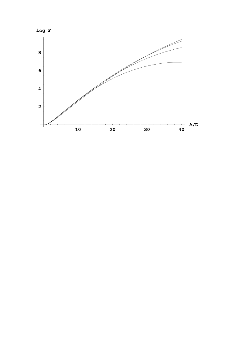
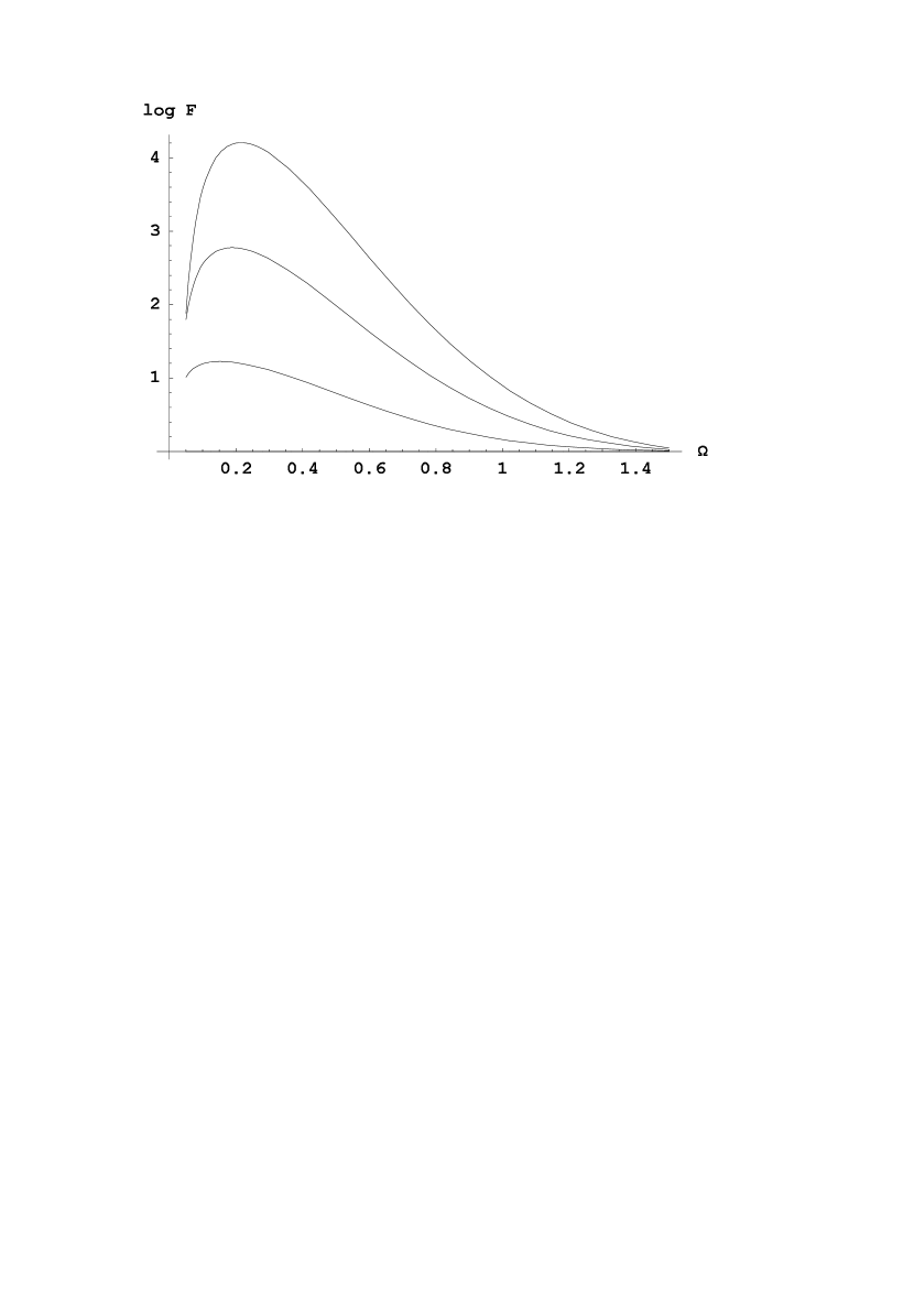
The dependencies of the prefactor on and are depicted in Fig.2 and Fig.3 respectively. They are obtained from (13) by numerical integration. For the potential the function is . We further use (14) for the estimate of the catalytic efficiency for the dynamical contribution (that is actually the prefactor) at enzyme action.
4 Solvent viscosity effect on the rate of enzymatic reaction
It is well known that enzymatic reaction rates exhibit unusual dependence on solvent viscosity [19]
| (15) |
where , that is generally of order of and is the cosolvent molecular weight. This dependence is the more pronounced the less is the cosolvent molecular weight, i.e., the higher its capacity to penetrate in protein interior [19]. The empirical law (15) is usualy verified in the range . As is commonly accepted the mechanism of this dependence should be somehow mediated by protein dynamics but its origin is still not clear.
Below we argue that the present approach can shed some light on the empirical relationship (15). We recall that for both -helix and anti-parallel -sheet the local potentials for the DB are of hard type [26] for which the frequency decreases with the decrease of the amplitude of oscillations. As a result we come to the following scenario. The increase of the viscosity leads to the increase of the probability for a cosolvent molecule to be located near the oscillating peptide group of the DB that restricts the amplitude of the oscillations simply by creating steric hindrance. The more the number of cosolvent molecules in the vicinity of the DB the less the amplitude of the oscillations. As a result of decreasing amplitude the frequency of the DB is decreased. Now we phenomenologically find the law for this decrease to obtain the required scaling of the reaction rate constant with the solvent viscosity (15).
In order to obtain from (14) the required empirical law (15) we should have
| (16) |
where is a constant. We take into account that and that frequency of the DB is some function (that is not kwown in the analytical form) of the amplitude of oscillations . In pure solvent without cosolvent molecules () we have some amplitude of the DB such that the frequency mathes the requirement of the resonant activation . Then we can determine the constant and write
| (17) |
This relationship implicitly defines the decrease of the amplitude of oscillations with the increase of solvent viscosity . Let us demonstrate it explicitly. We suppose that we are in the the region where so that . If we denote then we obtain from (17)
| (18) |
Taking into account that most probably (see next Sec.) we see that the solution of this equation is , i.e., indeed is sharply decreased copared with at large . The construction of a physical model for solvent viscosity effect on the amplitude of the DB and for the dependence of the frequency of the DB on the amplitude of oscillations is beyond the scope of the present paper and will be the subject of a separate investigation.
Concluding this Sec. the following comment should be done. Let us write the full expression for the logarithm of the escape rate obtained from (14) and (11)
| (19) |
This formula predicts two effects that at first glance can be detected in the experiment. First the factor leads to a deviation of the Arrhenius plot from the straight line. However the deviation is negligible because this factor is fully overshadowed by a much more significant dependence . Second the factor suggests that not only the prefactor, but also the exponent in the escape rate should scale with the viscosity of the solvent due to the dependence of on it. However this dependence also seems to be hardly observable because the driving amplitude is very small . In the next Sec. it will be argued that at present the traditional chemical enzymology leaves no reasons to expect the driving amplitude to noise intensity ratio to be very large. Most probably we have , i.e., at our value we obtain . To regret this value seems to be too small compared with for the dependence of the exponent on solvent viscosity to be noticed in the experiment. No matter how insignificant the effect is, this specific prediction remains the most direct way to verify the theory experimentally.
5 Discussion
We construct a realistic physical model in which the origin of the RPV at enzyme action is the oscillating electric field produced by dipole moments of peptide groups participating in long-lived localized vibrational modes (i.e., in a DB) of protein dynamics. We evaluate the magnitude of the oscillating electric field from peptide groups in two cases. In the first case the field is produced by a single peptide group that is the central one of a three-site DB. In this case we conclude that peptide group can provide the driving force amplitude to noise intensity ratio up to . For this purpose the DB should be favorably situated relative the reaction coordinate, i.e., not far and at favorable angle (see (8)). In the second case the field is produced by three peptide groups of a five-site DB. In this case we can attain up to .
The efficiency of the RPV as a dynamical contribution into enzyme catalytic efficiency is evaluated by us within the framework of the existing theories for the thermally activated escape in the presence of the driving force [39], [37]. It is interesting to note that revealing the role of driving at activated escape in biological systems is considered by the authors of the paper [32] as ”a fundamentally important and most challenging open scientific problem”. We compile the results of the theory in a simple phenomenological formula (14) that yields the required retardation of the exponential growth of the prefactor with the increase of (see Fig.2) in accordance with the results of [37]. Moreover in this formula the phenomenon of the so called ”resonant activation” (see Fig.3), i.e., the existence of an optimal frequency for reaction acceleration manifests itself. The notion of resonant activation is traditionally used for phenomena with the potential modulation by random fluctuations [47]. However recent numerical simulations of the Langevin equation [50] demonstrate that this phenomenon takes place also for deterministic driving and our result reproduces this computational finding. It should be stressed once more that the maximum of the frequency curves at in Fig.3 is not related to the phenomenon of stochastic resonance (defined by the requirement [35]) since the value of in our case is negligibly small ().
With the help of the formula (14) we conclude that the RPV produced by a three-site DB can provide reaction acceleration up to (see Fig.2 at and ). If we assume that the RPV is produced by a five-site DB we can get up to . In this case we can obtain enormous reaction acceleration up to (see Fig.2 at and ). Thus the present model has potential capacity to explain significant part of the total effect of enzyme efficiency. Moreover it gives an answer to the question why the frequency range of the RPV was chosen by the evolution of enzymes as an optimal one. This range (dimensionless ) turns out to be favorite because it matches the requirement of resonant activation where driving is most efficient for reaction acceleration (see Fig.3).
Also the formula (14) is shown to be compatible with experimental data for solvent viscosity effect on enzymatic reaction rates at a physically understandable assumption that the amplitude of oscillations in the DB decreases with the increase of solvent viscosity by the relationship (LABEL:eq22). The latter actually means that with the increase of the number of cosolvent molecules in the vicinity of the oscillating peptide group of the DB the amplitude of the oscillations is decreased because cosolvent molecules create steric hindrance for oscillations. For realization of this relationship a separate model should be constructed. The latter seems to be a feasible task and is planned for future work. Thus in our opinion the present approach sheds new light on the origin of the dependence of enzymatic reaction rates on solvent viscosity.
What kind of experiments can be interpreted with the help of the
present model? In its moderate form (reaction acceleration up to
by a three-site DB) it can be directly applied to
explaining the residual catalytic activity of mutant enzymes. It
is known that even at substitution of all catalytically active
groups in the enzyme active site by inactive ones by methods of
gene engineering (when one should expect no lowering of the
potential barrier) the mutants still exhibit residual catalytic
activity accurately [49], [48].
Also the known phenomenon of catalytic antibodies can be treated.
These proteins are actually not enzymes. They have no active site
and no catalytically active groups at all [48]. Still they
also exhibit catalytic activity accurately
[48]. The explanation of chemical enzymology is that they
nevertheless bind the transition state of substrate molecules
preferentially thus stabilizing it [48]. The present
model suggests another explanation, namely, being proteins they
contain secondary structure in which DBs producing the RPV can be
excited at substrate binding. Another case if the extreme
estimates of the present model (reaction acceleration up to
by a five-site DB) were necessitated and called
for by difficulties of traditional chemical enzymology. Such
enormous acceleration constitutes significant part of total
catalytic efficiency at enzyme action. Then the present model
would be applicable to interpreting much wider range of kinetic
data. We conclude that the present model for the dynamical
contribution into enzyme catalytic efficiency is able to describe
various situations from partial reaction acceleration up to major
part of the total effect and besides suggests a route to
revealing the origin of solvent viscosity effect on rate
constants. Whether or not nature makes use of such mechanism
remains the subject of further investigations.
Acknowledgements. The author is grateful to Prof. V.D. Fedotov, R.Kh. Kurbanov for helpful discussions. The work was supported by the grant from RFBR.
References
- [1] M. Karplus, J.A. McCammon, Protein structural fluctuations during a period of 100 ps, Nature (London) 277 (1979) 578-578.
- [2] G.R. Welch, B. Somogyi, S. Damjanovich, The role of protein fluctuations in enzyme action, Prog. Biophys., Mol. Biol. 39 (1983) 109-146.
- [3] M. Kurplus, J.A. McCammon, Dynamics of Proteins: Elements and Function, Annu.Rev.Biochem. 52 (1983) 263-300.
- [4] A. Warshel, Dynamics of enzymatic reactions, Proc.Natl.Acad.Sci.USA 81 (1984) 444-448.
- [5] C.R. Welsh, ed., The fluctuating enzyme, Wiley, N.Y., 1986.
- [6] E. Neria, M. Karplus, Molecular dynamics of an enzyme reaction: proton transfer in TIM, Chem.Phys.Lett. 267 (1997) 23-30.
- [7] K.O. Alper, M. Singla, J.L. Stone, C.K. Bagdassarian, Correlated conformational fluctuations during enzymatic catalysis: Implications for catalytic rate enhancement, Prot.Sci. 10 (2001) 1319-1330.
- [8] P.K. Agarwal, Role of protein dynamics in reaction rate enhancement by enzymes, J.Am.Chem.Soc. 127 (2005) 15248-15256.
- [9] A. Fersht, Structure and mechanism in protein science: a guide to enzyme catalysis and protein folding, Freeman, N.Y., 1999.
- [10] A. Warshel, Computer modeling of chemical reactions in enzymes and solutions, Wiley, N.Y., 1997.
- [11] A. Warshel, Electrostatic origin of the catalytic power of enzymes and the role of preorganized active sites, J.Biol.Chem. 273 (1998) 27035-27038.
- [12] W.W. Cleland, P.A. Frey, J.A. Gerlt, The low barrier hydrogen bond in enzymatic catalysis, J.Biol.Chem. 273 (1998) 25529-25532.
- [13] D. Antoniou, S.D. Schwartz, Internal enzyme motions as a source of catalytic activity: Rate-promoting vibrations and hydrogen tunneling, J.Phys.Chem. B105 (2001) 5553-5558.
- [14] M.J. Sutcliffe, N.S. Scrutton, A new conceptual framework for enzyme catalysis. Hydrogen tunneling coupled to enzyme dynamics in flavoprotein and quinoprotein enzymes, Eur.J.Biochem. 269 (2002) 3096-3102.
- [15] D. Antoniou, S. Caratzoulas, C. Kalyanarman, J.S. Mincer, S.D. Schwartz, Barrier passage and protein dynamics in enzymatically catalyzed reactions, Eur.J.Biochem. 269 (2002) 3103-3112.
- [16] M.J. Knapp, J.P. Klinman, Enviromentally coupled hydrogen tunneling. Linking catalysis to dynamics, Eur.J.Biochem. 269 (2002) 3113-3121.
- [17] J.S. Mincer, S.D. Schwartz, Protein promoting vibrations in enzyme catalysis. A conserved evolutionary motif, J.Proteome Res. 2 (2003) 437-439.
- [18] J.S. Mincer, S.D. Schwartz, A computational method to identify residues important in creating a protein promoting vibration in enzymes, J.Phys.Chem. B107 (2003) 366-371.
- [19] S. Yedgar, C. Tetreau, B. Gavish, D. Lavalette, Viscosity dependence of escape from respiratory proteins as a function of cosolvent molecular weight, Biophys.J. 68 (1995) 665-670.
- [20] N. More, R.M. Daniel, H.H. Petach, The effect of low temperatures on enzyme activity, Biochem. J. 305 (1995) 17-20.
- [21] R.M. Daniel, J.C. Smith, M. Ferrand, S. Hery, R. Dunn, J.L. Finney, Enzyme activity below the dynamical transition at 220 K, Biophys.J. 75 (1998) 2504-2507.
- [22] J.M. Bragger, R.V. Dunn, R.M. Daniel, Enzyme activity down to , BBA 1480 (2000) 278-282.
- [23] R.V. Dunn, V. Reat, J. Finney et al., Enzyme activity and dynamics: xylanase activity in the absence of fast anharmonic dynamics, Biochem. J. 346 (2000) 355-358.
- [24] E.T. Iben, D. Braunstein, W. Doster et al., Glassy behavior of a protein, Phys.Rev.Lett. 62 (1989) 1916-1919.
- [25] V.A. Kovarskii, Quantum processes in biological molecules. Enzyme catalysis, Usp.Phys.Nauk, 169 (1999) 889-908.
- [26] A.E. Sitnitsky, Discrete breathers in protein secondary structure, in: ”Soft condensed matter. New research.” Ed. K.I. Dillon, Nova Science Publishers Inc., NY, 2006; Los Alamos preprint datebase, arXiv:cond-mat/0306135.
- [27] A.J. Sievers, S. Takeno, Intrinsic localized modes in anharmonic cristals, Phys.Rev.Lett. 61 (1988) 970-974.
- [28] R.S. MacKay, S. Aubry, Proof of existence of breathers for time-reversible or harmonic networks of weakly coupled oscillators, Nonlinearity 7 (1994) 1623-1629.
- [29] S. Flach, C.R. Willis, Discrete breathers, Phys.Reports 295 (1998) 181-264.
- [30] A. Xie, A. van der Meer, R.H. Austin, Excited-state lifetimes of far-infrared collective modes in proteins, Phys.Rev.Lett. 88 (2002) 018102-1-018102-4.
- [31] C.R. Cantor, P.R. Schimmel, Biophysical Chemistry, Freeman, San Francisco, 1980, part 1.
- [32] M.I. Dykman, B. Golding, L.I. McCann et al., Activated escape of periodically driven systems, Chaos 11 (2001) 587-598.
- [33] P. Hänggi, P. Talkner, M. Borkovec, Fifty years after Kramers’ equation: reaction rate theory, Rev.Mod.Phys. 62 (1990) 251-341.
- [34] P. Jung, Periodically driven stochastic systems, Phys.Reports 234 (1993) 175-295.
- [35] L. Gammaitoni, P. Hänggi, P. Jung, F. Marchesoni, Stochastic resonance, Rev.Mod.Phys. 70 (1998) 223-287.
- [36] J. Lehmann, P. Reimann, P. Hänggi, Surmounting oscillating barriers, Phys. Rev. Lett. 84 (2000) 84-87.
- [37] J. Lehmann, P. Reimann, P. Hänggi, Surmounting oscillating barriers: Path-integral approach for weak noise, Phys.Rev. E62 (2000) 6282-6294.
- [38] J. Lehmann, P. Reimann, P. Hänggi, Activated escape over oscillating barriers: The case of many dimensions, Phys.stat.Sol.(b) 237 (2003) 53-64.
- [39] V.N. Smelyanskiy, M.I. Dykman, B. Golding, Time oscillations of escape rates in periodically driven systems, Phys.Rev.Lett. 82 (1999) 3193-3197.
- [40] V.N. Smelyanskiy, M.I. Dykman, H. Rabitz et al., Nucliation in periodically driven electrochemical systems, J.Chem.Phys. 110 (1999) 11488-11504.
- [41] R.S. Maier, D.L. Stein, Noise-activated escape from a sloshing potential well, Phys.Rev.Lett. 86 (2001) 3942-3946.
- [42] P. Talkner, Stochastic resonance in the semiadiabatic limit, New J.Phys. 1 (1999) 4.1-4.25.
- [43] P. Talkner, J.Luczka, Rate description of Fokker-planck processes with time dependent parameters, Phys.Rev.E 69 (2004) 046109.
- [44] D. Ryvkine, M.I. Dykman, B. Golding, Scaling and crossovers in activated escape near a bifurcation point, Phys. Rev. E 69 (2004) 061102.
- [45] M.I. Dykman, D. Ryvkine, Activated escape of periodically modulated systems, 94 (2005) 070602.
- [46] D. Ryvkine, M.I. Dykman, Noise-induced escape of periodically modulated systems: From weak to strong modulation, Phys.Rev E72 (2005) 011110.
- [47] P. Reimann, Thermally driven escape with fluctuating potentials: A new type of resonant activation, Phys.Rev.Lett. 74 (1995) 4576-4579.
- [48] C. Branden, J. Tooze, Introduction to protein structure, 2-nd ed., Garland Publishing Inc, N.Y., 1999.
- [49] P. Carter, J. Wells, Dissecting the catalytic triad of a serine protease, Nature (London) 332 (1988) 564-568.
- [50] Y. Zolotaryuk, V.N. Ermakov and P.L. Christiansen, Resonant enhancement of the jump rate in a double-well potential, J.Phys. A: Math.Gen. 37 (2004) 6043-6051.