Dielectrophoresis of nanoscale double-stranded DNA and humidity effects on its electrical conductivity
Abstract
Dielectrophoresis method for trapping and attaching nanoscale double-stranded DNA between nanoelectrodes was developed. The method gives a high yield of trapping single or a few molecules only which enables transport measurements at the single molecule level. Electrical conductivity of individual 140-nm-long DNA molecules was measured, showing insulating behaviour in dry conditions. In contrast, clear enhancement of conductivity was observed in moist conditions, relating to the interplay between the conformation of DNA molecules and their conductivity.
Controlled manipulation of single molecules is a prerequisite for fully understanding their properties as well as for realizing their potential in molecular electronics. At the present, the fabrication of single-molecule devices in nanoscale mostly relies on passive, uncontrollable methods of manipulation such as deposition of the molecules on the substrate or on the fabricated structure. Dielectrophoresis1,2 (DEP), an active manipulation method utilizing electro-magnetic fields, has been widely applied for microscale objects,3 e.g., DNA of bacteriophage lambda (-DNA).4-6 In nanoscale, however, Brownian motion poses a challenge: the few successful demonstrations are for trapping nanoscale objects,7,8 and for attaching DNA molecules between nanoelectrodes by DC-DEP.9 Concerning the intriguing question of DNA conductivity,9-18 there starts to be a consensus that double-stranded DNA (dsDNA) molecules exposed to untreated SiO2 or mica surfaces, in dry environment or vacuum, are insulating.19-23 However, the conductivity of DNA on specially treated surfaces,23,24 in solutions25-27 or inside dried films10 remains open. Also, the effect of humidity on the electrical conductivity of DNA films28,29 or constellations of DNA molecules30,31 has been discussed recently. The effects of the ambient conditions are related to the intimate connection between the conformation of the molecules and their conductivity.
In the present paper, we report a fully developed AC-DEP technique applicable for trapping, stretching and attaching nanoscale dsDNA molecules between nanoelectrodes. The technique has a high yield and allows transport measurements of single or a few molecules. Electrical conductivity of the trapped, 140 nm long dsDNA molecules was measured. Especially, the effect of humidity was investigated. While dsDNA in dry environment showed insulating behaviour, the molecules in moist conditions showed significantly lower resistances (linear resistance of the order of 100 M) providing the first observation of humidity effects for individual nanoscale DNA molecules.
We fabricated narrow finger-tip type gold electrodes, with a gap of about 100 nm, on a SiO2 substrate using standard electron beam lithography (Fig. 1; see EPAPS Ref. 32). We chose to use AC-DEP instead of DC to eliminate undesired electrophoretic effects and to enhance stretching of dsDNA molecules.4 Double-stranded 414 bp (140 nm) long DNA containing a thiol group (–SH) in both ends was fabricated and diluted in Hepes buffer. To optimize the process, we studied the DEP of fluorescent labeled DNA in situ under a confocal microscope [Fig. 1(a); see EPAPS Ref. 32 and the movies in Ref. 33]. The optimal DEP frequency was found to be 750 kHz combined with field strength of 107 V/m.
Electrical DC conductivity measurements of the DNA were done in room temperature (23∘C) both with relative air humidity of about 30 % (’dry’ environment) and of 80%-90% (’moist’ environment). Tens of samples containing DNA were measured in the dry environment, and they all showed insulating behaviour: - curves were linear at small voltages with resistance of about 10 T.

These resistance values for dry dsDNA on the SiO2 surface are in agreement with many recent observations by other groups.10,11,14,19-22,24,31 In contrast, in moist conditions, several samples showed clear increase in conductivity which was much higher than observed in the reference samples, for which control experiments were done using exactly the same procedure for the DEP and subsequent transport measurements, but using a buffer solution without DNA.
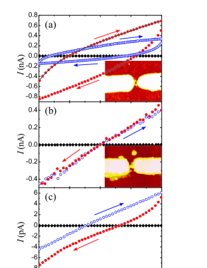
For instance, a sample showing conductivity in humid air was the one with three individual DNA molecules, Fig. 1(d) (Sample I). In dry environment, the resistance was 10 T. It dropped to 250 M after the sample had been half an hour in moist environment [red circles in Fig. 2(a)]. After that, the resistance slowly increased during the measurement, resulting to 700 M after three hours (blue open circles). This deterioration of conductivity during the measurements is probably due to disturbance of the DNA structure caused by gathering of contaminants from the moist air.32 After the measurement in moisture, the sample was dried with nitrogen and the resistance increased back to the original dry value. The sample was imaged with AFM right after these measurements confirming that at least two of the DNAs were still properly attached [inset in Fig. 2(a)]. The material between the electrodes was confirmed afterwards to be dsDNA using confocal microscopy with dsDNA-specific fluorescent labelling.32 Similar behaviour was observed also in a sample containing a bundle of DNA, Fig. 1(e) (Sample II). The resistance was a few T in dry environment and 40 G immediately after applying the moist conditions, but decreased to 250 M after the sample had been over ten hours at moist conditions [circles in Fig. 2(b)]. The increase in conductivity in this case was much slower than in the case of Sample I, furthermore, the resistance stayed the same during the measurements and did not increase as in the case of Sample I. The sample was finally dried and the resistance rose to a few T again. The AFM image in inset of Fig. 2(b) shows that the DNA bundle was still in place after the measurement.
The behaviour of Samples I and II, i.e., resistance dropping to hundreds of M in moist conditions, was observed in five different samples with single or a few DNAs. Such behaviour was never observed in the reference samples, containing no DNA. However, some of the samples containing DNA behaved in a similar way to reference samples, indicating that there is either no DNA properly attached to the electrodes or the DNA is not conducting, e.g., due to being severely deformed. - curves from one of the reference samples are shown in Fig. 2(c). They also show clear difference between the dry and moist environment measurements. However, in moist conditions, the minimum resistance observed for the reference samples was 7 G, and the resistance in dry environment was always around 10 T. The number of samples, the double check with AFM and confocal imaging, and the comparison to the reference samples using buffer without DNA provide, altogether, firm evidence for the strong effect of moisture on the electrical conductivity of single nanoscale DNA molecules. Note that the conductance can still be limited by the used hexanethiol-linkers reported resistance of .34
Even when the effect of humidity on the conductivity of individual DNA molecules is evident, the nature of the charge transport cannot be completely determined based on these experiments. In earlier experiments,28-31 the humidity enhanced conductivity of DNA has been explained by dipole relaxation losses of,29 or dissociation, i.e., proton transfer through,28,30 the hydrated water molecules. The first model applies only to AC conductivity. Since reduction-oxidation processes are negligible due to low voltages used in our experiments,27 buffer salts and the counterions do not contribute to the total steady-state DC-current. Instead, diffusion of the ions to the electrodes, especially in moist environment, causes extra capacitance as seen in Fig. 2. One more possibility is enhanced electron transport/transfer caused by humidity induced conformational changes in DNA structure. The direct electronic conductivity, by means of overlapping -orbitals of the base pairs along the molecular axis, is likely to be sensitive to the helical conformation of dsDNA (Refs. 35 and 36) (the contributions of protons or counterions might also be affected by the deformations). Also magnetic properties of -DNA are shown to depend on the conformation of dsDNA.37 The deformations of the structure can be due to, e.g., ambient conditions such as humidity, or interactions with the substrate surface.23 For instance, a single dsDNA on graphite appears in its natural B-DNA form at moist conditions, but collapses to a form resembling A-DNA (defected overlap of the -orbitals) when dried to the surface.38 [We observed, in dry conditions, reduced height of the dsDNA (1 nm compared to the expected 2 nm) corresponding to a deformed state.] Also the contribution of the positive counterions to the electrical conductivity via gating effect has been suggested.39
In our case, the slower time scale of the conductivity change for DNA bundle (Sample II) vs. single molecules suggests that proton transfer along the dissociated hydrated water layer is not dominant: it would be enough to have water present at the surface of the object, which should happen as fast for a bundle as for a single molecule. The slower conductivity change in Sample II could be a result of slower hydrations of DNA helices inside the bundle. Likewise, the bundle can also keep the moisture inside and protect inner molecules, thus making the B-state more stable, as observed in the measurements. In the samples with individual molecules, the assumed recovery of the helical conformation due to humidity was either much faster, e.g., Sample I, or not successful at all. Also the increase of conductance at moist conditions only in some of the samples suggests the charge transfer mechanism being highly sensitive to the conformation, rather than proton transfer through the water layer on a (deformed) molecule. Our results are consistent with the resistance values observed for -DNA (Ref. 25) and short duplexes27 in buffer. This suggests that the hydration layer around the dsDNA in high humidity environment enables similar behaviour than the buffer environment, e.g., maintaining the double helical conformation of B-DNA.
In summary, we have developed a nanoscale AC-DEP technique that has a high yield and provides a platform for reliable transport measurements at the single molecule level. We observed a remarkable increase in the electrical conductivity of 140 nm long (414 bp) dsDNA molecules with increasing humidity of ambient air, and the observation was confirmed by various reliability checks.32 Our results also suggest that the change is related to a humidity induced conformational change of the molecular structure and associated with a contribution from electron transfer. Further research is required, however, to identify in detail the contributions from electrons, water ions and counterions.
Acknowledgements. The authors thank J. A. Virtanen and M. S. Kulomaa for useful discussions and acknowledge the financial support by Academy of Finland (Project Nos. 205470, 53903), the Emil Aaltonen foundation and the National Graduate School in Informational and Structural Biology (V.P.H.).
References
- (1) H. A. Pohl, J. Appl. Phys. 22, 869 (1951); H. A. Pohl, Dielectrophoresis: The Behavior of Neutral Matter in Nonuniform Electric Fields (Cambridge University Press, Cambridge, UK, 1978).
- (2) For a review, see e.g., P. J. Burke, Encyclopedia of Nanoscience and Nanotechnology, Vol. 6, p. 623 (2004).
- (3) P. Debye, P. P. Debye, and B. H. Eckstein, Phys. Rev. 94, 1412 (1954); J. S. Batchelder, Rev. Sci. Instrum. 54, 300 (1983); F. F. Becker, X.-B. Wang, Y. Huang, R. Pethig, J. Vykoukal, P. R. C. J. Gascoyne, J. Phys. D 27, 2659 (1994).
- (4) M. Washizu and O. Kurosawa, IEEE Trans. Indust. Appl. 26, 1165 (1990); S. Suzuki, T. Yamanashi, S. Tazawa, O. Kurosawa, M. Washizu, ibid. 34, 75 (1998).
- (5) C. L. Asbury and G. van den Engh, Biophys. J. 74, 1024 (1998); S. Tsukahara, K. Yamanaka, and H. Watarai, Chem. Lett. 3, 250 (2001); W. A. Germishuizen, C. Wälti, R. Wirtz, M. B. Johnston, M. Pepper, A. G. Davies, A. P. J. Middelberg, Nanotechnology 14, 896 (2003); L. Zheng, J. P. Brody, and P. J. Burke, Biosensors Bioelectron. 20, 606 (2004).
- (6) B. Hartzell, B. McCord, D. Asare, H. Chen, J. J. Heremans, V. Soghomonian, Appl. Phys. Lett. 82, 4800 (2003);
- (7) M. Washizu, S. Suzuki, O. Kurosawa, T. Nishizaka, T. Shinohara, IEEE Trans. Indust. Appl. 30(4), 835 (1994); T. Schnelle, S. Muller, S. Fiedler, G. Shirely, K. Ludwig, A. Hermann, G. Fuhr, Naturwissenschaften, 83, 172 (1996); A. Bezryadin, C. Dekker, and G. Schmid, Appl. Phys. Lett. 71, 1273 (1997); N. G. Green, H. Morgan, and J. J. J. Milner, Biochem. Biophys. Methods 35, 89 (1997); X. Q. Chen, T. Saito, H. Yamada, K. Matsushige, Appl. Phys. Lett. 78, 3714 (2001).
- (8) C.-F. Chou, J. Tegenfeldt, O. Bakajin, S. S. Chan, E. C. Cox, N. Darnton, T. Duke, and R. H. Austin, Biophys. J. 83, 2170 (2002); L. Ying, S. S. White, A. Bruckbauer, L. Meadows, Y. E. Korchev, and D. Klenerman, Biophys. J. 86, 1018 (2004).
- (9) D. Porath, A. Bezryadin, S. De Vries, and C. Dekker, Nature 403, 635 (2000).
- (10) Y. Okahata, T. Kobayashi, K. Tanaka, and M. Shimomura, J. Am. Chem. Soc. 120, 6165 (1998); Y. Okahata, T. Kobayashi, H. Nakayama, and K. Tanaka, Supramol. Sci. 5, 317 (1998).
- (11) E. Braun, Y. Eichen, U. Sivan, and G. Ben-Yoseph, Nature 391, 775 (1998).
- (12) S. O. Kelley and J. K. Barton, Science 283, 375 (1999).
- (13) H.-W. Fink and C. Schönenberger, Nature 398, 407 (1999).
- (14) P. J. de Pablo, F. Moreno-Herrero, J. Colchero, J. G mez-Herrero, P. Herrero, A. M. Baro, P. Ordejon, J. M. Soler, and E. Artacho, Phys. Rev. Lett. 85, 4992 (2000).
- (15) L. Cai, H. Tabata, and T. Kawai, Appl. Phys. Lett. 77, 3105 (2000).
- (16) A. Yu. Kasumov, M. Kociak, S. Gu ron, B. Reulet, V. T. Volkov, D. V. Klinov, and H. Bouchiat, Science 291, 280 (2001).
- (17) K.-H. Yoo, D. H. Ha, J.-O. Lee, J. W. Park, J. Kim, J. J. Kim, H.-Y. Lee, T. Kawai, and H. Y. Choi, Phys. Rev. Lett. 87, 198102 (2001).
- (18) A. Rakitin, P. Aich, C. Papadopoulos, Yu. Kobzar, A. S. Vedeneev, J. S. Lee, and J. M. Xu, Phys. Rev. Lett. 86, 3670 (2001).
- (19) A. J. Storm, J. van Noort, S. de Vries, and C. Dekker, Appl. Phys. Lett. 79, 3881 (2001).
- (20) Y. Zhang, R. H. Austin, J. Kraeft, E. C. Cox, and N. P. Ong, Phys. Rev. Lett. 89, 198102 (2002).
- (21) M. Bockrath, N. Markovic, A. Shepard, m. Tinkham, L. Gurevich, L. P. Kouwenhoven, M. W. Wu, and L. L. Sohn, Nano Lett. 2, 187 (2002).
- (22) C. Gómez-Navarro, F. Moreno-Herrero, P. J. de Pablo, J. Colchero, J. Gómez-Herrero, and A. M. Baró, Proc. Natl. Acad. Sci. U.S.A. 99, 8484 (2002).
- (23) A. Yu. Kasumov, D. V. Klinov, P.-E. Roche, S. Guéron, and H. Bouchiat, Appl. Phys. Lett. 84, 1007 (2004).
- (24) T. Heim, D. Deresmes, and D. Vuillaume, J. Appl. Phys. 96, 2927 (2004).
- (25) P. Tran, B. Alavi, and G. Grüner, Phys. Rev. Lett. 85, 1564 (2000).
- (26) E. M. Boon and J. K. Barton, Curr. Opin. Struct. Biol. 12, 320 (2002).
- (27) B. Xu, P. Zhang, X. Li, and N. Tao, Nano Lett. 4, 1105 (2004).
- (28) Y. Otsuka, H.-Y. Lee, J.-H. Gu, J.-O. Lee, K.-H. Yoo, H. Tanaka, H. Tabata, and T. Kawai, Jpn. J. Appl. Phys. 41, 891 (2002).
- (29) M. Briman, N. P. Armitage, E. Helgren, and G. Grüner, Nano Lett. 4, 733 (2004).
- (30) D. H. Ha, H. Nham, K.-H. Yoo, H.-M. So, H.-Y. Lee, and T. Kawai, Chem. Phys. Lett. 355, 405 (2002).
- (31) H. Kleine, R. Wilke, Ch. Pelargus, K. Rott, A. Pühler, G. Reiss, R. Ros, and D. Anselmetti, J. Biotechnol. 112, 91 (2004).
- (32) See EPAPS Document No. E-APPLAB-87-010543 for details of the experiments. This document can be reached via a direct link in the online article’s HTML reference section or via the EPAPS homepage (http://www.aip.org/pubservs/epaps.html)
-
(33)
http://www.phys.jyu.fi/research/electronics/research/
depmovies.html - (34) B. Xu and N. J. Tao, Science 301, 1221 (2003) and references therein.
- (35) D. D. Eley and D. I. Spivey, Trans. Faraday Soc. 58, 411 (1962).
- (36) For a review, see e.g., R. G. Endres, D. L. Cox,and R. R. P. Singh, Rev. Mod. Phys. 76, 195 (2004).
- (37) S. Nakamae, M. Cazayous, A. Sacuto, P. Monod, and H. Bouchiat, Phys. Rev. Lett. 94, 248102 (2005).
- (38) M. H. Zareie and P. B. Lukins, Biochem. Biophys. Res. Commun. 303, 153 (2003).
- (39) R. N. Barnett, C. L. Cleveland, A. Joy, U. Landman, and G. B. Schuster, Science 294, 567 (2001).