Comparative Raman Studies of Sr2RuO4, Sr3Ru2O7 and Sr4Ru3O10
Abstract
The polarized Raman spectra of layered ruthenates of the Srn+1RunO3n+1 () Ruddlesden-Popper series were measured between 10 and 300 K. The phonon spectra of Sr3Ru2O7 and Sr4Ru3O10 confirmed earlier reports for correlated rotations of neighboring RuO6 octahedra within double or triple perovskite blocks. The observed Raman lines of or symmetry were assigned to particular atomic vibrations by considering the Raman modes in simplified structures with only one double or triple RuO6 layer per unit cell and by comparison to the predictions of lattice dynamical calculations for the real and structures. Along with discrete phonon lines, a continuum scattering, presumably of electronic origin, is present in the , and , but not in the and spectra. Its interference with phonons results in Fano shape for some of the lines in the and spectra. The temperature dependencies of phonon parameters of Sr3Ru2O7 exhibit no anomaly between 10 and 300 K where no magnetic transition occur. In contrast, two lines in the spectra of Sr4Ru3O10, corresponding to oxygen vibrations modulating the Ru-O-Ru bond angle, show noticeable hardening with ferromagnetic ordering at 105 K, thus indicating strong spin-phonon interaction.
pacs:
78.30.Hv, 63.20.Dj,75.30.DS, 75.50.EeI Introduction
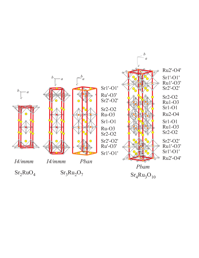
The properties of layered ruthenates Srn+1RunO3n+1 ), known as the Ruddlesden-Popper series, exhibit strong dependence on the number of RuO6 octahedral layers. Sr2RuO4 () is -wave superconductor,maeno1 ; braden1 Sr3Ru2O7 () is nearly ferromagnetic (enhanced paramagnetic) metal,ikeda1 whereas Sr4Ru3O10 ( K) is a ferromagnetic metal.crawford1 ; cao1 There are indications that the variations with of the magnetic and transport properties of Srn+1RunO3n+1 are partly related to the structural distortions in Sr3Ru2O7 and Sr4Ru3O10. While the structure of Sr2RuO4 is tetragonal (, No.139, Fig.1) and the Ru-O-Ru angle in the plane is 180∘, the structures of Sr3Ru2O7 (, No.50) and Sr4Ru3O10 (, No. 55) are orthorhombic due to correlated rotations about the -axis of the neighboring corner-sharing octahedra within each layer of the double or triple perovskite blocks.ikeda1 ; huang1 ; crawford1 ; cao1 These rotations result in decrease of the Ru-O-Ru angle in the plane to 166∘ for Sr3Ru2O7 and 169∘ for the outer layers and 158∘ for the middle layers of Sr4Ru3O10, respectively. The Sr-based ruthenates, however, are less distorted than corresponding Ca-based compounds. Indeed, besides being rotated around the axis, the RuO6 octahedra in Ca2RuO4 (, No.61)braden1 and Ca3Ru2O7 (, No.36) cao2 are also tilted around an axis lying in the RuO2 plane.
The coupling among the charge, lattice and spin degrees of freedom in (Ca,Sr)n+1RunO3n+1 compounds has been subject of several magnetotransportikeda1 ; cao1 , pressureshaked1 ; snow1 and Ramansnow1 ; liu1 ; sakita1 ; rho1 studies. The polarization-, temperature-, pressure- and substitution-dependent Raman spectra allowed observation of the two-magnon scattering, opening of the spin gap, and pressure- and substitution-induced variations in metal-insulator transition in Ca2RuO4snow1 , Ca2-xSrxRuO4rho1 and Ca3Ru2O7liu1 ; snow1 , as well as spin gap and strong direction-dependent electron-phonon interaction in Sr2RuO4sakita1 . The latter studies illustrated the ability of Raman scattering to provide information on the interplay of spin, charge, and lattice degrees of freedom. To our knowledge, there are yet no reports on the Raman spectroscopy of double-layer Sr3Ru2O7 and triple-layer Sr4Ru3O10. The Raman spectra of these materials and their variations with temperature are of definite interest as they contain information about the local structure, electron-phonon, spin-phonon interactions and their variations with the number of RuO6 layers. An essential precondition for correct analysis and understanding of complex structure-properties relationships is the assignment of the Raman lines to particular atomic motions.
In this work we present results of comparative polarization- and temperature-dependent Raman studies of Sr2RuO4, Sr3Ru2O7 and Sr4Ru3O10 between 10 and 300 K. The spectra of Sr3Ru2O7 and Sr4Ru3O10 provide clear evidence for a structure containing correlated rotations of RuO6 octahedra within perovskite blocks. On the basis of their symmetry, considering corresponding modes in simplified tetragonal structures, containing RuO6 rotations, and by comparison to the predictions of lattice dynamical calculations (LDC) for the real orthorhombic and structures, the observed Raman lines of Sr3Ru2O7 and Sr4Ru3O10 are assigned to definite phonon modes. Except for phonon lines, a continuum scattering, presumably of electronic origin, with components of ( and ) and () symmetry is present in the whole temperature range. Its interference with phonons results in Fano shape for some of the lines in the and spectra. Phonon anomalies related to magnetic ordering at T K are observed in the temperature-dependent spectra of Sr4Ru3O10.
II Samples, Experimental and Lattice Dynamical Calculations
Rectangular platelet-like single crystals of Srn+1RunO3n+1 () with typical size 110.2 mm3 were grown using SrCO3 and RuO2 as starting materials and SrCl2 as a flux. The compositions Sr2RuO4, Sr3Ru2O7, and Sr4Ru3O10 were obtained by varying the SrCO3:SrCl2 ratio and temperature profiles. The temperature profile was basically as follows: (1) Temperature increase to 1400-1500∘C in 7 hours and then constant temperature for 25 hours; (2) Cooling down to 1250-1350∘C at a rate of 2 deg/hour; (3) Further cooling to room temperature in 1 hour.
The x-ray diffraction showed that the lattice parameters of all three compounds corresponded to the ones known from the literature. It was also established and further confirmed by Raman polarization selection rules that the large surfaces of the crystal platelets were parallel to the (001) plane and their edges were along either {100} or {110} directions.
Raman spectra were collected using Jobin-Yvon HR640 spectrometer equipped with microscope (100 or 50 objective, focus spot size 1-3 m), notch filters and liquid-nitrogen-cooled charge-coupled device (CCD) detector. The He-Ne (632.8 nm) and Ar+ (514.5 nm and 488.0 nm) laser lines were used for excitation. The lack of spurious signals from impurity phases was verified by the reproducibility of the spectra and their strict polarization. Given the crystallographic directions were known, measurements could be done in several exact backward scattering configurations: , , , , , , , , . The first and forth letters in these notations stay for the directions of incident and scattered light, whereas their polarizations are denoted by the second and third letters, respectively. As and are indistinguishable, and are interchangeable. The same is valid for and . Further, the short notations , , , and will also be used.
The lattice dynamical calculations were done using a shell model described in detail in Ref.popov1 . This model gives an adequate description of the vibrations in perovskitelike structures because it accounts for their predominant ionicity. The ionic interactions are represented by long-range Coulomb potentials and short-range repulsive potentials of the Born-Mayer form where and are constants and is the interionic separation. The deformation of the electron charge density of the ions is described in the dipole approximation considering each atom as consisting of a point charged core and a concentric spherical massless shell with charge . Each core and its shell are coupled together with a force constant giving rise to the free ionic polarizability . The model parameters for the strontium, ruthenium, and oxygen ions and their interaction potentials are taken from a previous study of simpler compounds with perovskitelike structurepopov1 ; iliev1 .
III Results and Discussion
III.1 Sr2RuO4
The polarized Raman spectra of Sr2RuO4 at room temperature as obtained with 488.0 nm excitation are shown in Fig.2. Three of the four () Raman allowed phonons are observed at 200 cm-1 (, Sr vibrations along ), 247 cm-1 (, apex oxygen vibrations in the plane), and 545 cm-1 (, apex oxygen vibrations along ), in consistence with earlier reports of Udagawa et al.udagawa1 and Sakita et al.sakita1 . The Raman phonon intensities exhibit clear resonant behavior. For example in the -polarized spectra the ratio is 19.3 for 632.8 nm (1.96 eV), 4.2 for 514.5 nm (2.41 eV), and 3.5 for 488.0 nm (2.54 eV) excitations, respectively. In the polarized spectra the corresponding values are 2.1, 1.5, and 4.2. For all three excitation energies used, the and spectra were practically identical.

Except for the phonon lines, an electronic background was present in all spectra. The electronic Raman scattering for incident polarization parallel to the plane has previously been reported by Yamanaka et al.yamanaka1 , who observed structureless continuum with , and components of comparable intensity. Our measurements have shown, however, that the component of the continuum is much weaker than those of and symmetry.
III.2 Sr3Ru2O7
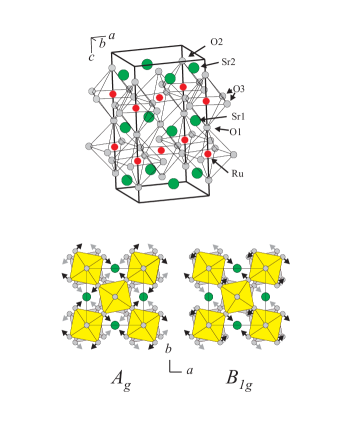
The orthorhombic structure of Sr3Ru2O7 can be obtained from the idealized structure (similar to Sr2RuO4, see also Fig.1), by ordered counter-phase rotations of RuO6 octahedra around the -axis. The and axes of the structure are rotated by 45 degree with respect to those of the tetragonal one and the and -parameters are larger by factor . While ten -point phonon modes () are Raman-allowed in the tetragonal structure of Sr3Ru2O7, much more modes () are Raman-allowed in the structure due to the doubling of unit cell, absence of the cell centering and appearance of new modes related to RuO6 rotations. One should not expect, however, observation of such a large number of Raman lines, as to each , or tetragonal mode one can juxtapose a pair of two , two or orthorhombic modes, which involve practically the same atomic vibrations and have very close frequencies (see Table I for LDC of the and modes). Experimentally, these pairs will be observed as a single line.
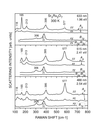
Polarized Raman spectra of Sr3Ru2O7 measured at room temperature with 633 nm, 515 nm and 488 nm laser line excitation are shown in Fig. 3. Five lines of symmetry are clearly pronounced in the and spectra at 118, 165, 212, 395, and 577 cm-1. Another two lines of symmetry are seen in the spectra at 306 and 377 cm-1, but no lines of detectable intensity are observed in the () spectra where are allowed the modes of () symmetry. The intensity of the Raman lines changes with excitation energy due to alteration of the resonance conditions. The relative intensities of the line at 395 cm-1 and the two lines, however, remain nearly the same. This suggests that the three modes involve motions of same type of atoms, different from those with main contribution to the mode at 577 cm-1. The number of Raman lines in the spectra of Sr3Ru2O7 is noticeably lower than in the corresponding spectra of Ca3Ru2O7.liu1 This had to be expected as the structure of Ca3Ru2O7 is more strongly distorted and the number of fully symmetrical modes allowed in , and configurations is higher.
A simple approach to the assignment of the Raman lines is based on the reasonable assumption for weak interaction between the double Ru-O slabs, the only important distortion being the rotation of the RuO6 octahedra. Within such assumption, instead of the real structure, one can consider a simplified structure with elementary cell containing only one double layer (Fig.4). This structure is tetragonal (, No.125, Z=2) with the same and parameters as the real one, but twice shorter parameter. The normal mode analysis gives Raman modes. Therefore one expects in the and spectra five Raman lines of symmetry ( in tetragonal notations), which is exactly the case. In the spectra one expects observation of four modes ( in tetragonal notations), two of them involving mainly oxygen vibrations.
The Raman mode frequencies in ionic materials, such as transition metal oxides, are determined by the mass, charge, and bond lengths of participating atoms as well by the type of atomic motions (stretching, bending or rotational). Based on comparison to other perovskitelike oxides, the modes involving mainly vibration of heavier Ru and Sr or rotational vibrations of oxygens are expected in the frequency range below 250 cm-1, the bending oxygen modes - between 200 and 500 cm-1 and stretching oxygen modes - above 500 cm-1. Our LDC for the structure (Table I) predict that the modes below 250 cm-1 are strongly mixed, each involving vibrations along of Ru and Sr2 as well as RuO6 rotations around . In the structure, however, the rotational motions (see Fig.4), are not Raman active. Therefore, the 165 cm-1 and 213 cm-1 lines, which are close to LDC frequencies predicted for both and structures, can tentatively be assigned to mixed vibrations of Ru and Sr along . The line at 118 cm-1 is close to the LDC() frequency of 117 cm-1, which has no partner in the LDC() data, is assigned to mainly RuO6 rotations. The ”soft”-mode temperature behavior of the latter line is also typical for a rotational mode. As to the two high-frequency modes, with great certainty, confirmed by LDC, they correspond to out-of-plane in-phase vibrations of O3 (395 cm-1) and stretching vibrations of O2 (577 cm-1). The latter frequency is higher that that of the corresponding apex oxygen vibrations in Sr2RuO4 (545 cm-1), which can be explained accounting the bond-length changes. Indeed, it is plausible to assume that the force constants between O and the ion M follows the simple relation, where and are the charges of the oxygen and cation, respectively, and is the oxygen-cation bond length. This relation is valid for harmonic ionic crystals and shown to apply for perovskitelike transition metal oxides.kakihana1 ; hadjiev1 . Taking into account that and restricting interactions to only nearest-neighbors (Ru and Sr), one obtains for the O2 stretching vibrations
| (1) |
where , , and the values of and are respectively 2.016 Å and 2.459 Å for Sr3Ru2O7huang1 and 2.059 Å and 2.440 Å for Sr2RuO4neumeier1 . Using these values one obtains , which is close to the experimental ratio of 1.059.
There is little doubt too, that the two experimentally observed lines in the spectra correspond to the two oxygen modes of the distorted structure. The main atomic motions are out-of-phase vibrations along of O3 (306 cm-1) and ”scissors-like” bendings of O3 parallel to the plane (377 cm-1). The shape of the latter mode is also shown in Fig.4.
Along with the phonon lines, a structureless background of ( and ), and () symmetry, but not of ( or ) symmetry, is also present in the spectra. This is illustrated in Fig.5 for room temperature spectra taken with 633 nm excitation. The observation of relatively strong -polarized continuum was somewhat unexpected as the ARPES measurementspuchkov1 and band structure calculationssingh1 provide evidence for quasi-two-dimensional Fermi-surface sheets, in consistence with reports for large anisotropy of the electrical resistivity ( at 300 K)ikeda1 . The continuum-phonons interference is clearly pronounced only for incident radiation parallel to the plane through Fano shape of the 213 cm-1 line in the spectra and the 377 cm-1 line in the spectra (Fig.5). For a phonon coupled to electronic background, the Fano profile is generally used to describe the line shape, where , is the ”bare” phonon frequency, is the linewidth, and is the asymmetry parameter, reflects the electron-phonon interaction . While for an uncoupled phonon , the increase of electron-phonon interaction increases . The values of , , and as obtained from the Fano fit of the experimental line profiles are listed in Fig.5.
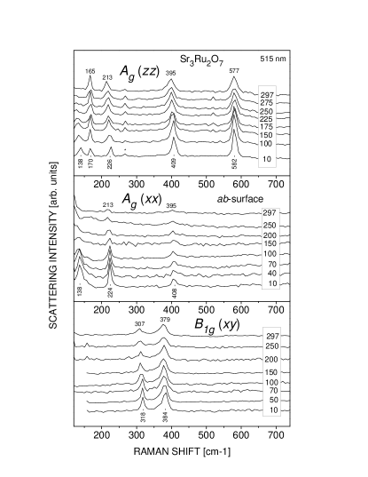
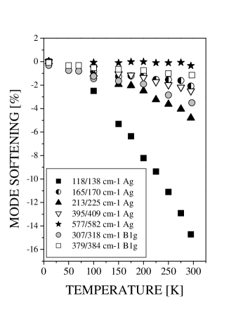
Fig.6 shows the temperature-dependent , and Raman spectra of Sr3Ru2O7 between 10 K and 300 K. The variations of phonon frequencies and line widths with temperature exhibit no anomalies. The relative changes of frequency for all observable modes are summarized in Fig.7. The low frequency mode shows ”soft”-mode behavior, its frequency decreasing by between 10 and 300 K, while the frequency of the highest mode remains practically unchanged. The continuum scattering in the , and spectra only slightly decreases with lowering temperature, in contrast to the case of Ca3Ru2O7, where a rapid suppression of the electronic background of symmetry has been observed below the metal-insulator transition at K.liu1 The interaction between the continuum and phonons also decreases with lowering temperature, as evidenced from Fig.5, where are compared the values at 10 and 300 K.
| Mode | LDC | Exp | Atomic | Mode | LDC | Mode |
| 300K/10K | motions | |||||
| 117 | 118/138 | RuO6 rot | ||||
| 179 | 165/170 | Ru(), Sr2() | 180 | |||
| 210 | 213/225 | Sr2(), Ru() | 214 | |||
| 223 | 213/225 | |||||
| 250 | ||||||
| 254 | ||||||
| 402 | 395/409 | O3() | 512 | |||
| 402 | 395/409 | |||||
| 520 | ||||||
| 522 | ||||||
| 580 | 577/581 | O2() | 589 | |||
| 580 | 577/581 | |||||
| 108 | ||||||
| 109 | ||||||
| 162 | ||||||
| 162 | ||||||
| 181 | ||||||
| 181 | ||||||
| 337 | 306/316 | O3() | 310 | |||
| 337 | 306/316 | |||||
| 378 | 377/380 | O3() | ||||
| 378 | 377/380 | |||||
| 466 | ||||||
| 466 | ||||||
| 521 | ||||||
| 521 | ||||||
| 726 | ||||||
| 726 | ||||||
III.3 Sr4Ru3O10
The structure of Sr4Ru3O10 has been refined as .crawford1 From symmetry considerations in total 96 () modes are Raman allowed. Like in the case of Sr3Ru2O7, however, most modes are practically degenerated in pairs and lower number of Raman lines is expected in the spectra. Fig.8 shows the , , and spectra obtained at room temperature with 633 nm, 515 nm, and 488 nm excitation. The temperature variations of the spectra between 10 and 300 K are given in Fig.9. Except for line shifts and appearance of additional weak lines, the spectral profiles and their dependence on scattering configuration and excitation wavelength resembles that of Sr3Ru2O7. Lines of symmetry are observed at room temperature 129, 163, 195-203, 360-365, 533, 582-585, 619 and 745 cm-1. The broad structure near 200 cm-1 in the spectra appears to be a superposition of two lines. This becomes evident at low temperatures where these two line are well separated. Like in the case of Sr3Ru2O7, only two lines of symmetry are pronounced and the intensity of the and lines is below the detection limit.

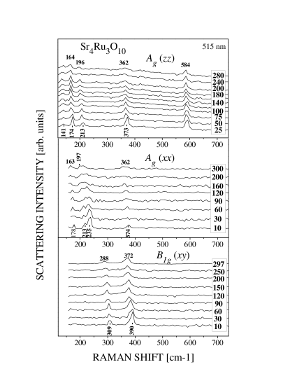
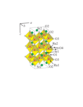

To assign the Raman lines to particular phonon modes let us again consider the Raman modes in a simplified distorted tetragonal structure and compare the experimental frequencies to the prediction of lattice dynamical calculations for the real structure of Sr4Ru3O10. The simplified structure (No.127, Z=2), shown in Fig.10, has only one triple layer in the unit cell. It is characterized by the same equal and parameters as the real structure, but twice shorter parameter. From symmetry considerations modes are Raman active. The modes, allowed in and spectra, correspond to modes in orthorhombic , whereas the tetragonal modes, allowed in the spectra, correspond to the orthorhombic modes. Compared to the simplified structure of Sr3Ru2O7, there are three new modes corresponding to: (i) Rotations of middle RuO6 octahedra; (ii) Vibrations along of internal apex oxygen atoms (O1); (iii) Vibrations along of internal Sr1 atoms. The LDC predict close frequencies for the rotational and Sr1 modes and our assignment of the new polarized line, seen at low temperature at 235 cm-1 and also having a ”soft”-mode temperature behavior, to rotational vibrations of middle RuO6 octahedra is only tentative. There is little doubt that the new lines at 745 cm-1, seen in the spectra with 488 nm excitation, correspond to the vibrations along of the inner O1 atoms. Using Eq.(1) with the experimental values of Å , Å and Å , Å one obtains frequency of O1 vibrations along should be by factor 1.35 higher than that of the outer apex oxygens (O2). This is in good agreement with the experimentally observed ratio of 745:584 = 1.28. A comparison of phonon frequencies predicted by LDC with experimental data is given in Table II.
| Mode | LDC | Exp | Atomic | Mode | Mode | LDC | Exp | Atomic | Mode |
|---|---|---|---|---|---|---|---|---|---|
| 300K/10K | motions | 300K/10K | motions | ||||||
| 90 | 129/140 | outer RuO6 rot in | 147 | ||||||
| 150 | 163/178 | Ru1()Sr2() | 151 | ||||||
| 173 | 175 | ||||||||
| 190 | 198/214 | Sr2()Ru1( | 176 | ||||||
| 198 | 185 | ||||||||
| 210 | 188 | ||||||||
| 231 | 213/235 | middle RuO6 rot in | 327 | 288/307 | O3() | ||||
| 234 | 327 | ||||||||
| 286 | 354 | 373/388 | O3() | ||||||
| 288 | 356 | ||||||||
| 309 | 464 | ||||||||
| 313 | 464 | ||||||||
| 358 | 361/372 | O3() | 476 | ||||||
| 364 | 476 | ||||||||
| 512 | 533/ | O3() | 482 | ||||||
| 514 | 482 | ||||||||
| 552 | 584/588 | O2() | 708 | ||||||
| 562 | 708 | ||||||||
| 678 | 745/ | O1() | |||||||
| 678 |
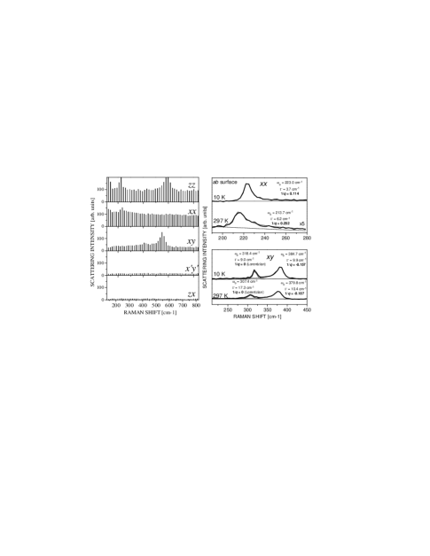
The left panel of Fig.11 illustrates the presence in the , and spectra of Sr4Ru3O10 of scattering continuum. Like in the case of Sr3Ru2O7, some lines exhibit clear Fano shape for light polarization parallel to the plane. This is shown in more detail in the right panels of Fig.11.
The ferromagnetic ordering at T=105 K has a moderate, but clearly pronounced effect on phonon parameters and electron-phonon interaction. The temperature dependencies of the position (Fig.12a) and width (Fig.12b) of the Fano shaped line, corresponding to O3 vibrations in the plane, change their slope near TC. The changes of (Fig.12c) are even more pronounced, providing evidence that the electron-phonon coupling decreases in the ferromagnetic phase. Weaker changes near TC of the parameters of the second line, corresponding to O3 vibrations in direction, are also observed (Figs.12d and 12e).
The magnetic ordering may affect phonon frequency through different mechanisms: exchange striction, baltensperger1 ; udagawa1 dependence of the spin energy on ion displacementsbaltensperger1 ; udagawa2 ; chen1 , variations of the density of itinerant carriers near TM.iliev1 Although consistent with observation of anomaly in , the latter mechanism, proposed to explain the anomalous hardening near TC of several Raman modes in ferromagnetic SrRuO3, is irrespective to the bond lengths and band angles being modulated and seems to be of less importance in Sr4Ru3O10. The observation of detectable anomaly near TC only for modes, which modulate the Ru1-O3-Ru1 angle, rather favors direct interaction of these phonons with the magnetization.baltensperger1 ; chen1 .

IV Conclusions
We studied in detail the polarized Raman spectra of Sr2RuO4, Sr3Ru2O7 and Sr4Ru3O10 with particular attention to the two latter compounds as their phonon spectra have not been reported so far. All observed Raman lines are of either or symmetry. They have been assigned to definite atomic vibrations by: (1) considering the Raman active modes in simplified tetragonal and structures, which contain only one double or triple RuO6 layers per unit cell and account for the rotational distortions; (2) comparison to the predictions of lattice dynamical calculations for the real orthorhombic double layer and triple layer structures. Except for the discrete phonon lines, a continuum background, presumably of electronic origin, is present in the , and , but not in the and spectra. The interaction of and continuum with the modes involving atomic motions in the plane results in Fano shape of corresponding Raman lines. While no anomaly in phonon parameters of Sr3Ru2O7 is seen between 10 and 300 K, where no magnetic transition occur, an anomaly is observed near ferromagnetic transition at 105 K in Sr4Ru3O10.
Acknowledgements.
This work is supported in part by the state of Texas through the Texas Center for Superconductivity and Advanced Materials, by NSF grant no. DMR-9804325, the T.L.L. Temple Foundation, the J. J. and R. Moores Endowment, and at LBNL by the Director, Office of Energy Research, Office of Basic Energy Sciences, Division of Materials Sciences of the US Department of Energy under contract no. DE-AC03-76SF00098. J.B. acknowledges financial support from the Swedish Superconductivity Consortium.References
- (1) Y. Maeno, T. M. Rice, and M. Sigrist, Phys. Today 54 (1), 42 (2001).
- (2) M. Braden, Y. Sidis, P. Bourges, P. Pfeuty, J. Kulda, Z. Mao, and Y. Maeno, Phys. Rev. B 66, 064522 (2002).
- (3) S.-I. Ikeda, Y. Maeno, S. Nakatsuji, M. Kosaka, and Y. Uwatoko, Phys. Rev. 62, R6089 (2000).
- (4) Q. Huang, J. W. Lynn, R. W. Erwin, J. Jarupatrakorn, and R. J. Cava, Phys. Rev. B 58, 8515 (1998).
- (5) M. K. Crawford, R. L. Harlow, W. Marshall, Z. Li, G. Cao, R. L. Lindstrom, Q. Huang, and J. W. Lynn, Phys. Rev. B 65, 214412 (2002).
- (6) G. Cao, L. Balicas, W. H. Song, Y. P. Sun, Y. Xin, V. A. Bondarenko, J. W. Brill, S. Parkin, and X. N. Lin, Phys. Rev. B 68, 174409 (2003).
- (7) G. Cao, K. Abbound, S. McCall, J. E. Crow, and R. P. Guertin, Phys. Rev. B 62, 998 (2000).
- (8) H. Shaked, J. D. Jorgensen, S. Short, O. Chmaissem, S.-I. Ikeda, and Y. Maeno, Phys. Rev. B 62, 8725 (2000).
- (9) C. S. Snow, S. L. Cooper, G. Gao, J. E. Crow, H. Fukazawa, S. Nakatsuji, and Y. Maeno, Phys. Rev. Lett. 89, 226401 (2002).
- (10) H. L. Liu, S. Yoon, S. L. Cooper, G. Cao, and J. E. Crow, Phys. Rev. B 60, R6980 (1999).
- (11) S. Sakita, S. Nimori, Z. Q. Mao, Y. Maeno, N. Ogita, and M. Udagawa, Phys. Rev. B 63, 134520 (2001).
- (12) H. Rho, S. L. Cooper, S. Nakatsuji, H. Fukazawa, and Y. Maeno, Phys. Rev. B 68, 100404 (2003).
- (13) V. N. Popov, J. Phys.: Condens. Matter 7, 1625 (1995).
- (14) M. N. Iliev, A. P. Litvinchuk, H.-G. Lee, C. L. Chen, M. L. Dezaneti, C. W. Chu, V. G. Ivanov, M. V. Abrashev, and V. N. Popov, Phys. Rev. B 59, 364 (1999).
- (15) M. Udagawa, T. Minami, N. Ogita, F. Nakamura, T. Fujita, J. G. Bednorz, and F. Lichtenberg, Physica B 219&220, 222 (1996).
- (16) A. Yamanaka, N. Asayama, M. Sasada, K. Inoue, M. Udagawa, S. Nishizaki, Y. Maeno, and T. Fujita, Physica C 263, 516 (1996).
- (17) M. Kakihana, S-G. Eriksson, L. Börjesson, L. G. Johansson, C. Ström, and M. Käll, Phys. Rev. B 47, 5359 (1993).
- (18) Y. K. Atanassova, V. G. Hadjiev, P. Karen, and A. Kjekshus, Phys. Rev. B 50, 586 (1994).
- (19) J. J. Neumeier, M. F. Hundley, M. G. Smith, J. D. Thompson, C. Allgeier, H. Xie, W Yelon, and J. S. Kim, Phys. Rev. B 50, 17910 (1994).
- (20) A. V. Puchkov, Z.-X. Shen, and G. Cao, Phys. Rev. B 58, 6671 (1998).
- (21) D. J. Singh and I. I. Mazin, Phys. Rev. B 63, 165101 (2001).
- (22) W. Baltensperger and J. S. Helman, Helv. Phys. Acta 41, 668 (1968).
- (23) M. Udagawa, K. Kohn, N. Koshizuka, T. Tsushima, and K. Tsushima, Solid State Commun. 16, 779 (1975).
- (24) X. K. Chen, J. C. Irwin, and J. P. Franck, Phys. Rev. B 52, R13130 (195).