Surface freezing and a two-step pathway of the isotropic-smectic phase transition in colloidal rods
Abstract
We study the kinetics of the isotropic-smectic phase transition in a colloidal rod/polymer mixture by visualizing individual smectic layers. First, we show that the bulk isotropic-smectic phase transition is preceded by a surface freezing transition in which a quasi two-dimensional smectic phase wets the isotropic-nematic interface. Next, we identify a two step kinetic pathway for the formation of a bulk smectic phase. In the first step a metastable isotropic-nematic interface is formed. This interface is wetted by the surface induced smectic phase. In the subsequent step, smectic layers nucleate at this surface phase and grow into the isotropic bulk phase.
pacs:
64.70.Md 82.70.DColloids with hard core repulsive interactions are often studied due to the simplicity and generality of their intermolecular potential. As a result of these studies, the equilibrium phase diagram of hard rods and spheres is well understood at the present time Frenkel et al. (1988); Vroege and Lekkerkerker (1992); Pusey and van Megen (1986); Adams et al. (1998). However, much less is known about the kinetic pathways of phase transitions in these systems ten Wolde and Frenkel (1997); Zhu et al. (1997). Direct visualization of colloids in a system undergoing phase transition have provided a powerful tool to study general aspects of phase transition kinetics Gasser et al. (2001); Anderson and Lekkerkerker (2002). In this paper we study the kinetics of the isotropic-smectic phase transition by directly visualizing individual smectic layers in a phase separating sample. As a model system of colloidal rods we use a monodisperse suspension of fd virus Dogic and Fraden (2001). We elucidate a kinetic pathway of unexpected complexity. The existence of surface freezing and a metastable isotropic-cholesteric phase transitions is discovered and their influence on the kinetic pathway is discussed. Because the behavior of the fd/Dextran mixture is determined by steric interactions and since all molecules including low molecular weight thermotropics have a steric core the results reported in this paper are likely to be quite general. In addition, our results might be pertinent to understanding the dynamics of amphiphilic membranes Lipowsky (1995), 2D smectic systems Harrison et al. (2000), surface freezing and wetting transitions Wu et al. (1993); Ocko et al. (1986); Boon and Ross (2001) and self assembled nano-structures Lee et al. (2002).
It has been known for a long time that surface freezing/melting can dramatically alter the nucleation rate and the kinetic pathway of a phase transition. On one hand, most substances exhibit surface melting. In this case a liquid surface wets the crystalline bulk phase. It follows that crystals melt from the surface inwards and therefore it is difficult to prepare a superheated metastable solid van der Veen (1999); Cahn (1986). On the other hand, surface freezing is observed in very few systems, most notably thermotropic liquid crystals, alkanes and surfactant mesophases Wu et al. (1993); Ocko et al. (1986); Lang (1999). Upon supercooling these materials, the ordered phase nucleates at the frozen interface and propagates towards the bulk phase. Therefore, it is difficult to supercool liquids that exhibit surface freezing Sloutskin et al. (2001).
Another factor that can affect the nucleation rate of a transitions is the presence of metastable phases Sirota and Herhold (1999); ten Wolde and Frenkel (1997). For example, recent simulations predict that the free energy barrier for the formation of protein crystals is greatly reduced when a metastable gas-liquid phase transition is located in a vicinity of a stable liquid-solid phase boundary ten Wolde and Frenkel (1997). In this case the nucleation of protein crystals proceeds in two steps. In the first step a dense metastable droplet associated with the gas-liquid phase transition is formed, while in the subsequent step the protein crystal nucleates within this droplet. In this paper we show that both surface freezing and metastable phases are important for understanding the kinetics of the isotropic-smectic phase transition.
Bacteriophage fd is a semi-flexible virus with contour length of 880 nm, diameter of 7 nm and persistence length of 2200nm. It was prepared as previously described and dialyzed against buffer of known ionic strength (190 mM Nacl,10 mM Tris, pH = 8.10). The phase diagram of the rod-polymer mixture was measured according to the published procedure Dogic and Fraden (2001). All the samples are prepared in a metastable/unstable isotropic phase by shear melting any existing structure and samples are placed into rectangular capillaries (VitroCom, Mountain Lakes, NJ). Nucleation and growth of the order phase is observed with an optical microscope (Zeiss AxioPlan2) equipped with DIC optics. All images are recorded with a cooled CCD camera (AxioCam Zeiss)
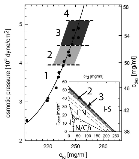
At zero polymer concentration fd is a good model system of hard rods and forms a stable isotropic (I), cholesteric (Ch) and smectic (S) phases with increasing concentration in agreement with theoretical predictions Dogic and Fraden (1997); Frenkel et al. (1988). Equilibrium I-S phase transition is observed in a mixture of rod-like fd viruses and non-adsorbing polymer Dextran. The phase diagram of this mixture is shown in the inset of Fig. 1. Adding non-adsorbing polymer to fd suspension produces effective attractive interactions between fd rods Asakura and Oosawa (1954). The main consequence of this attractive potential on the phase behavior of a rod like system is to widen the I-Ch coexistence concentrations with the polymer preferentially partitioning into the isotropic phase Bolhuis et al. (1997). Since the interactions in the fd/polymer mixtures are temperature independent, all phase transitions are entropically driven. In the first part of the paper we describe the equilibrium structures related to the surface freezing observed in region 2 of the phase diagram. In the second part of the paper we describe one of the kinetic pathways of phase separation observed in region 3.
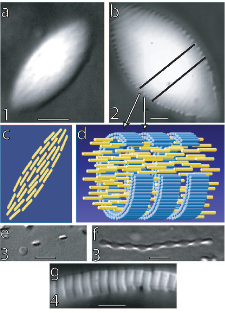
At rod concentrations below 235 mg/ml (region 1 in Fig. 1a and b), nematic droplets (tactoids) form in an isotropic background (Fig. 2a). Polarization microscopy indicates that the configuration of rods in the nematic tactoid is as shown in Fig. 2c. When confined to a small volume the cholesteric order is not able to develop; therefore we observe only unwound nematic phase within a individual tactoid. At higher rod concentrations (region 2 in Fig. 1) we observe droplets that have the same anisotropic shape. Microscopy indicates that the interior of these droplets is still nematic. However, each droplet has a corrugated I-N interface where the length of each ridge along the droplet’s long axis is approximately one virus long. As the tactoids coalesce and increase in size, the surface corrugations are always confined to a narrow layer of well defined thickness located at the I-N interface. This implies that the formation of corrugations is a purely surface effect. These observations lead us to conclusion that there exists a surface-induced quasi 2D smectic phase that wets the I-N interface. The ridges observed at the interface are individual layers of the surface-induced smectic phase. A schematic representation of a section of a corrugated tactoid is shown in Fig. 2d. The surface smectic phase is observed above an fd concentration of 235 mg/ml while the bulk I-S phase transition (region 4) is observed at 255 mg/ml.
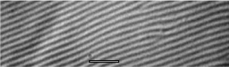
After a few hours, the fd/Dextran mixture prepared in region 2 completely phase separates with denser nematic tactoids coalescing and settling to the bottom of the sample. In this case a macroscopic I-N interface is formed. This makes it possible to focus on the interface and directly observe the surface induced smectic phase (Fig. 3). We conclude our description of the system in region 2 by noting that there are no theoretical predictions of the surface-induced smectic phase in rod/polymer mixture. We expect that such phase is a result of non-monotonic density profiles across the I-N interface Shundyak and van Roij (2002). Additionally, in the fd/polymer system rods in the surface frozen layer lie in the plane of the interface. This is in contrast to molecular systems which exhibit surface freezing where anisotropic molecules are either tilted or perpendicular to the interface Wu et al. (1993); Ocko et al. (1986).
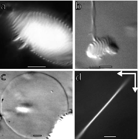
We now turn our attention to region 3 of the phase diagram. Right after mixing the sample, in addition to the formation of nematic droplets with a surface smectic, we observe self-assembly of rods into disk-like or ribbon-like structures (Fig. 2e and f). The thickness of the disk corresponds to the length of a single rod. When viewed from above a disk shows no birefringence while from the side it shows maximum birefringence when oriented at 45o with respect to the polarizer and analyzer (Fig. 4d). Therefore, polarization microscopy shows that disks are composed of a monolayer of aligned rods in the smectic-A configuration. We call these self-assembled disks colloidal membranes because of their similarity to amphiphilic membranes. Small homogeneously nucleated membranes (Fig. 2e) grow by coalescing laterally to form large 40 diameter isolated membranes (Fig. 4c) Dogic and Fraden (2001). This suggests that an isolated colloidal membrane and not a bulk smectic phase is the equilibrium structure in region 3. Polarization microscopy indicates that twisted ribbons are identical to disks except that they have a twist along their long axis due to the chiral nature of fd Dogic and Fraden (2000). We expect that the free energy difference between these two morphologies is small and will examine their relative stability elsewhere.
Real space images enables us to study the kinetic pathway for the formation of colloidal membranes. They can either homogeneously nucleate from the metastable isotropic suspension or can heterogeneously nucleate at the surface-induced smectic phase (Fig. 4a and b). A colloidal membrane nucleated at the interface grows into the isotropic phase either as a twisted ribbon or a flat disk. Over a period of a few days twisted ribbons can reach a lengths of several hundreds microns. Fluorescence images indicate that there are no rods in the isotropic solution. Therefore colloidal membranes (ribbons) must elongate due to rods that diffuse from a metastable nematic phase through a surface smectic to a more stable colloidal membrane. The fact that there is a transport of rods across the interface shows that the colloidal membranes are structures with lower free energy than the nematic phase or bulk smectic phase. At lower degrees of supercooling we mostly observe heterogenous surface induced nucleation instead of homogeneous nucleation of colloidal membranes. This shows that a two-step kinetic pathway has a lower nucleation barrier for the formation of colloidal membranes. To summarize, the phase separation in region 3 of the phase diagram proceeds in two steps. In the first step on timescale of seconds to minutes we observe the formation of nematic tactoids with surface smectic phase identical to those observed in region 2. However, these tactoids are metastable. In the second slow step on a time scale of hours to months we observe the nucleation of colloidal membranes at the surface frozen smectic phase and their subsequent growth into the isotropic phase.
A few comments are in order regarding the structures observed in region 3. First, to our knowledge this is the first time that non-amhiphilic objects with very simple excluded volume interactions have been self assembled into 2D membrane-like (Fig. 4c) and 1D polymer like structures (Fig. 2f) Tkachenko (2002). We speculate that these structures are stabilized by protrusion like fluctuations Israelechvili (1991). Second, it seems plausible that isolated colloidal membranes observed in region 3 are highly swollen lamellar phases previously observed in mixtures of nematic fd and hard spheres Adams et al. (1998). The swelling of the lamellar phase is predicted theoretically, but has yet to be observed in experiments Koda et al. (1996). Third, as the osmotic pressure is increased there is a transition to region 4 in which small colloidal membranes irreversibly stack up on top of each other to form elongated filaments (Fig. 2g). The nature of the transition from isolated membranes to a smectic phase remains unexplored. Fourth, we observe metastable nematic droplets wetted by a more stable surface induced smectic phase. Usually the reverse effect is observed pathways where a stable nucleus is wetted by a metastable phase ten Wolde et al. (1995); ten Wolde and Frenkel (1997)
In conclusion, there are two important results that can be deduced from our experiments. The first surprising result is that a rod/polymer mixture exhibits surface freezing in which a quasi 2D smectic phase wets the I-N interface. This effect occurs at rod concentration of 235 mg/ml while bulk I-S phase transition occurs at 255 mg/ml. To our knowledge this is the first time that the surface freezing has been directly visualized in a system whose phase behavior is dominated by entropic repulsive interactions. The second result of this work is to demonstrate the relationship between the surface freezing and the bulk isotropic-smectic phase transition. A complex two step kinetic pathway for the nucleation of the smectic phase out of the isotropic solution has been identified. In the first step a metastable nematic droplet with a surface frozen smectic phase nucleates in the isotropic solution. In the next step isolated monolayers (colloidal membranes) of smectic phase nucleate at the surface smectic phase and subsequently grow into the isotropic phase. Due to the simplicity and generality of the excluded volume interactions which dominate the phase behavior of fd/Dextran mixture, the results presented here should be relevant to a much wider class of systems than those studied here.
I wish to thank Seth Fraden, Gerhard Gompper, Arjun Yodh, Tom Lubensky, Daniel Chen, Peter Lang and Pavlik Lettinga for useful discussions. I am particulary indebted to Jan Dhont for his hospitality at FZ-Juelich and the Alexander von Humboldt foundation for financial support. Part of this work was done at Brandeis University where this research was supported by the NSF-DMR grant to Seth Fraden.
References
- Frenkel et al. (1988) D. Frenkel, H. Lekkerkerker, and A. Stroobants, Nature 332, 822 (1988).
- Vroege and Lekkerkerker (1992) G. J. Vroege and H. N. W. Lekkerkerker, Repts. on Prog. Phys. 8, 1241 (1992).
- Pusey and van Megen (1986) P. N. Pusey and W. van Megen, Nature 320, 340 (1986).
- Adams et al. (1998) M. Adams, Z. Dogic, S. L. Keller, and S. Fraden, Nature 393, 349 (1998).
- ten Wolde and Frenkel (1997) P. R. ten Wolde and D. Frenkel, Science 277, 1975 (1997).
- Zhu et al. (1997) J. Zhu, M. Li, R. Rogers, W. Meyer, R. H. Ottewill, W. B. Russel, and P. M. Chaikin, Nature 387, 883 (1997).
- Gasser et al. (2001) U. Gasser, E. R. Weeks, A. Schofield, P. N. Pusey, and D. A. Weitz, Science 292, 258 (2001).
- Anderson and Lekkerkerker (2002) V. J. Anderson and H. N. W. Lekkerkerker, Nature 416, 811 (2002).
- Dogic and Fraden (2001) Z. Dogic and S. Fraden, Phil. Trans. R. Soc. Lond. A. 359, 997 (2001).
- Lipowsky (1995) R. Lipowsky, in Generic interactions of flexible membranes., edited by R. Lipowsky and E. Sackmann (Elsevier, 1995), pp. 521–602.
- Harrison et al. (2000) C. Harrison, D. H. Adamson, Z. Cheng, J. M. Sebastian, S. Sethuraman, D. A. Huse, R. A. Register, and P. M. Chaikin, Science 290, 1558 (2000).
- Wu et al. (1993) X. Z. Wu, E. B. Sirota, S. K. Sinha, B. M. Ocko, and M. Deutsch, Phys. Rev. Lett. 70, 958 (1993).
- Ocko et al. (1986) B. M. Ocko, A. Braslau, P. S. Pershan, J. Als-Nielsen, and M. Deutch, Phys. Rev. Lett. 57, 94 (1986).
- Boon and Ross (2001) D. Boon and D. Ross, Rep. Prog. Phys. 64, 1085 (2001).
- Lee et al. (2002) S. W. Lee, C. B. Mao, C. E. Flynn, and A. M. Belcher, Science 296, 892 (2002).
- van der Veen (1999) J. F. van der Veen, Surf. Sci. 433-435, 1 (1999).
- Cahn (1986) R. W. Cahn, Nature 323, 668 (1986).
- Lang (1999) P. Lang, J. Phys. Chem. B 103, 5100 (1999).
- Sloutskin et al. (2001) E. Sloutskin, E. B. Sirota, H. Kraack, B. M. Ocko, and M. Deutsch, Phys. Rev. E 64, 031708 (2001).
- Sirota and Herhold (1999) E. B. Sirota and A. B. Herhold, Science 283, 529 (1999).
- Dogic and Fraden (1997) Z. Dogic and S. Fraden, Phys. Rev. Lett. 78, 2417 (1997).
- Asakura and Oosawa (1954) S. Asakura and F. Oosawa, J. Chem. Phys. 22 (1954).
- Bolhuis et al. (1997) P. G. Bolhuis, A. Stroobants, D. Frenkel, and H. N. W. Lekkerkerker, J. Chem Phys 107, 1551 (1997).
- Shundyak and van Roij (2002) K. Shundyak and R. van Roij, Phys. Rev. Lett. 88, 205501 (2002).
- Dogic and Fraden (2000) Z. Dogic and S. Fraden, Langmuir 16, 7820 (2000).
- Tkachenko (2002) A. V. Tkachenko, Phys. Rev. Lett. 89, 148303 (2002).
- Israelechvili (1991) J. Israelechvili, Intermolecular and Surafce Forces (Academic Press, London, 1991), 2nd ed.
- Koda et al. (1996) T. Koda, M. Numajiri, and S. Ikeda, J. Phys. Soc. Jpn. 65, 3551 (1996).
- ten Wolde et al. (1995) P. R. ten Wolde, M. J. Ruiz-Montero, and D. Frenkel, Phys. Rev. Lett. 75, 2714 2717 (1995).