Phase Behavior of Columnar DNA Assemblies
Abstract
The pair interaction between two stiff parallel linear DNA molecules depends not only on the distance between their axes but on their azimuthal orientation. The positional and orientational order in columnar B-DNA assemblies in solution is investigated, based on the DNA-DNA electrostatic pair potential that takes into account DNA helical symmetry and the amount and distribution of adsorbed counterions. A phase diagram obtained by lattice sum calculations predicts a variety of positionally and azimuthally ordered phases and bundling transitions strongly depending on the counterion adsorption patterns.
pacs:
82.35.Rs, 64.70.-p, 87.14.Gg, 82.70.DdDNA is a polyelectrolyte molecule. In aqueous electrolyte solutions, cations along its helices dissociate from it and dissolve into the mixture, leaving behind negative charges that reside on the phosphates of the DNA backbone. According to the Manning condensation theory, a fraction of the cations condenses into the Bjerrum layer near the molecular surface manning:biophys:78 . If some of the ions specifically adsorb onto DNA, its surface could be almost fully neutralized wison:79 or even overcharged pelta:96 . Far from its axis, DNA can be apprehended as a charged cylinder. If the charge were continuously smeared, there would be only an electrostatic repulsion between two molecules, exponentially screened by the electrolyte. However, the net distribution of charge on the molecules is not homogeneous and this can dramatically alter the interaction potential at intermediate distances. Indeed, in order to condense DNA in an aggregate, one has either to apply osmotic stress rau:etal:pnas:84 or use condensing agents, such as salts with Mn2+, Cd2+, spermidin, protamine or cobalt hexammine bloomflield:96 cations. These cations are known to specifically adsorb on DNA, predominantly in the DNA grooves tamir:etal:93 . Other counterions, such as, e.g., Ca2+ or Mg2+, that have strong affinity to phosphates and adsorb preferentially on the strands do not induce DNA aggregation. Obviously, one effect of these specifically adsorbing counterions is the reduction of the net charge on the DNA. However, were this to be the only effect, it would have been hard to explain the observed sensitivity to the sort of counterions of DNA condensation bloomflield:96 and of the mesomorphism of resulting aggregates podgornik:cocis:84 .
Recently a new explanation of the features of DNA aggregation was suggested kl:prl:99 resting on a Debye-Bjerrum theory of electrostatic interaction between helical macromolecules kl:JCP:97 . The theory offered first a formalism for a description of interaction between cylindrical molecules (with parallel axes) for arbitrary surface charge distributions on the molecules kl:JCP:97 . Then it explored its consequences for helical charge distributions, including those typical for double stranded B- and A-forms of DNA kl:JCP:97 ; kl:PNAS-BJ-PRL . Various patterns of adsorbed counterions, including those spiraling through DNA major and minor groves, were considered. Thus, the effect of helically structured separation between negative and positive charges on each molecule was rationalized, explaining, in particular, a stronger DNA-DNA attraction in the presence of counterions preferentially adsorbing into the major groove. A number of applications of the theory kl:PNAS-BJ-PRL proved to be in line with experimental observations, and the main properties of the calculated interaction potential were verified by computer simulations allahyarov:loewen:pre:00 .
Let us draw a plane perpendicular to the parallel axes of the molecules. For each molecule draw a vector joining the axes where the strand sinden hits the plane and call it ‘spin’. The angle between the two spins, , may be called the angle of mutual azimuthal orientation of the two molecules. A remarkable effect of DNA double strandedness is a peculiar dependence of the interaction potential on . To a good approximation the -dependent part of the potential reads kl:JCP:97 , where and are positively-definite functions of the interaxial separation , with dominating at large . For this potential, the optimum angle is , with the Heaviside step function . In other words, there are two symmetrical nonzero values of the angle at distances smaller than a critical distance at which , and zero value of the angle at large distances. The ratio diminishes with decreasing and the absolute value of the angle grows. The values of the functions and depend on the parameters of the DNA helical structure and distribution of adsorbed ions, but typically the absolute value of the optimum angle varies between and .
Thus the problem of statistical properties of columnar aggregates of long DNA molecules can be mapped on a 2d-problem of XY-spins interacting via such an unusual potential. Since and exponentially decay with , the dominant role is played by nearest neighbor interactions. While the -case is compatible with simple a hexagonal lattice, the case results into frustrations of positional and orientational order strey:etal:prl:00 . Due to the coupling between the positional and orientational variables in the interaction (‘ coupling’), one may expect most peculiar positional and spin structures in the aggregate, a feature known as mesomorphism of DNA assemblies podgornik:cocis:84 .
In this work, we analyze the statistical properties of such assemblies in aqueous solutions. We calculate phase diagrams that depend on the DNA- and salt-concentrations, and on the counterion adsorption pattern. To investigate the stability of various phases, we carry out lattice-sum calculations for interacting DNA molecules and supplement them with the entropic and cohesive contributions from the ions of the solution. The so-obtained variational Helmholtz free energy is finally minimized among the candidate phases and the equilibrium states are obtained. Treating DNA molecules as rigid is justified as long as their contour length does not exceed the persistence length . The axes of the molecules remain parallel (to the -axis) as long as the nearest-neighbor distance in the aggregate remains below . To model the interaction, we envision the molecules as long cylinders, carrying helical, continuous line charges on their surface. Each DNA-double helix carries the negative charge of phosphates with surface charge density plus a compensating positive charge coming from the adsorbed counterions. Let be the degree of charge compensation, , , and be the fractions of condensed counterions in the major and the minor grooves, and on the two strands, respectively (). The mobile counterions in solution screen the Coulomb interactions between the helices, causing at large separations an exponential decay of the latter with the Debye screening length . Solvent screening is accounted for by its dielectric constant . For DNA structural parameters we take the B-DNA values: pitch () and the hard-core radius . For the pair interaction potential, we take the form kl:prl:99 ():
where is a vertical displacement, equivalent to a ‘spin angle’ foot . Here, ( at physiological ionic strength), is the azimuthal half-width of the minor groove, and . is given by
| (2) |
with the modified Bessel functions and . The primes denote derivatives. The precise form of the -dependence is affected by the distributions , of the condensed counterions kl:prl:99 . Keeping only the -term in the sum of Eq. (LABEL:interaction) returns a pair potential depending on only, as it corresponds to the approximation of continuously charged cylinders. Truncating the infinite sum of Eq. (LABEL:interaction) at is sufficient for full convergence of the sum, for all cases studied here.

For all case studies of this work, the pair potential is greater than , thus we focus on the ground state-analysis of the basic structures of the assembly. To this end, we considered the five two-dimensional Bravais lattices, i.e., the hexagonal (HEX), square (SQ), rectangular (REC), rhombic (RHO) and oblique (OBL) lattices. In order to explore the ordered spin structures, we constructed a certain spin pattern on the elementary plaquette of every lattice and repeated it along the lattice directions. If all site-site interactions in the Hamiltonian are contained within the elementary geometrical cell (plaquette) of the lattice, then the exact ground state can be obtained as follows. The energy of this plaquette must be minimized with respect to the spin angles, and then the optimized spin pattern on the plaquette must be repeated throughout the lattice. We have interactions of higher-order-neighbors in our model but the exponential decay of the -dependent prefactors guarantees that the nearest-neighbor interaction dominates. We have kept up to 10 neighboring shells in the calculations of the lattice sums, that turn out to be sufficient for convergence.
In Fig. 1 we show schematically the algorithms employed for the generation of the ordered spin structures. Choosing the orientation of one of the spins as reference (), we are left with two free orientations per plaquette for the HEX-lattice and three for the REC- and SQ-lattices. The lattice is filled by successive mirror-reflections of the cells across their edges, as shown in Figs. 1(a) and (b). As far as the RHO- and OBL-lattices are concerned, the procedure involving three free spin angles per plaquette does not generate identical plaquettes upon reflection: in these lattice types there is a short and a long diagonal which exchange their roles upon reflection. We employ two complementary algorithms for generating ordered magnetic structures on these lattices: first, we place spins of orientations and along the cell edges and along the long diagonal and use the successive reflection algorithm. This guarantees that all pairs of spins across all diagonals will have relative angles , see Fig. 1(c). Alternatively, we place along the long diagonal a spin with an angle and subsequently we increase the spin angle along the horizontal direction by an amount of , and along the oblique direction by an amount of for every step. We generate thus structures in which all spins along the short diagonals have an angle and all spins along the long diagonals an angle , as shown in Fig. 1(d).
The interaction between any two molecules is , where is the persistence length and is given by Eq. (LABEL:interaction). It was found that the energy needed to destroy the translational or orientational order must be more than several at room temperature, hence the lattice-sum calculations provide the representative thermodynamic states. The 2d DNA-concentration was varied within , the upper limit corresponding to the close-packed configuration in a HEX-lattice. For every density, minimizations of the lattice energy with respect to the plaquette sets , the size ratios (for the REC-lattice) and/or the geometrical angle (RHO- and OBL-lattices), Fig. 1, were carried out. This way the optimized lattice-sum energy, , was obtained, where stands for the lattice type and denotes the configuration of the spins in the system.
To access the full thermodynamics of the DNA solution-salt mixture, we have to add the contributions to the free energy arising from the counter- and co-ions, (numbers and concentrations , respectively.) The effect of these degrees of freedom is to add an extensive term to the free energy of the system graf:pre:99 , , where are entropic contributions from the kinetic part of the Hamiltonian with the thermal de Broglie wavelengths of the counter- and co-ions, and
| (3) |
is a cohesive term. In Eq. (3), is the electron charge, is the uncompensated DNA-charge, and , with the salt concentration . Finally, is the volume of the system and for monovalent salt ions. The Helmholtz free energy is .
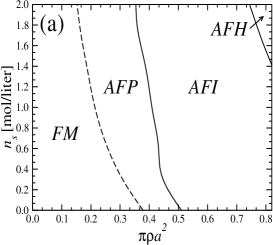
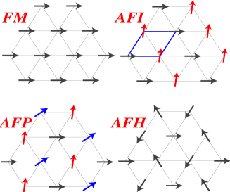
When counterions are condensed on strands, i.e., and , the DNA-DNA interaction is purely repulsive. The system is found to crystallize into the HEX lattice at all DNA-densities but a large variety of orientational (magnetic) structures occur, as a result of the frustration of the system. The structures are shown in Fig. 2 and the phase diagram of the DNA-salt mixture in Fig. 3(a). The phase denoted FM is a simple ferromagnetic phase, in which all DNA-molecules have the same azimuthal orientation. The phase denoted AFI displays antiferromagnetic-Ising type ordering, with half DNA-molecules having a given orientation angle if they lie on one of the sublattices and a different orientation on the other. The AFP phase has a three-state antiferromagnetic Potts yeomans:book type of ordering, with 1/3 of the spins pointing in a reference direction , 1/3 in the angle and 1/3 in the angle . Note that the angle grows with DNA concentration. Finally, the AFH-phase has the orientational ordering of the two-dimensional antiferromagnetic Heisenberg model, with spins residing in the three sublattices of the hexagonal lattice having mutual orientational angles of to one another. The transition between the FM and AFP phases is second-order but the AFP AFI and AFI AFH transitions are first order. Referring to Fig. 3(a), we see that the FM phase is stable at low DNA-concentrations. Indeed, for such average intermolecular separations the optimal orientation angle between the molecules is zero. The nontrivial phases arise at higher densities of the aggregates. Similar mesophases were found recently within the framework of a phenomenological Landau theory lorman:prl:01 .
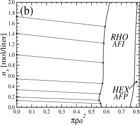
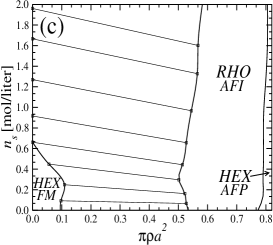
When counterions condense in grooves, an attraction between the DNA-molecules arises, since the possibility of having positively charged parts of one molecule approaching close to negatively charged parts of the other through an appropriate mutual orientation opens up. This leads to broad phase coexistence lines between dense DNA-aggregates and DNA-free solutions. This is demonstrated in Fig. 3(b) for the case , and for . Lowering , i.e., increasing the Coulomb repulsion, opens up a hexagonal phase at small DNA- and electrolyte-concentrations, see Fig. 3(c). In the one-phase region, a rhombic phase shows up for moderate to high densities and, due to packing constraints, a HEX crystal appears at very high DNA-concentrations. A strong qualitative difference in the macroscopic behavior of columnar DNA assemblies arises, depending on whether the counterions condense on strands or in grooves. In the former case, all transitions are ‘magnetic’ in nature. In the latter, DNA-bundling takes place and rhombic lattices are stabilized.
The predictions of the theory ask for experimental verification. Such a task is not easy, since the reliable data up to date refer only to highly concentrated phases grimm:physicab:91 , where the number of the basic assumptions inherent to the form of the pair potential may be questioned (the Debye-Bjerrum approximation, independence of solvent dielectric constant on the aggregate density, effects of nonlocal polarizabilty, etc.). Such a verification might become possible with the increase of experimental resolution in X-ray diffraction, which could open the way for the study of less dense aggregates. The predicted specific effect of cation adsorption on the phase diagram is particularly challenging. Since the adsorption isotherms and the distributions of the adsorbed ions between the minor and major grooves are poorly known, one should concentrate here on the qualitative effects, i.e., the (dis)appearance of mesophases triggered by the presence of different DNA condensing counterions.
The authors are thankful to A. Cherstvy, S. Leikin, A. Parsegian, and A. Esztermann for numerous useful discussions and acknowledge financial support through the DFG, grants LO 418/6 and KO 139/4.
References
- (1) G. S. Manning, Q. Rev. Biophys. 11, 179 (1978).
- (2) R. W. Wison and V. A. Bloomfield, Biochem. 18, 2192 (1979); J. Widom and R. L. Baldwin, J. Mol. Biol. 144, 431 (1980); P. G. Heath and J. M. Schurr, Macromolecules 25, 4149 (1992).
- (3) J. Pelta, F. Livolant, and J.-L. Sikorav, J. Biol. Chem. 271, 5656 (1996).
- (4) D. C. Rau, B. Lee, and V. A. Parsegian, Proc. Nat. Acad. Sci. USA 81, 2621 (1984).
- (5) V. A. Bloomfield, Curr. Opin. Struct. Biol. 6, 334 (1996).
- (6) A. H. A. Tajmir-Riahi et al., J. Biomol. Struc. Dyn. 11, 83 (1993); I. Fita et al., J. Mol. Biol. 167, 157 (1983); N. V. Hud et al., Biochem. 33, 7528 (1994); X. Shui et al., Biochem. 37, 8341 (1998).
- (7) R. Podgornik, H. H. Strey, and V. A. Parsegian, Curr. Opin. Colloid Interface Sci. 3, 534 (1984).
- (8) A. A. Kornyshev and S. Leikin, Phys. Rev. Lett. 82, 4138 (1999).
- (9) A. A. Kornyshev and S. Leikin, J. Chem. Phys. 107, 3556 (1997); Erratum, ibid. 108, 7035 (1998).
- (10) A. A. Kornyshev and S. Leikin, Proc. Nat. Acad. Sci. USA 95, (1998); Biophys. J. 75, 2513 (1998); Phys. Rev. Lett. 86, 3666 (2001).
- (11) E. Allahyarov and H. Löwen, Phys. Rev. E 62, 5542 (2000).
- (12) R. R. Sinden, DNA Structure and Function (Academic, New York, 1994).
- (13) H. H. Strey et al., Phys. Rev. Lett. 84, 3105 (2000).
- (14) The ‘two-cosine approximation’, , follows from Eq. (LABEL:interaction) by truncating the sum at . The truncated form already captures the main effect of the interaction, i.e., the frustration of the optimal azimuthal angle.
- (15) H. Graf and H. Löwen, Phys. Rev. E 57, 5744 (1998); ibid. 59, 1932 (1999).
- (16) J. M. Yeomans, Statistical Mechanics of Phase Transitions (Clarendon, Oxford, 1992).
- (17) V. Lorman, R. Podgornik, and B. Žekš, Phys. Rev. Lett. 87, 218101 (2001).
- (18) R. Langridge et al., J. Mol. Biol. 2, 19 (1960); S. D. Dover, J. Mol. Biol. 110, 699 (1977); H. Grimm and A. Ruprecht, Physica B 174, 291 (1991).