[1,2,3]\fnmRainer A. \surBöckmann \equalcontThese authors contributed equally to this work.
1]Computational Biology, Department of Biology, Friedrich-Alexander-Universität Erlangen-Nürnberg, Erlangen, Germany 2]Erlangen National High-Perfomance Computing Center (NHR@FAU), Erlangen, Germany 3]FAU Profile Center Immunomedicine (FAU I-MED), Erlangen, Germany *]rainer.boeckmann@fau.de
Revisiting Lipid Nanoparticle Composition and Structure: A Critical Take on Simulation Approaches
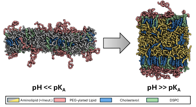
The impact of pH on the structural organization of lipid nanoparticles (LNPs) has been well-documented, with both all-atom and coarse-grained simulations revealing a pH-dependent phase transition [1, 2, 3, 4]. Specifically, at low pH (where aminolipids are fully protonated), LNP-mimetic systems exhibit a lipid bilayer phase, while at high pH, they transition to an LNP-like structure (Fig. 1). This structure comprises a hydrophobic core primarily made up of aminolipids and cholesterol, surrounded by a monolayer of helper lipids (e.g., DSPC and PEGylated lipids), cholesterol, and a few aminolipids [1, 3, 5, 6].
A recent study published in PNAS [7] challenges this understanding by employing direct coexistence (DC) simulations. Rather than simulating complete LNPs, the authors modeled smaller cross-sectional systems with a hydrophobic core sandwiched between lipid monolayers in an aqueous environment — an approach similar to previous studies [4, 1, 6]. However, unlike prior results, these DC simulations indicated minimal structural changes between neutral and acidic pH (see Fig. 5 of [7]), with protonated lipids remaining uniformly distributed within the core (as shown in Fig. 2B of [7]), leading to charge densities of approximately 0.4-0.5 e/nm3. This charge distribution implies a significant electrostatic energy of approximately kJ/mol for LNPs of 100-200 nm in size (as in the LNP1 and LNP2 systems described in [7], dielectric constant assumed). This level of energy is roughly 20–140 times the equivalent of TNT.
Such a charge distribution seems improbable and may stem from the chosen simulation ensemble. By fixing the monolayer area while coupling only the -direction (normal to the surface) to environmental pressure ( ensemble), the study’s setup restricts the system’s ability to undergo structural modifications that would naturally arise from the protonation of all aminolipids (from a core-monolayer phase to a lipid bilayer) or from changes in LNP formulation. This constraint likely hinders an unbiased exploration of the LNP core-shell architecture and establishes an energetic barrier that inhibits RNA escape under neutral pH conditions, a phenomenon suggested from previous unbiased simulations [1, 3]. Moreover, differences observed between the two LNP formulations, such as DSPC-water micellar structures below the LNP surface, seem predetermined by the systemic bias introduced by this setup and highly sensitive to system configuration. Therefore, the simulations by Garaizar et al. [7] may not provide conclusive insights into the role of varying LNP compositions on LNP activity.
References
- \bibcommenthead
- [1] Trollmann, M. F. W. & Böckmann, R. A. mRNA lipid nanoparticle phase transition. Biophys. J. 121, 3927–3939 (2022).
- [2] Philipp, J. et al. pH-dependent structural transitions in cationic ionizable lipid mesophases are critical for lipid nanoparticle function. Proc. Natl. Acad. Sci. U. S. A. 120, e2310491120 (2023).
- [3] Paloncýová, M. et al. Atomistic insights into organization of RNA-loaded lipid nanoparticles. J. Phys. Chem. B 127, 1158–1166 (2023).
- [4] Ramezanpour, M. et al. Ionizable amino lipid interactions with POPC: implications for lipid nanoparticle function. Nanoscale 11, 14141–14146 (2019).
- [5] Yanez Arteta, M. et al. Successful reprogramming of cellular protein production through mRNA delivered by functionalized lipid nanoparticles. Proc. Natl. Acad. Sci. U. S. A. 115, E3351–E3360 (2018).
- [6] Kjølbye, L. R. et al. Martini 3 building blocks for lipid nanoparticle design. ChemRxiv (2024).
- [7] Garaizar, A. et al. Toward understanding lipid reorganization in RNA lipid nanoparticles in acidic environments. Proc. Natl. Acad. Sci. U. S. A. 121, e2404555121 (2024).