IGUANe: a 3D generalizable CycleGAN for multicenter harmonization of brain MR images
Abstract
In MRI studies, the aggregation of imaging data from multiple acquisition sites enhances sample size but may introduce site-related variabilities that hinder consistency in subsequent analyses. Deep learning methods for image translation have emerged as a solution for harmonizing MR images across sites. In this study, we introduce IGUANe (Image Generation with Unified Adversarial Networks), an original 3D model that leverages the strengths of domain translation and straightforward application of style transfer methods for multicenter brain MR image harmonization. IGUANe extends CycleGAN architecture by integrating an arbitrary number of domains for training through a many-to-one strategy. During inference, the model can be applied to any image, even from an unknown acquisition site, making it a universal generator for harmonization. Trained on a dataset comprising T1-weighted images from 11 different scanners, IGUANe was evaluated on data from unseen sites. The assessments included the transformation of MR images with traveling subjects, the preservation of pairwise distances between MR images within domains, the evolution of volumetric patterns related to age and Alzheimer’s disease (AD), and the performance in age regression and patient classification tasks. Comparisons with other harmonization and normalization methods suggest that IGUANe better preserves individual information in MR images and is more suitable for maintaining and reinforcing variabilities related to age and AD. Future studies may further assess IGUANe in other multicenter contexts, either using the same model or retraining it for applications to different image modalities.
keywords:
brain MRI , harmonization , multisite , brain age , Alzheimer , image synthesis1 Introduction
In the last few years, MRI data from multiple acquisition sites have become more accessible, leading to an increase in sample sizes and, consequently, enhanced statistical power in brain MRI studies. However, heterogeneities related to technical factors, including manufacturer, field strength, sequence design, and RF coil, have been reported [9, 33]. These differences can impact statistical analyses, even when efforts have been made to standardize acquisition protocols [41, 61]. Technical variabilities in medical images can also limit the generalizability of predictive models based on machine learning [3, 76].
Various retrospective harmonization methods have been proposed to face this problem. Two broad categories exist: (i) statistical and machine learning methods harmonizing image features, and (ii) deep learning models processing MR data at the voxel level. The first category is mainly composed of methods derived from ComBat [8, 23, 53]. Efficiency was demonstrated for specific applications but these methods require different harmonization procedures for each set of features and are not well-suited for processing whole brain images. In the second category, certain approaches have been designed for specific prediction tasks [19, 32, 70]. Therefore, they require a training procedure for each application and are limited to prediction contexts.
Researchers have addressed these limitations by focusing on image-to-image translation (IIT). Some models are based on supervised learning, proving efficient with small datasets but requiring traveling subjects [17, 65, 66]. In contrast, many recently proposed deep learning models operate in an unsupervised manner, enabling harmonization without the need of traveling subjects. However, certain limitations persist, hindering widespread adoption by end-users: potential loss of biological information, insufficient validation on external datasets, important computational resource requirements, the lack of readily usable code for method reuse, and the independent processing of 2D slices from the same volume [13, 35].
In this work, we propose IGUANe (Image Generation with Unified Adversarial Networks), a 3D unsupervised IIT model designed for inter-site harmonization of structural brain MR images. It extends the CycleGAN framework with a many-to-one strategy that enables the harmonization of any MR image after training with a large multicenter dataset. Our experiments, conducted on T1-weighted (T1w) brain MR images using a diverse training set from various studies, showcase the model’s ability to generalize to unseen sites. Validations and comparisons with different methods focus on image similarity metrics, conservation and reinforcement of biological patterns, and prediction tasks. The online availability of both code and trained models allows for application to any T1w brain images and training of new models for harmonization of other image types.
2 Related works
In this section, we focus on unsupervised IIT employed for harmonization of structural brain MR images.
2.1 Unsupervised frameworks
The first category of unsupervised IIT is domain translation, CycleGAN [77] being one of the most renowned models. Studies have demonstrated their efficacy in harmonizing brain MR images [10, 21, 29, 49, 51, 57]. However, CycleGAN requires training for every pair of sites and may not fully leverage complementary information from all sites. Other domain translation models have addressed this issue [3, 22, 25, 44].
The second category of unsupervised IIT is style transfer, focusing on image-level style rather than domain-level characteristics [26]. For instance, the model of Zuo et al. [80] is trained with assumptions about MRI volumes: slices at different locations share the same contrast but have different anatomic information. Similar assumptions guide other methods, supplemented by self-supervised learning through pairing different MRI contrasts [79] or creating pairings with image processing functions [7]. The simplified version of StarGAN v2 proposed by Liu et al. [43] does not use domain information.
2.2 Evaluation metrics
The use of image similarity metrics on datasets with traveling subjects is a prevalent practice in assessing image-to-image translation models for harmonization [7, 25, 43, 44, 49, 55, 57, 79, 80]. These datasets, providing ground truth for harmonization, are well-suited for evaluation. However, these validations are often constrained to a small number of subjects scanned at a few sites. Consequently, they are supplemented by assessments of similarities between sites before and after harmonization. This includes site prediction [7, 49], visualization of MRI data decomposed in two-dimensional spaces [7, 43, 49, 57], quantification of differences in feature distributions across sites [44, 57] and comparison of intensity histograms [7, 57]. However, these evaluations don’t account for biological patterns and are applicable only to MR images that can be clustered into domains (e.g. sites). To address these limitations, assessments of the performances of predictive models [3, 7, 10, 25, 43, 49, 55, 57] and the examination of the evolution of specific biological patterns with harmonization [43, 57] have been proposed. It is noteworthy that experiments on the generalization of harmonization models to unseen sites have been limited to visual assessment and image similarity metrics with traveling subjects [7, 25, 43].
3 Materials and Methods
3.1 MRI datasets
We used T1w brain MR images from public databases for this study, limiting inclusion to participants aged between 18 and 80 years. Metadata enabling the identification of the used images are available in our online repository (section 3.5). The characteristics of each dataset are given below.
Training dataset
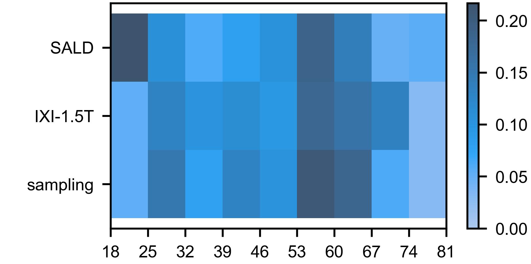
In this collection, we included MR images from eight studies: SALD [73], IXI333https://brain-development.org/ixi-dataset/ accessed 2022-01-15, OASIS-3 [42], NKI-RS [50], NMorphCH444http://otto.fsm.northwestern.edu/ accessed 2020-01-15, AIBL [20], HCP Young Adult555https://www.humanconnectome.org/study/hcp-young-adult accessed 2020-01-15 and the International Consortium for Brain Mapping666https://ida.loni.usc.edu/ accessed 2023-03-12 (ICBM). All participants were healthy controls. MR images were acquired using eleven different machines, referred to as domains in our harmonization model (section 3.2). Each participant was exclusively present in a single domain. Demographic and scanner information is given in Table 1.
| Study | Scanner model | Field strength, Tesla | Nb of MR images / nb of participants | Age, years# | Males, % |
|---|---|---|---|---|---|
| SALD | Siemens Magnetom TrioTim | 3 | 494/494 | 38 | |
| IXI | Philips Intera | 1.5 | 305/305 | 48 | |
| IXI | Philips Intera | 3 | 176/176 | 43 | |
| OASIS-3 | Siemens Magnetom TrioTim | 3 | 857/350 | 33 | |
| OASIS-3 | Siemens Biograph mMR PET-MR | 3 | 412/311 | 46 | |
| NKI-RS | Siemens Magnetom TrioTim | 3 | 249/247 | 40 | |
| NMorphCH | Siemens Magnetom TrioTim | 3 | 141/44 | 53 | |
| AIBL | Siemens Magnetom TrioTim | 3 | 489/280 | 46 | |
| HCP | Siemens Connectome Skyra | 3 | 402/402 | 39 | |
| ICBM | Siemens Sonata | 1.5 | 677/135 | 48 | |
| ICBM | Philips ACS III | 1.5 | 145/145 | 56 | |
| # Age is expressed as mean standard deviation. | |||||
Traveling subject dataset
The SRPBS traveling subject MRI dataset [64] includes brain MR images from 9 healthy male participants (age range 24-32 years). In this study, we used a total of 97 images acquired with 11 different machines (GE, Siemens and Philips manufacturers).
Generalization dataset
We included MR images from five studies that were unseen during the training phase: the Alzheimer’s Disease Neuroimaging Initiative777https://adni.loni.usc.edu/ accessed 2023-03-12 (ADNI), the Mind Clinical Imaging Consortium (MCIC) database [30], PPMI [47], COBRE [1] and ABIDE888https://fcon_1000.projects.nitrc.org/indi/abide/ accessed 2023-03-12. All participants were healthy controls. Demographic and scanner information is given in Table 2.
| Study | Manufacturer# | Field strength, Tesla# | Nb of MR images / nb of participants | Age, years† | Males, % |
| ADNI | GE (115); Philips (109); Siemens (104) | 3 (199); 1.5 (129) | 328/216 | 45 | |
| MCIC | Siemens | 1.5 (192); 3 (52) | 244/89 | 67 | |
| PPMI | Siemens (202); Philips (39) | 3 (215); 1.5 (24); unknown (2) | 241/141 | 64 | |
| COBRE | Siemens | 3 | 227/91 | 73 | |
| ABIDE | Siemens (105); Philips (34) | 3 | 139/139 | 88 | |
| # The number of MR images is indicated in brackets if there are several options. | |||||
| † Age is expressed as mean standard deviation. | |||||
Clinical dataset
We used data from ADNI, AIBL and MIRIAD [45] to investigate the impact of harmonization on patterns related to Alzheimer’s disease. We specifically selected participants diagnosed as cognitively normal (CN) or with Alzheimer’s disease (AD). To conduct a CN/AD classification (section 3.4.4), we divided the collection in four sets: AD_train for training, AD_test for testing, AD_GE for generalization to MR images from GE manufacturer (no GE MR image is in the AD_train, AD_test nor Training datasets) and MIRIAD for generalization to MR images from another study. Importantly, none of the participants in the AD_train dataset were included in the other three datasets. Additionally, no subject or scanner was present during the training of the harmonization model. Demographic and scanner information is detailed in Table 3.
| Dataset | Manufacturer# | Field strength, Tesla# | Nb of MR images / nb of participants | CN/AD, %† | Age, years‡ | Males, % | ||
| CN | AD | CN | AD | |||||
| AD_train | Siemens (1286); Philips (489) | 3 (1132); 1.5 (643) | 1775/546 | 50/50 | 44 | 53 | ||
| AD_test | Siemens (518) Philips (169) | 3 (317); 1.5 (270) | 687/237 | 50/50 | 49 | 49 | ||
| AD_GE | GE | 3 (642); 1.5 (653) | 1295/275 | 50/50 | 46 | 53 | ||
| MIRIAD | GE | 1.5 | 652/64 | 33/67 | 46 | 41 | ||
| # The number of MR images is indicated in brackets if there are several options. | ||||||||
| † CN: cognitively normal; AD: Alzheimer’s disease | ||||||||
| ‡ Age is expressed as mean standard deviation. | ||||||||
3.2 IGUANe model
In this section, we present the main characteristics of the IGUANe model. Additional information is given in A.
3.2.1 MRI preprocessing
To eliminate the most trivial technical variabilities in the data and facilitate harmonization, we preprocessed the MR images as follows: (i) skull-stripping with HD-BET [36], (ii) bias correction with N4ITK [67], (iii) linear registration to 1 mm3 MNI space with FSL-FLIRT [38] (six degrees of freedom), (iv) cropping to 160 x 192 x 160 voxels, and (v) division of intensities by the median brain intensity. This last step standardizes the median intensity inside the brain while keeping the background at 0. We found it to be more robust to outliers than the classical rescaling approach.
3.2.2 Universal generator
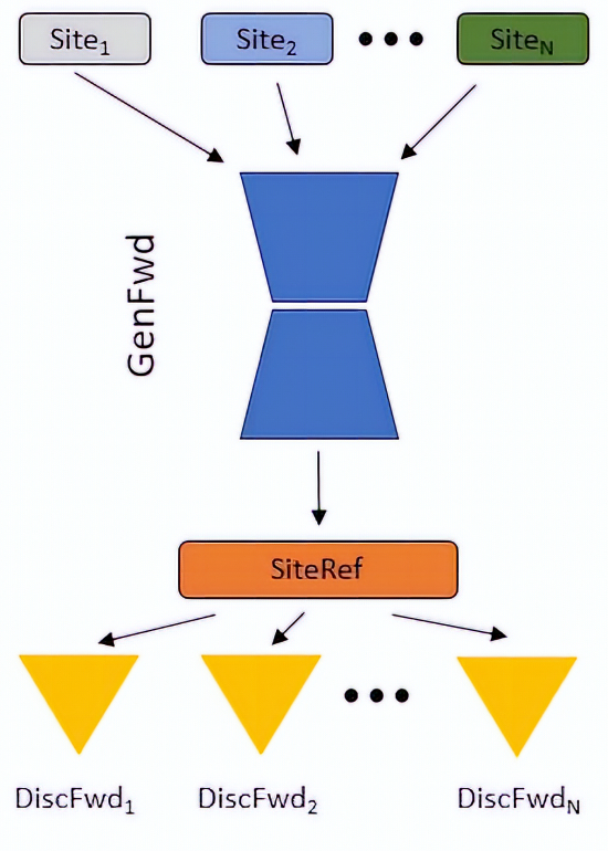
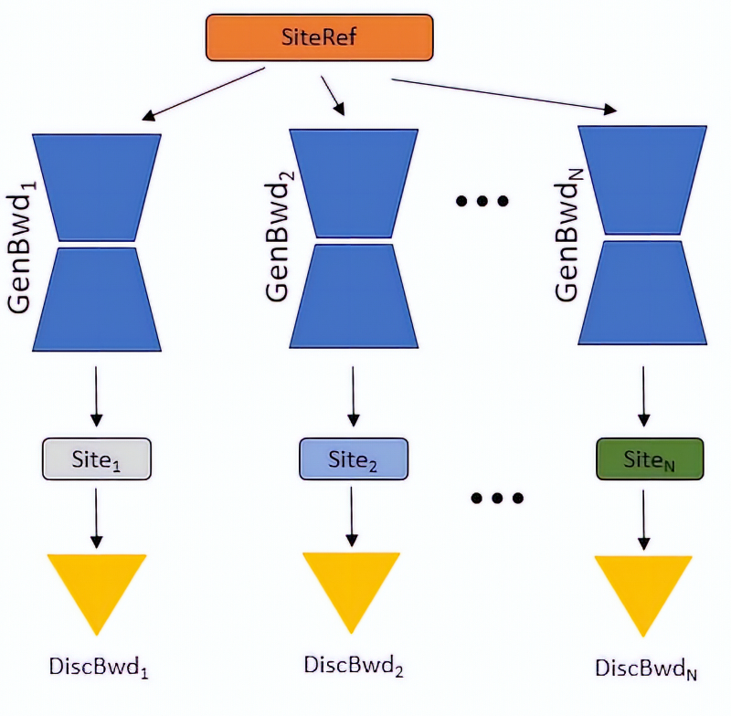
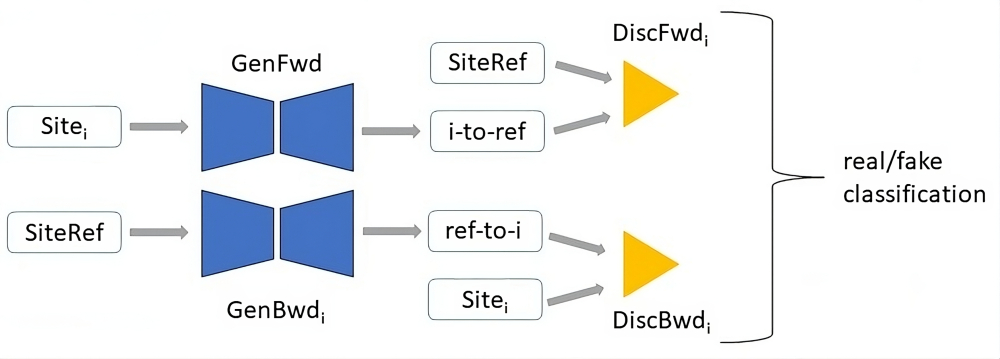
The IGUANe framework is an extension of CycleGAN [77] and has been inspired by the many-to-one strategy by Gao et al. [25]. The objective is to train a generator, , to translate images from source sites (, , …, ) to a reference site, . To be able to apply cycle-consistency constraints [77], backward generators (, , …, ) are set up to translate images from to each source site. For adversarial training, forward discriminators (, , …, ) learn to distinguish real images from harmonized images generated by from each source site. Similarly, backward discriminators (, , …, ) learn to distinguish real images from each source site from images harmonized with the backward generators.
The modules of the framework are illustrated in Fig. 1. During inference, is used to harmonize MR images. The assumption is that a sufficiently diverse training set in terms of acquisition sites and biological characteristics will enable the harmonization of any MR image, including those with an unknown acquisition site.
3.2.3 Training procedure
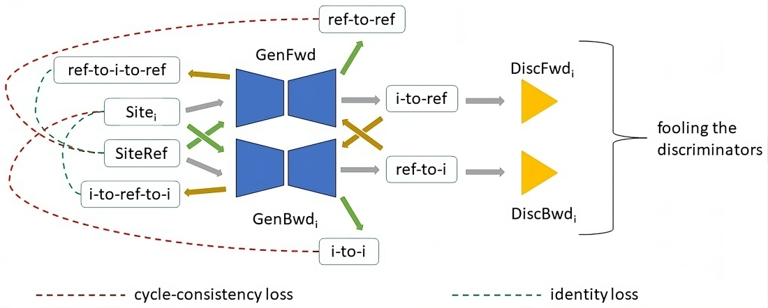
The training procedure is illustrated in Fig. 2. Mean square error is used as the adversarial loss [46]. To generate realistic MR images, is successively trained adversarially with the forward discriminators. The backward generators are trained in the same way with each backward discriminator. To preserve original image information and regulate the generator trainings, cycle-consistency and identity losses are used [77]. The cycle-consistency losses are calculated by having collaborate with each backward generator through translation and inverse translation in both directions.
3.2.4 Network architectures
The generator architectures follow the one described by Dewey et al. [17], incorporating multiple skip connections to favor the conservation of anatomical details. A notable difference is that 3D convolutions are used in IGUANe, as opposed to 2D, to be able to process whole brain images. Additionally, for enhanced preservation of the original images’ content, the task of the generator networks has been modified towards residual learning, which means that the generator output is added to the input image to obtain the final harmonized image [5]. The last activation function before the addition is tanh, to enable both negative and positive residuals. During inference, negative voxels in the output volume are clipped to the background value.
The discriminators are patchGAN classifiers [37] with 3D convolutions and a receptive field of voxels.
Given that the model is tailored to process skull-stripped MR images (section 3.2.1), a substantial portion of the images is background. By default, the background is associated with the minimal intensity value in the image. In IGUANe, the background is updated and set to the median brain intensity before feeding the images to the generators and discriminators. The aim is to assign a more neutral value to a portion of the image that, despite its large size, should not influence the harmonization process. Additionally, the original brain mask is applied after each image translation to guide the generators to focus on brain intensities during training [57].
3.2.5 Implementation
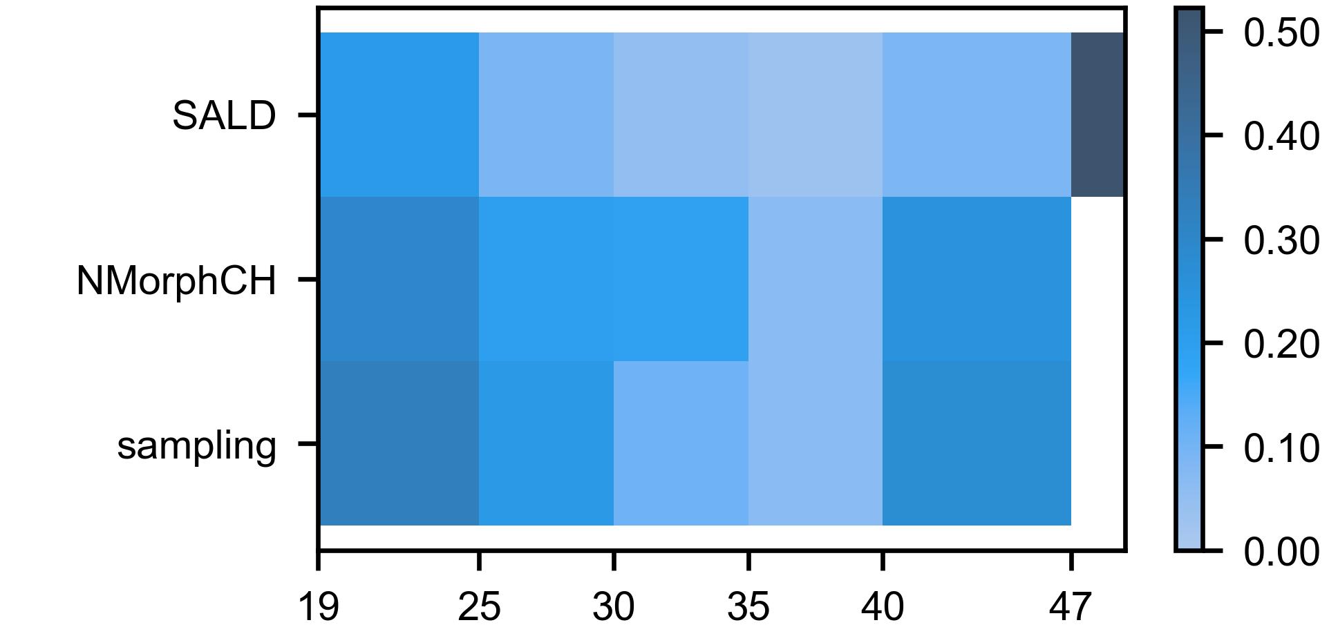
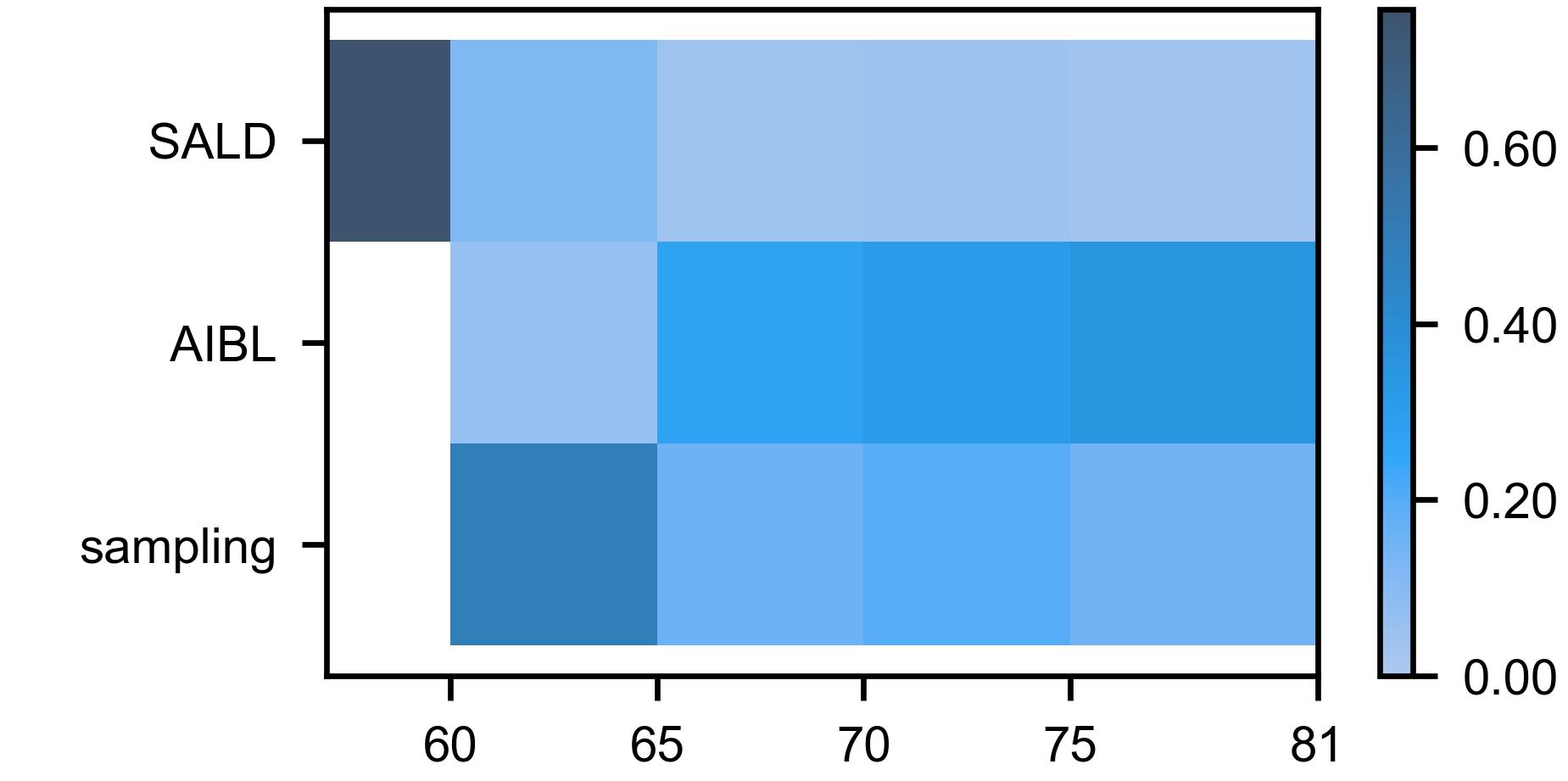
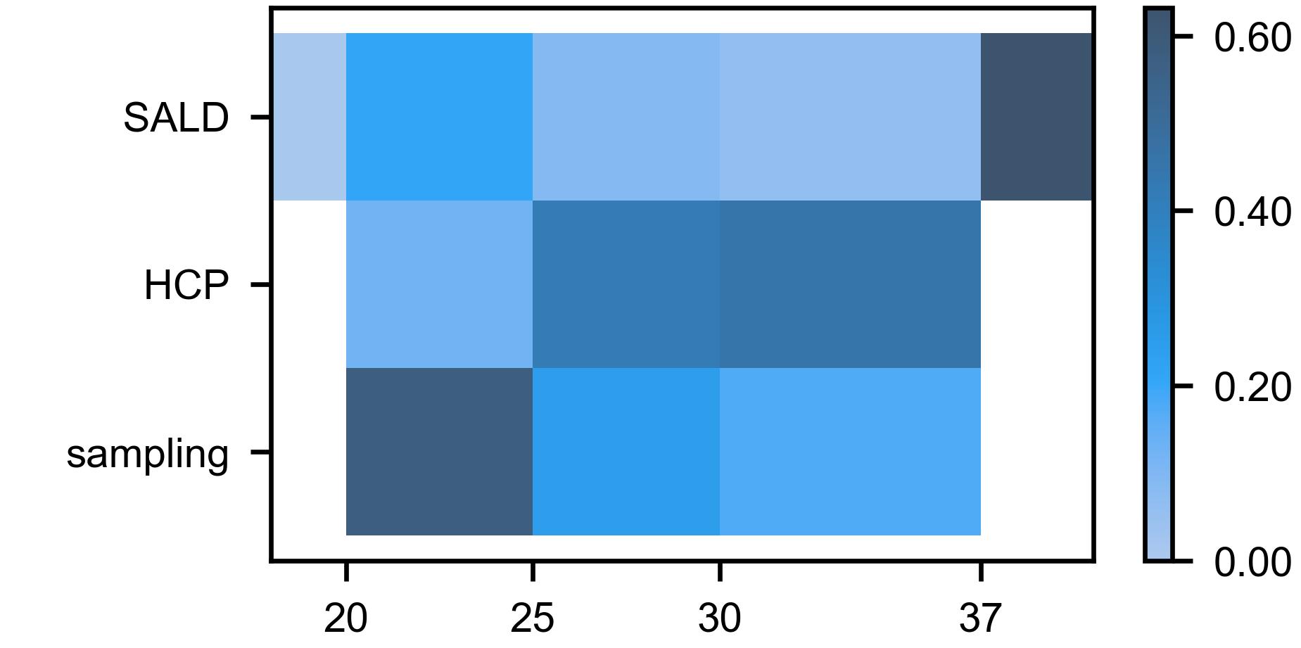
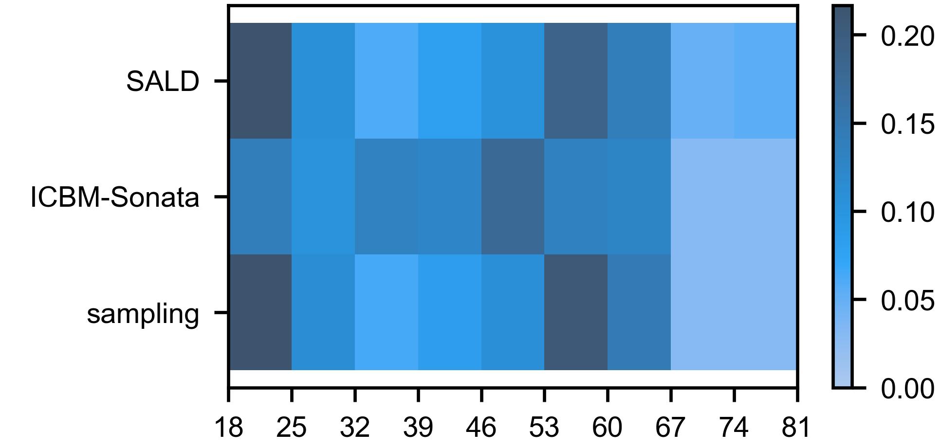
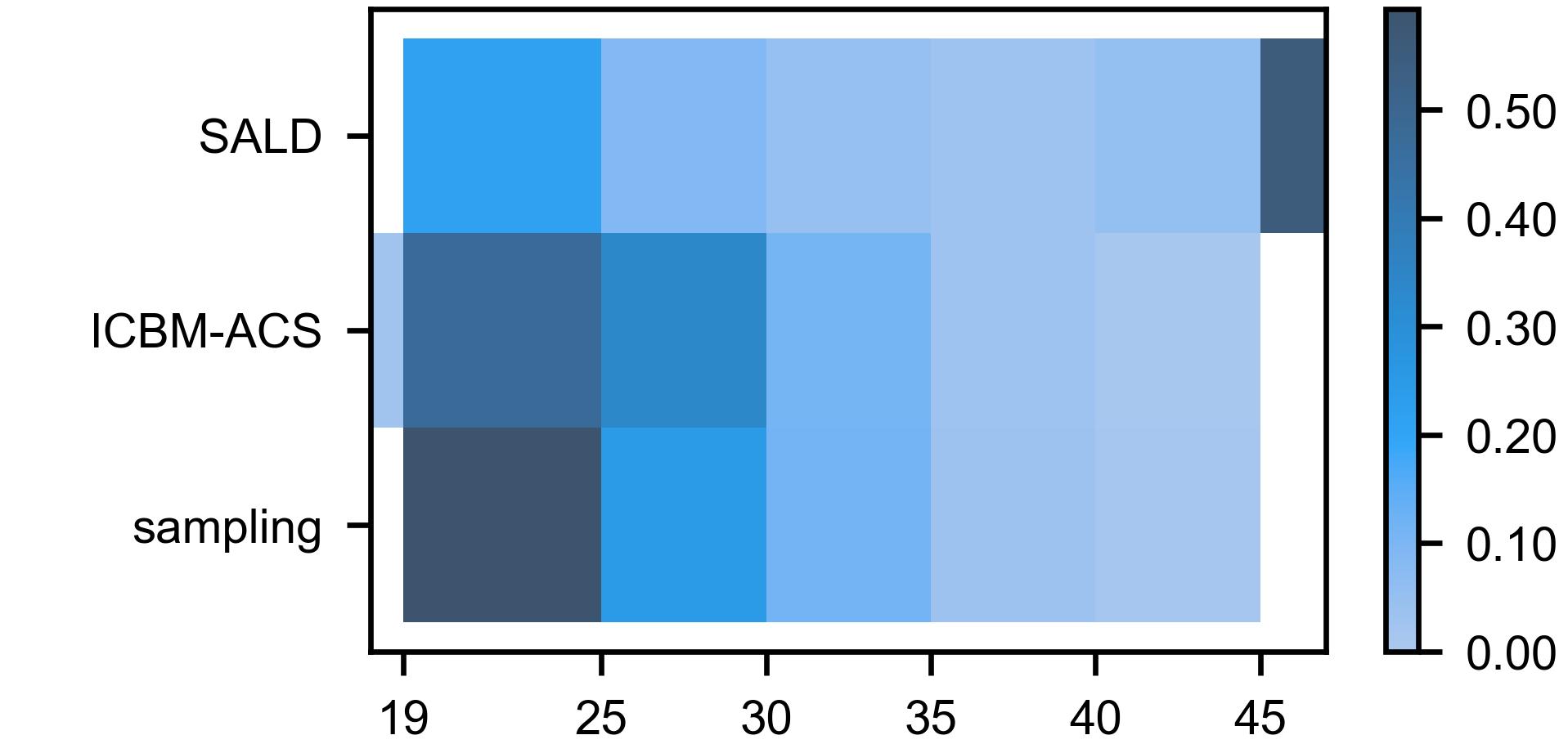
We trained IGUANe using the Training dataset, choosing SALD as the reference site due to its large number of MR images and a wide age range (19-80 years). The remaining ten sites in the Training dataset served as the source sites. Given the age imbalance between SALD and each source site (Table 1), we implemented a sampling strategy for each source site to achieve a balanced age distribution [57] with SALD. A probability distribution, based on participant ages, was thus effective for sampling MR images during the training sub-steps of each source site. The probability distributions used in this study can be visualized in B.
To further promote the preservation of biological information, we set up a validation procedure that evaluated the current model every 5 epochs and saved the best version. For this purpose, two deep learning models pretrained for age and sex prediction using MR images from were applied to a subset of MR images from each source site harmonized towards SiteRef. The network architecture described by Cole et al. [14] was used.
In this study, training IGUANe involved optimizing 11 generators and 20 discriminators simultaneously, imposing an important computational resource cost. To save GPU memory and speed-up the computations, a mixed precision policy was adopted [48], allowing training on an NVIDIA Quadro RTX 6000 GPU with 24 GB of memory.
3.3 Comparison with reference methods
3.3.1 Normalization techniques
We implemented two widely adopted approaches in MR multicenter studies: histogram matching (HM) [3, 24, 51, 56, 74] with the SALD dataset for establishing the standard scale to which the intensity histograms were matched, and WhiteStripe normalization (WS) [24, 25, 74]. We used the algorithms of Shah et al. [60] and Shinohara et al. [62] for HM and WS, respectively. For both methods, we preprocessed the MR images as done for IGUANe (section 3.2.1) and used the brain masks to compute the statistics of intensity distributions.
3.3.2 Style transfer approaches
We compared the performance of IGUANe to two style transfer approaches using the code and pretrained model available online: STGAN [43]999https://github.com/USCLoBeS/style_transfer_harmonization accessed 2023-07-15 and CALAMITI [79, 78]101010https://iacl.ece.jhu.edu/index.php?title=CALAMITI accessed 2023-07-27.
For STGAN, we used the reference image from the online repository for the style code extraction and as the harmonization target. Additionally, we performed intensity rescaling, not explicitly mentioned in the paper but found effective in the online code, as part of the preprocessing steps described in the study.
For CALAMITI, the reference image from the online repository served as the harmonization target. We followed the preprocessing steps given online. We used FSL-FLIRT with six degrees of freedom and a 0.8 mm3 MNI template for registration. After the WS normalization, the last preprocessing step of CALAMITI, we scaled/shifted the MR intensities to align with the mean and the standard deviation of the normal appearing white-matter of the reference image (further details in C).
3.4 Experiments
3.4.1 Harmonization on traveling subjects
We used the images from the Traveling subject dataset to assess the ability of harmonization models to transform images from one site to their equivalent in another.
For each subject, we computed a structural similarity index (SSIM) [71] for each image pair, corresponding to each site pair. The intensity range for SSIM was set as the 99th percentile of voxel intensities from the two MR images. Since SSIM is designed for non-negative images [31, 71], we added constant intensities before performing computations on certain datasets (D).
For each harmonization method, we assessed the statistical significance of the differences in SSIM before and after harmonization using a two-tailed Wilcoxon signed-rank test for clustered data [58], treating each subject as a cluster. We then corrected the 5 p-values using the Benjamini-Hochberg procedure.
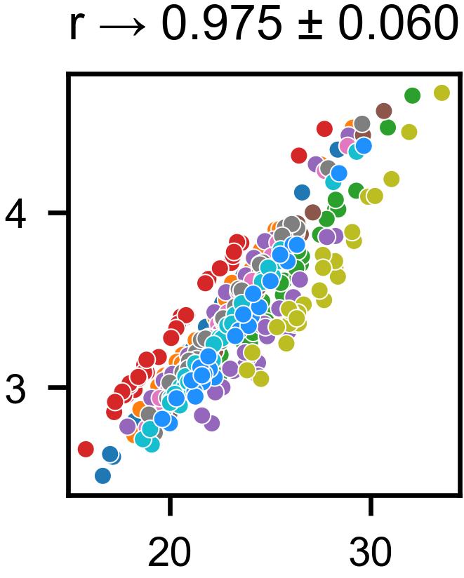
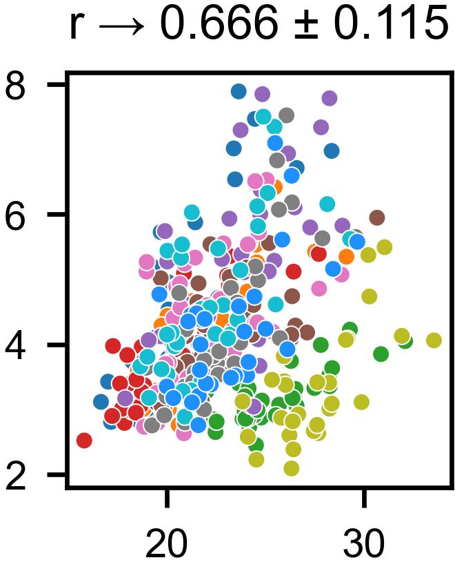
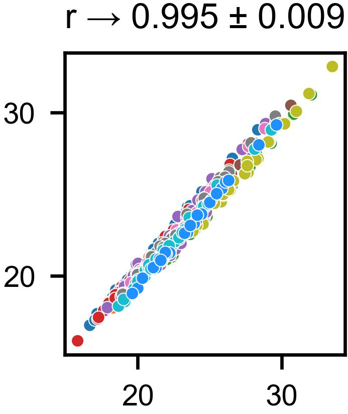
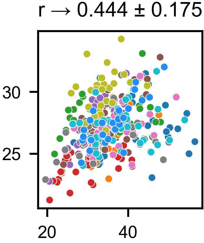
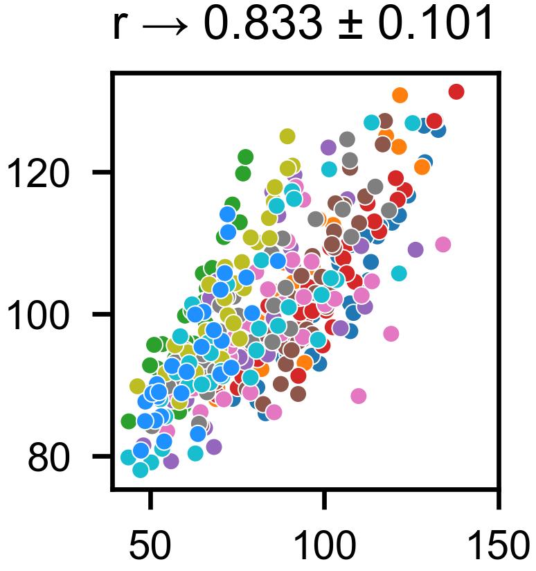
We assessed the preservation of inter-subject differences after the harmonization process [43]. Within each site, we computed the Euclidean distance for each image pair (corresponding to each subject pair) before and after harmonization. Next, we computed, for each site, the Pearson correlation coefficient between the distances before and after harmonization.
3.4.2 Correlation between age and gray-matter volume
In healthy adults, brain aging is characterized by a constant decrease in gray-matter (GM) volume [28, 34, 72].To quantify this linear trend, we segmented the MR images of the Generalization dataset using SPM12 software. We then computed the Pearson correlation coefficient between GM volume (divided by the total intracranial volume) and age. Additionally, we applied a linear least-squares regression to quantitatively assess the GM loss with age as the independent variable.
For each harmonization method, we assessed the statistical significance of the differences in correlations before and after harmonization using a two-tailed Steiger’s test [63] applied to the weighted correlation coefficients [16], where each participant’s weights sum to 1. We used the number of participants as sample size for the Steiger’s test. We then corrected the 5 p-values using the Benjamini-Hochberg procedure.
3.4.3 Hippocampal volumes: case/control effect size
The accelerated loss of hippocampal volume is a well-known pattern in Alzheimer’s disease [59]. To investigate this pattern, we randomly selected 250 CN and 250 AD participants from the Clinical dataset (mean age 71.34 and 71.90 in the CN and AD groups, respectively). Then, we segmented the MR images using SynthSeg [6] and computed Cohen’s d scores between the hippocampal volumes in the CN and the AD groups.
3.4.4 Prediction tasks
We assessed the impact of harmonization on two prediction tasks: brain age and CN/AD classification. Brain age prediction involves training a model to predict an individual’s age from brain MRI data and has been widely investigated with deep learning models [14, 27, 39]. Similarly, these approaches have been applied to classification of AD [2, 69].
We implemented brain age prediction and CN/AD classification based on MR images, using the network architecture proposed by Cole et al. [14]. For training the age prediction models, we randomly sampled 2178 MR images from the Training dataset, maintaining the proportion from each site. Evaluations were performed on MR images from the Generalization dataset. The CN/AD classifiers were trained using the AD_train dataset and evaluated on the AD_TEST, AD_GE and MIRIAD datasets. Further details are given in E.
We excluded STGAN and CALAMITI from these experiments due to the larger size of the MR images (almost ), which would have implied much larger models, making training challenging. Thus, we trained and evaluated a brain age model and a CN/AD classifier on the preprocessed MR images (IGUANe preprocessing) as well as on the MR images after IGUANe, HM and WS harmonization. This resulted in the training and evaluation of 4 brain age models and 4 CN/AD classifiers.
For each harmonization method, we assessed the statistical significance of the differences in age prediction errors before and after harmonization using a two-tailed Wilcoxon signed-rank test for clustered data [58], treating each subject as a cluster. We then corrected the 3 p-values with the Benjamini-Hochberg procedure.
3.5 Data and code availability
The IGUANe’s code is available in an online repository: https://github.com/RocaVincent/iguane_harmonization.git, along with the model weights and code for the prediction tasks. Metadata about the MR images in the various public datasets we used is also provided.
4 Results
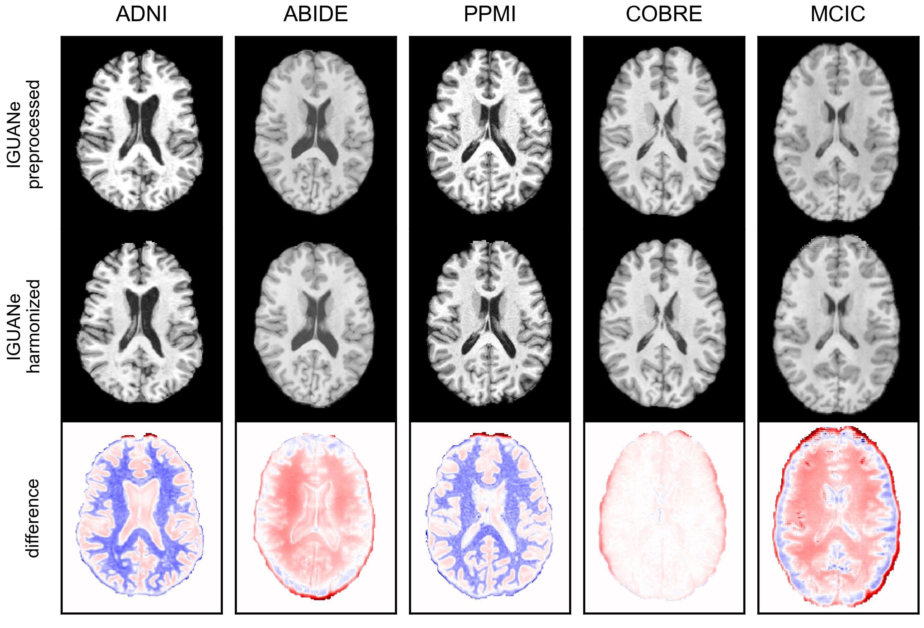
4.1 Visual assessment of harmonization
The effect of IGUANe harmonization is illustrated in Fig. 3 using MR images from the Generalization dataset. Since we designed the model for application to T1w images from any acquisition source, with a primary goal of preserving anatomical information, visualizing contrast changes may not be obvious. However, the difference maps reveal that IGUANe can apply various modifications to input contrasts based on the input image. For example, IGUANe reduced the GM/white-matter contrast in the ADNI and PPMI images, while it increased it in the ABIDE and MCIC images.
4.2 Traveling subjects evaluations
Table 4 presents the SSIM values obtained from MR images of the same subject in the Traveling subject dataset. IGUANe harmonization showed negligible change, while SSIM slightly increased with HM, WS, and STGAN. A significant decrease in SSIM was observed with CALAMITI.
| HM | WS | IGUANe | STGAN | CALAMITI | ||
|---|---|---|---|---|---|---|
| SSIM# | preprocessed | |||||
| harmonized† | ** | * | ** | * | ||
| # SSIM is expressed as mean standard deviation. | ||||||
| † Asterisks indicate significant Wilcoxon signed-rank tests (*: p0.05; **: p0.01; ***: p0.001). | ||||||
On the other hand, the intra-site correlation between the image distances before and after harmonization was much greater with IGUANe compared to the other methods (Fig. 4).

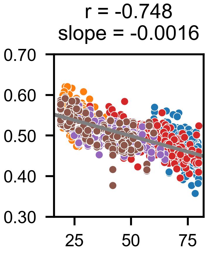
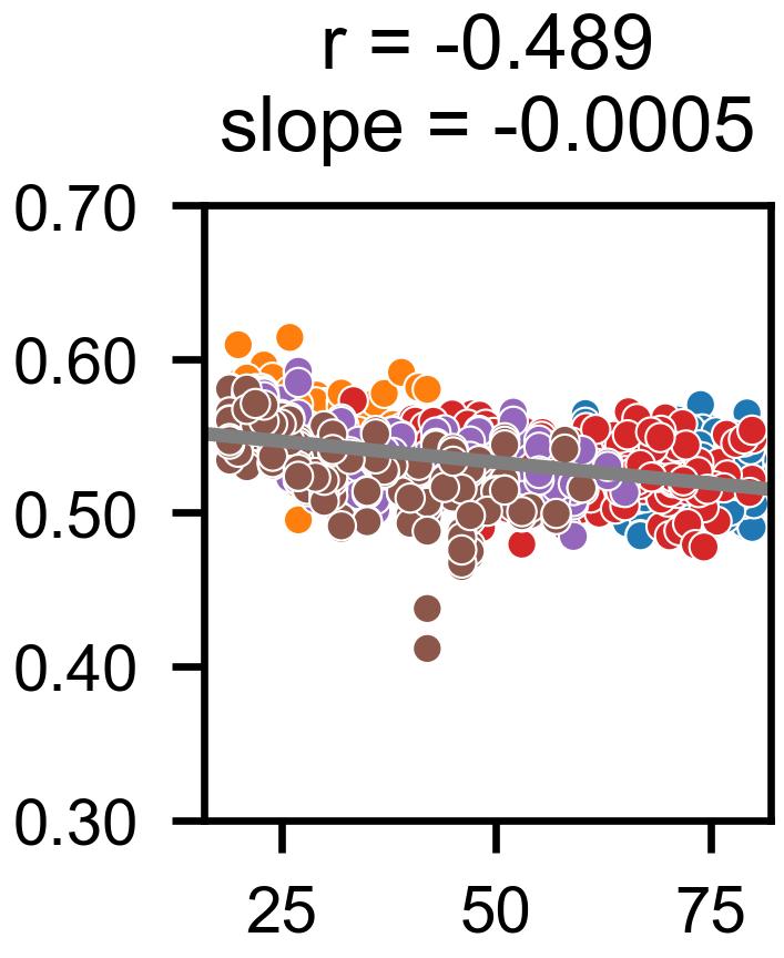
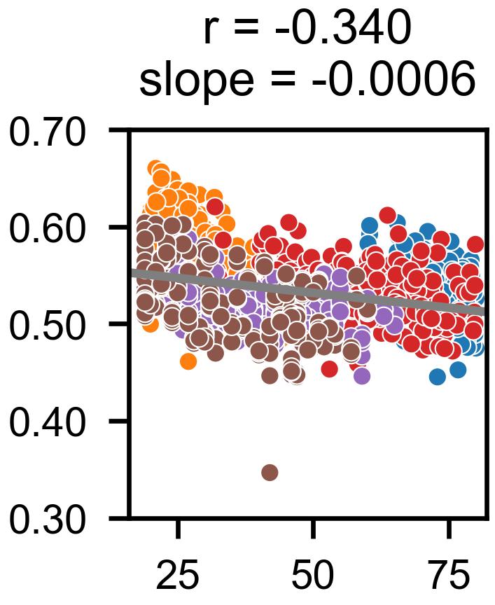
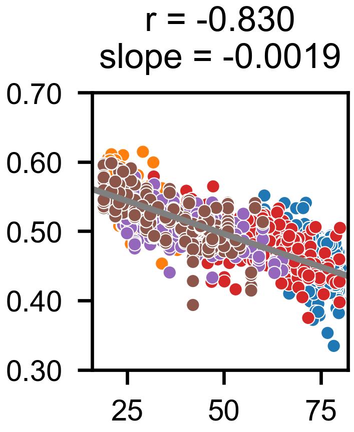
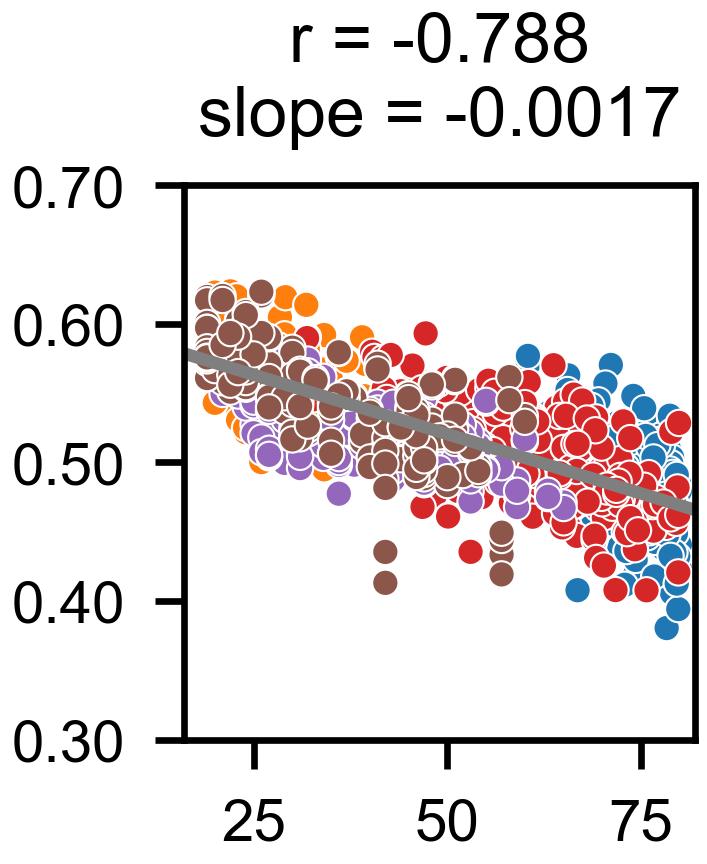
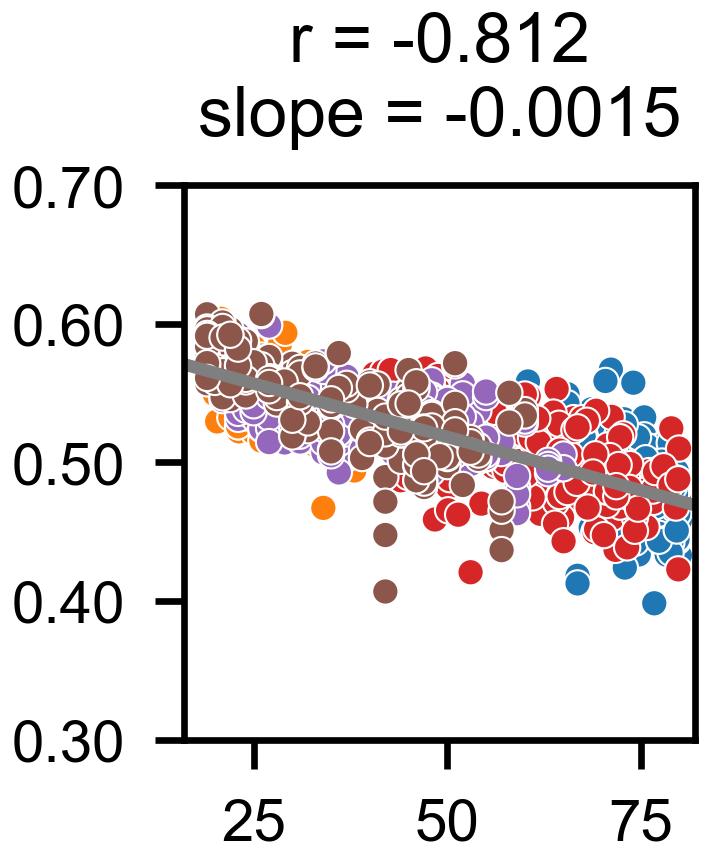
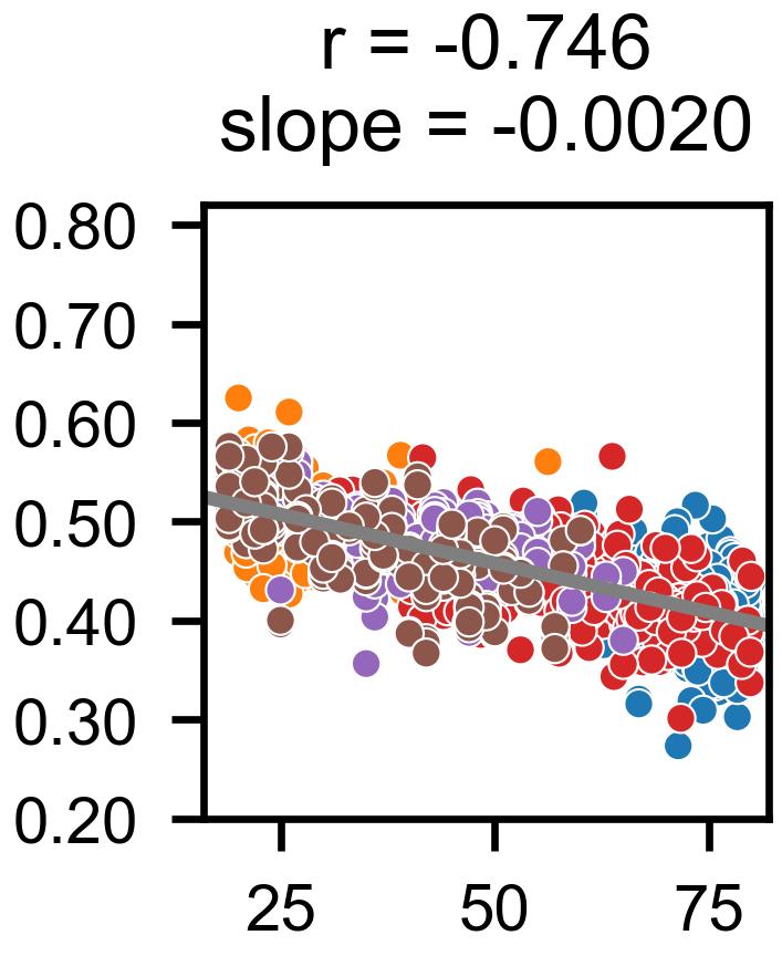
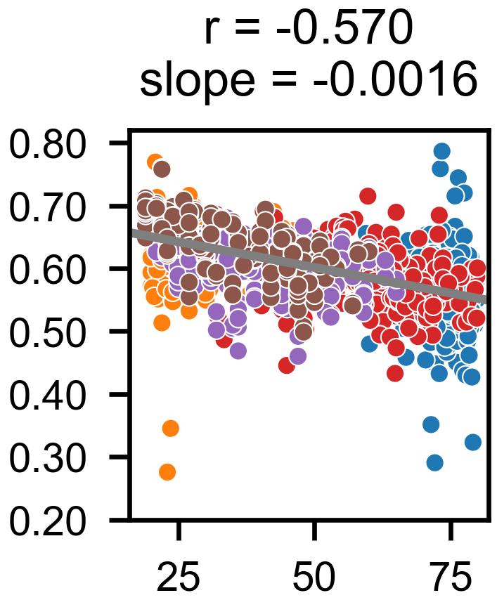
4.3 Correlation between age and gray-matter volume
In Fig. 5, it can be seen that the stronger negative correlation was achieved with IGUANe harmonization. Furthermore, IGUANe strengthened the regression slope (Fig. 5(c)), indicating a reinforcement of the GM loss pattern with harmonization. While STGAN also yielded a high correlation (Fig. 5(d)), it was less significant compared to IGUANe, and the regression slope was reduced. In contrast, HM, WS and CALAMITI diminished linearity and weakened the regression slope (Fig. 5(a), 5(b) and 5(e), respectively). Notably, the linear correlation following STGAN preprocessing was stronger than those observed after the others.

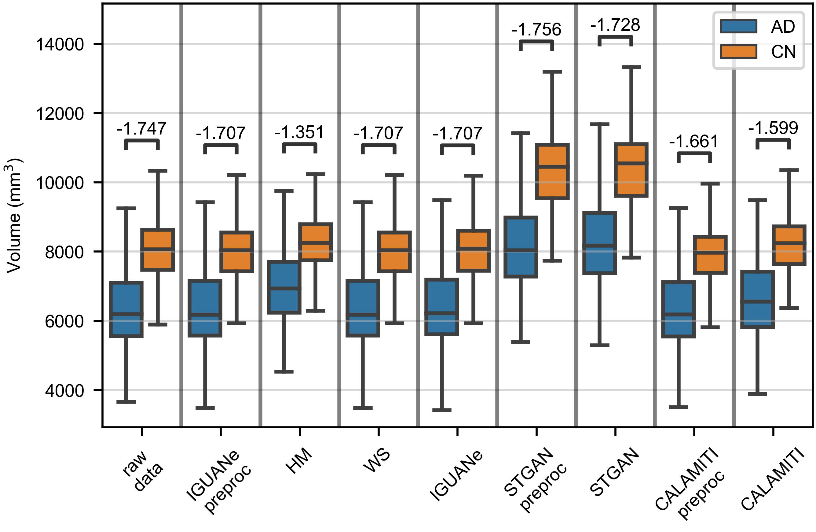
4.4 Comparison of hippocampal volumes
Several observations emerge from the comparisons between hippocampal volumes of AD and CN participants in the Clinical dataset (Fig. 6). Firstly, the measures were higher for MR images preprocessed with STGAN compared to all other measures. Secondly, IGUANe and WS better preserved the case/control distinction than STGAN, CALAMITI and HM (especially the latter). Lastly, excluding STGAN, which produced different results, the most substantial effect size was obtained from raw data for both the left and right hippocampus.
4.5 Brain age prediction
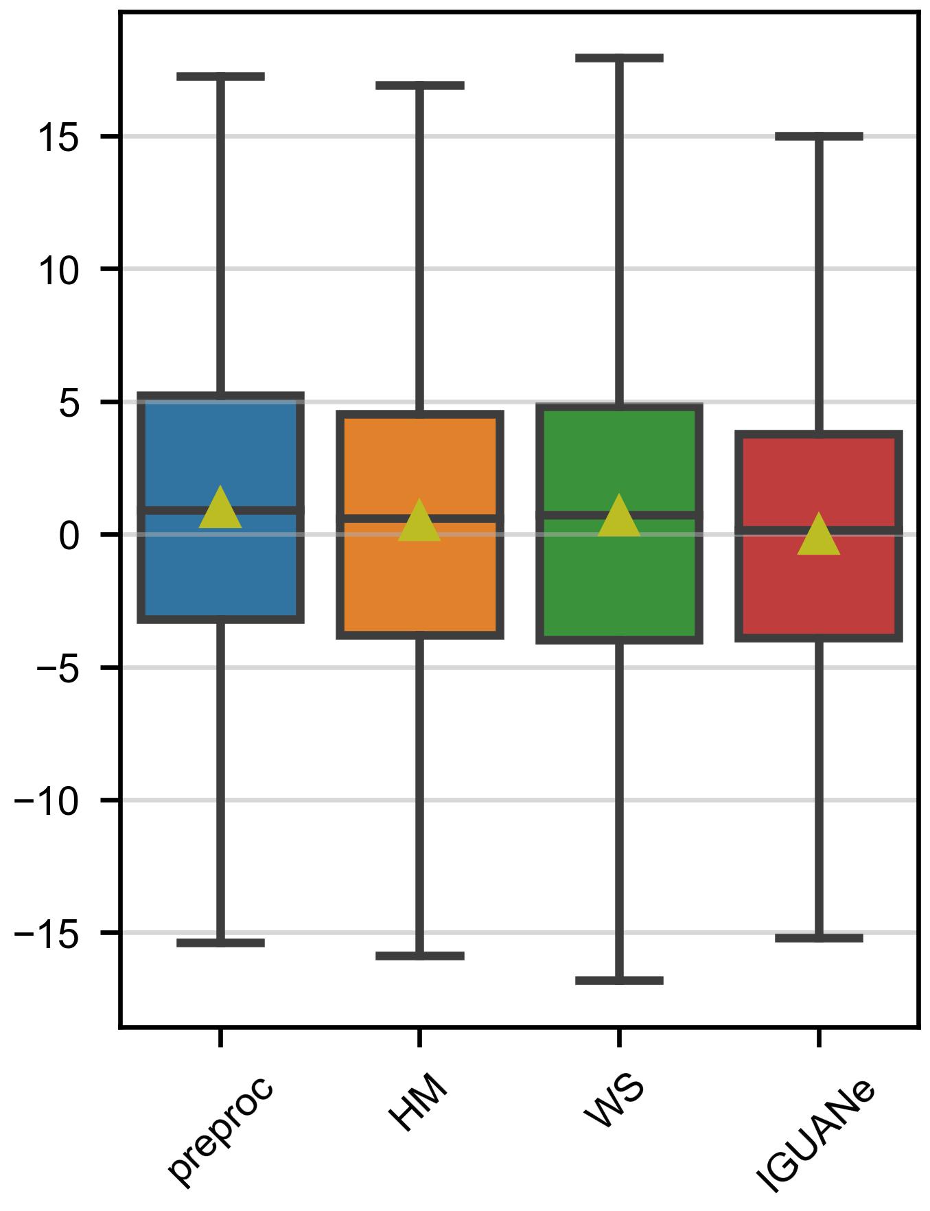
In Fig. 7(a), it can be seen that brain age prediction was improved with IGUANe (mean absolute error (MAE) decreased from 4.92 to 4.63). HM did not change the precision (MAE = 4.92), while WS led to a significant increase in errors (MAE = 5.42). A slight over-estimation pattern was observed in preprocessed data and after HM and WS, whereas the predicted age differences were more centered around zero after IGUANe (Fig. 7(b)).
4.6 Classification of healthy and Alzheimer participants
The CN/AD classification results are presented in Table 5. Across the three evaluation datasets, IGUANe achieved the highest accuracy and AUC score, except for the AUC score in the MIRIAD dataset. Overall, IGUANe demonstrated the best accuracy and AUC score, while only marginal improvements were observed with the other two methods.
| preproc# | HM | WS | IGUANe | |||||
| accuracy | AUC | accuracy | AUC | accuracy | AUC | accuracy | AUC | |
| AD_TEST† | 0.843 | 0.901 | 0.809 | 0.926 | 0.834 | 0.924 | 0.872 | 0.938 |
| AD_GE† | 0.828 | 0.894 | 0.837 | 0.920 | 0.835 | 0.902 | 0.839 | 0.921 |
| MIRIAD† | 0.880 | 0.977 | 0.920 | 0.984 | 0.931 | 0.992 | 0.933 | 0.977 |
| GLOBAL† | 0.845 | 0.919 | 0.850 | 0.939 | 0.858 | 0.934 | 0.871 | 0.942 |
| #preprocs refers to the images obtained after the IGUANe preprocessing. | ||||||||
| † Bold font indicates the best accuracies and AUC scores. | ||||||||
5 Discussion
In this study, we developed IGUANe, a model for inter-site harmonization capable of processing structural MR images from any site. Following a training phase with a dataset including 11 scanners for a total of 4347 T1w brain images, we applied IGUANe to data from additional studies without any fine-tuning. Various experiments were conducted to assess the quality of the generated images and the preservation of reinforcement of biological patterns. Comparisons with two intensity normalization techniques and two style transfer methods demonstrate the robustness of the proposed approach.
5.1 IGUANe model
Similar to StarGAN [11] and StarGAN v2 [12], IGUANe extends the CycleGAN framework and adopts a unified training procedure for harmonizing images across multiple domains in parallel. However, unlike these two methods, IGUANe employs a learning approach based on domain pairs, enabling the use of bias sampling strategies to balance biological covariates. This design choice prevents the suppression of some biological information. In this study, we implemented this strategy using age, allowing us to use training sets with different age distributions (section 3.2.5). Notably, the model by Gao et al. [25], which partially inspired IGUANe, does not support such flexibility, as it employs only one discriminator for the reference domain.
The network architectures in IGUANe are 3D and process whole brain images. To our knowledge, with the exception of Roca et al. [57], almost all existing unsupervised harmonization models have been designed for processing slices or low-volume patches. The resultant loss of spatial and contextual information in this approaches is often either unjustified or dictated by technical limitations [18, 43, 49, 51, 79]. Another originality in IGUANe is the application of residual learning. For inter-site harmonization, models can focus on learning contrast modifications rather than reproducing the entire anatomical information. Additionally, the setting of a neutral value for the background during training enhances the images generated with IGUANe; this approach was similarly used by Robinson et al. [55] in their model.
5.2 Signal quality
The visualization of harmonized MR images (section 4.1) and the SSIMs obtained with the traveling subjects (section 4.2) indicate that IGUANe introduces minimal changes to the input images. In contrast, the other tested methods are less conservative and managed to slightly increase the SSIMs, except CALAMITI. However, the analysis of inter-subject differences (section 4.2) suggests that IGUANe better maintains these differences, and the observed SSIM increase with other methods may be partly due to over-homogenization.
Liu et al. [43] demonstrated effective preservation of inter-subject variabilities with STGAN, which contrasts with our results. This disparity may stem from their experiment being based on only 10 input MR images, all acquired with a Siemens 3 Tesla from the same study included in the training of STGAN. Our findings also highlight that voxel-level similarity metrics are significantly influenced by image preprocessing steps, such as resampling and skull-stripping. These factors should be taken into consideration when comparing methods with distinct preprocessing approaches. Our results align with previous studies that have underscored the limitations of SSIM in medical imaging [52, 54].
5.3 Patterns of brain aging
In contrast, the analysis of the correlation between age and GM volume has the advantage of assessing potential reinforcements of a specific pattern of brain aging. IGUANe exhibited a strong linearity and a steep regression slope compared to other approaches (section 4.3), suggesting that IGUANe could serve as a preliminary step in studies relying on automated segmentation software.
We further evaluated the preservation of aging patterns through brain age prediction. Brain age prediction is commonly employed to assess harmonization by training a model on MR images from one site and applying it to MR images from others [3, 43]. However, this setting is not common, and recent studies establishing brain age models often utilize large multicenter training sets, rendering models robust to site effects [4, 15, 27]. In our case, employing such a training set (section 3.4.4), we demonstrated that even in this configuration, performances were enhanced after IGUANe harmonization, while they remained similar with HM and diminished with WS (section 4.5. It is important to note that these positive results should be tempered, as brain age prediction was optimized during the training phase of IGUANe harmonization using a subset of the training set.
5.4 Patterns of Alzheimer’s disease
Despite exclusively including healthy participants in the training set of IGUANe to avoid confounding effects and overcorrection, the results indicate that IGUANe not only preserved age-related variabilities in healthy populations but also patterns associated with Alzheimer’s disease. Similar to the findings of Liu et al. [43] with STGAN, IGUANe demonstrated the ability to retain differences in hippocampal volumes between CN and AD participants (section 4.4). Moreover, IGUANe improved CN/AD classification, both with data originating from distributions similar to the classifier training set and with data acquired from unseen MRI scanners (GE manufacturer) and/or in different studies (section 4.6).
5.5 Comparison with other harmonization methods
Numerous methods have been proposed for harmonizing MR images from unseen sites, but the lack of standardized practices to ensure method reproducibility poses a challenge for meaningful comparisons. We opted to compare our approach with two others, STGAN and CALAMITI, for which both the code and the trained model were available. The issue of reproducibility has also been highlighted by Hu et al. [35] in the context of harmonization models based on deep learning.
The results obtained with CALAMITI may be influenced by potential challenges related to method reproducibility, as indicated by certain ambiguities identified in the preprocessing steps (section 3.3.2 and C). It is noteworthy that while Zuo et al. [79] used fine-tuning to adapt CALAMITI to new sites, their observation of favorable outcomes even without fine-tuning appears to differ from our findings.
5.6 Limitations and perspectives
In this study, our focus was on the harmonization of T1w images, a sequence widely used in research studies and clinical practice. The simplicity of IGUANe’s training and application, which exclusively requires T1w images, is noteworthy. Nevertheless, assessing the model’s ability to harmonize other MRI modalities could be valuable. Several options can be explored, including training a distinct model for each image type, amalgamating all images during the training phase of a single model, or incorporating input/output channels in the network architectures for the parallel harmonization of multiple sequences. Dewey et al. [17] found that the latter approach improved their harmonization model.
To further assess the generalizability of our approach, additional experiments involving other pathologies could be conducted. For instance, exploring whether the harmonization of MR images with brain lesions would require retraining with more representative populations to prevent alteration of lesion characteristics is an avenue worth investigating.
We used SynthSeg for the segmentation of MR images and the measurement of hippocampal volumes (section 3.4.3). The comparisons between CN and AD participants (section 3.4.3) suggest that the segmentations are more consistent before any image processing. This observation is not unexpected, considering that SynthSeg has been designed to operate on any MRI contrast with domain independence [6]. To further explore the effect of harmonization, contrast-dependent tools such as FreeSurfer could be tested.
6 Conclusion
This study introduces IGUANe, an unsupervised generative model designed for the inter-site harmonization of structural brain MR images. We present a model trained on a large multicenter dataset of T1w images, capable of seamlessly harmonizing images from any site. The IGUANe framework incorporates adversarial training among multiple learning modules and is specifically designed for harmonization without overcorrection. Our experimentations, conducted on diverse cohorts from multiple studies not seen during the training phase, demonstrate that IGUANe enhances aging patterns and differences between CN and AD participants. They also highlight the robustness of our approach compared to other harmonization methods, whether in terms of segmentation consistency or prediction performances. IGUANe holds promise for future multicenter studies, providing a tool for harmonizing images without the requirement of a new training phase.
Acknowledgements
Data were provided in part by OASIS-3: Longitudinal Multimodal Neuroimaging: Principal Investigators: T. Benzinger, D. Marcus, J. Morris; NIH P30 AG066444, P50 AG00561, P30 NS09857781, P01 AG026276, P01 AG003991, R01 AG043434, UL1 TR000448, R01 EB009352. In the OASIS-3 study, AV-45 doses were provided by Avid Radiopharmaceuticals, a wholly owned subsidiary of Eli Lilly.
Data used in preparation of this article were obtained in part from the Neuromorphometry by Computer Algorithm Chicago (NMorphCH) dataset. As such, the investigators within NMorphCH contributed to the design and implementation of NMorphCH and/or provided data but did not participate in analysis or writing of this report.
Data collection and sharing for this project was funded in part by NIMH grant R01 MH056584.
Data were provided in part by the Human Connectome Project, WU-Minn Consortium (Principal Investigators: David Van Essen and Kamil Ugurbil; 1U54MH091657) funded by the 16 NIH Institutes and Centers that support the NIH Blueprint for Neuroscience Research; and by the McDonnell Center for Systems Neuroscience at Washington University.
Data collection and sharing for this project was provided in part by the International Consortium for Brain Mapping (ICBM; Principal Investigator: John Mazziotta, MD, PhD). ICBM funding was provided by the National Institute of Biomedical Imaging and BioEngineering. ICBM data are disseminated by the Laboratory of Neuro Imaging at the University of Southern California.
Data collection and sharing for this project was funded in part by ADNI (National Institutes of Health Grant U01 AG024904) and DOD ADNI (Department of Defense award number W81XWH-12-2-0012). ADNI is funded by the National Institute on Aging, the National Institute of Biomedical Imaging and Bioengineering, and through generous contributions from the following: AbbVie, Alzheimer’s Association; Alzheimer’s Drug Discovery Foundation; Araclon Biotech; BioClinica, Inc.; Biogen; Bristol-Myers Squibb Company; CereSpir, Inc.; Cogstate; Eisai Inc.; Elan Pharmaceuticals, Inc.; Eli Lilly and Company; EuroImmun; F. Hoffmann-La Roche Ltd and its affiliated company Genentech, Inc.; Fujirebio; GE Healthcare; IXICO Ltd.; Janssen Alzheimer Immunotherapy Research & Development, LLC.; Johnson & Johnson Pharmaceutical Research & Development LLC.; Lumosity; Lundbeck; Merck & Co., Inc.; Meso Scale Diagnostics, LLC.; NeuroRx Research; Neurotrack Technologies; Novartis Pharmaceuticals Corporation; Pfizer Inc.; Piramal Imaging; Servier; Takeda Pharmaceutical Company; and Transition Therapeutics. The Canadian Institutes of Health Research is providing funds to support ADNI clinical sites in Canada. Private sector contributions are facilitated by the Foundation for the National Institutes of Health (www.fnih.org). The grantee organization is the Northern California Institute for Research and Education, and the study is coordinated by the Alzheimer’s Therapeutic Research Institute at the University of Southern California. ADNI data are disseminated by the Laboratory for Neuro Imaging at the University of Southern California.
The imaging data and demographic information was collected and shared in part by the Mind Research Network supported by the Department of Energy under Award Number DE-FG02-08ER64581.
Data used in the preparation of this article were obtained in part from the Parkinson’s Progression Markers Initiative (PPMI) database (https://www.ppmi-info.org/access-data-specimens/download-data). For up-to-date information on the study, visit www.ppmi-info.org.
PPMI – a public-private partnership – is funded by the Michael J. Fox Foundation for Parkinson’s Research and funding partners, including 4D Pharma, Abbvie, AcureX, Allergan, Amathus Therapeutics, Aligning Science Across Parkinson’s, AskBio, Avid Radiopharmaceuticals, BIAL, BioArctic, Biogen, Biohaven, BioLegend, BlueRock Therapeutics, BristolMyers Squibb, Calico Labs, Capsida Biotherapeutics, Celgene, Cerevel Therapeutics, Coave Therapeutics, DaCapo Brainscience, Denali, Edmond J. Safra Foundation, Eli Lilly, Gain Therapeutics, GE HealthCare, Genentech, GSK, Golub Capital, Handl Therapeutics, Insitro, Janssen Neuroscience, Jazz Pharmaceuticals, Lundbeck, Merck, Meso Scale Discovery, Mission Therapeutics, Neurocrine Biosciences, Neuropore, Pfizer, Piramal, Prevail Therapeutics, Roche, Sanofi, Servier, Sun Pharma Advanced Research Company, Takeda, Teva, UCB, Vanqua Bio, Verily, Voyager Therapeutics, the Weston Family Foundation and Yumanity Therapeutics.
Data was downloaded in part from the COllaborative Informatics and Neuroimaging Suite Data Exchange tool (COINS; http://coins.mrn.org/dx) and data collection was performed at the Mind Research Network, and funded by a Center of Biomedical Research Excellence (COBRE) grant 5P20RR021938/P20GM103472 from the NIH to Dr. Vince Calhoun.
Data used in the preparation of this article were obtained in part from the MIRIAD database. The MIRIAD investigators did not participate in analysis or writing of this report. The MIRIAD dataset is made available through the support of the UK Alzheimer’s Society (Grant RF116). The original data collection was funded through an unrestricted educational grant from GlaxoSmithKline (Grant 6GKC).
Appendix A Supplementary details of the IGUANe implementation
A.1 MRI cropping
We cropped the MR images so that their dimensions went from 182 218 182 to 160 192 160 (section 3.2.1). To this end, we computed for each MR image the number of background slices (with only zeros) on each of the six volume sides and cropped it according to the results. Images without enough background slices were excluded (e.g. 1.8% in the Training dataset). Nonetheless, it should be noted that the trained model that we used and that we made available online (section 3.5) can be applied to any image with dimensions divisible by 16 (e.g. 192 224 192). It can therefore be applied easily to any MR image with zero padding.
A.2 Scaling/shifting of MRI intensities
After preprocessing, the background and the median brain intensity are at 0 and 1, respectively (section 3.2.1). The intensities are then multiplied by 500 to set the median brain intensity to 500.
Before the images are fed to IGUANe, the intensities are divided by 500 and decreased by 1 so that the background and the median brain intensity are at -1 and 0, respectively. After inference, the intensities are incremented by 1 and then multiplied by 500 to restore a similar intensity scale.
For the evaluations relying on deep learning models (section 3.4.4), the intensities are also divided by 500 and decreased by 1 (not for HM- and WS-normalized images, see E).
A.3 Generator loss
A voxel-wise mean absolute difference is used for the cycle loss. Given an image and the cycled image , the formula is the following: with the number of voxels.
The identity loss is computed in the same way. Given an image and the image translated into the same domain, we have .
The global loss for each generator is then computed in the following way: with the adversarial loss. We set to 30.
A.4 Discriminator architecture
Let , , integers. CxSyKz a Convolution-InstanceNormalization-LeakyReLU block with filters, kernels and strides . The discriminator is composed of 4 consecutive blocks:
A.5 Training details
In this work, we trained IGUANe for 100 epochs of 200 steps (the procedure of a training step is given in section 3.2.3). We used an Adam optimizer [40] with a linear learning rate decay from 0.0002 to 0.00002. We set the batch size to 1 for the generators (one image for each of the two domains at each sub-step) and to 2 for the discriminators (two images for each of the two domains at each sub-step).
We implemented a data-augmentation that consisted in a random translation ( 5 voxels) along the three orthogonal axes and a random rotation ( 10 °) along a randomly selected orthogonal plane (rotation applied with a probability 1/2).
We randomly selected 44 participants from each source site of the Training dataset for the validation procedure (44 corresponds to the set with the least number of participants, i.e. NMorpchCH). Using the accuracy of sex prediction and the coefficient of determination (R2) of age prediction, we defined the following metric to determine and keep the best model: 0.75 R2 + 0.25 accuracy. Details about the predictive models used in this study are given in E.
Appendix B Sampling probabilities for IGUANe training
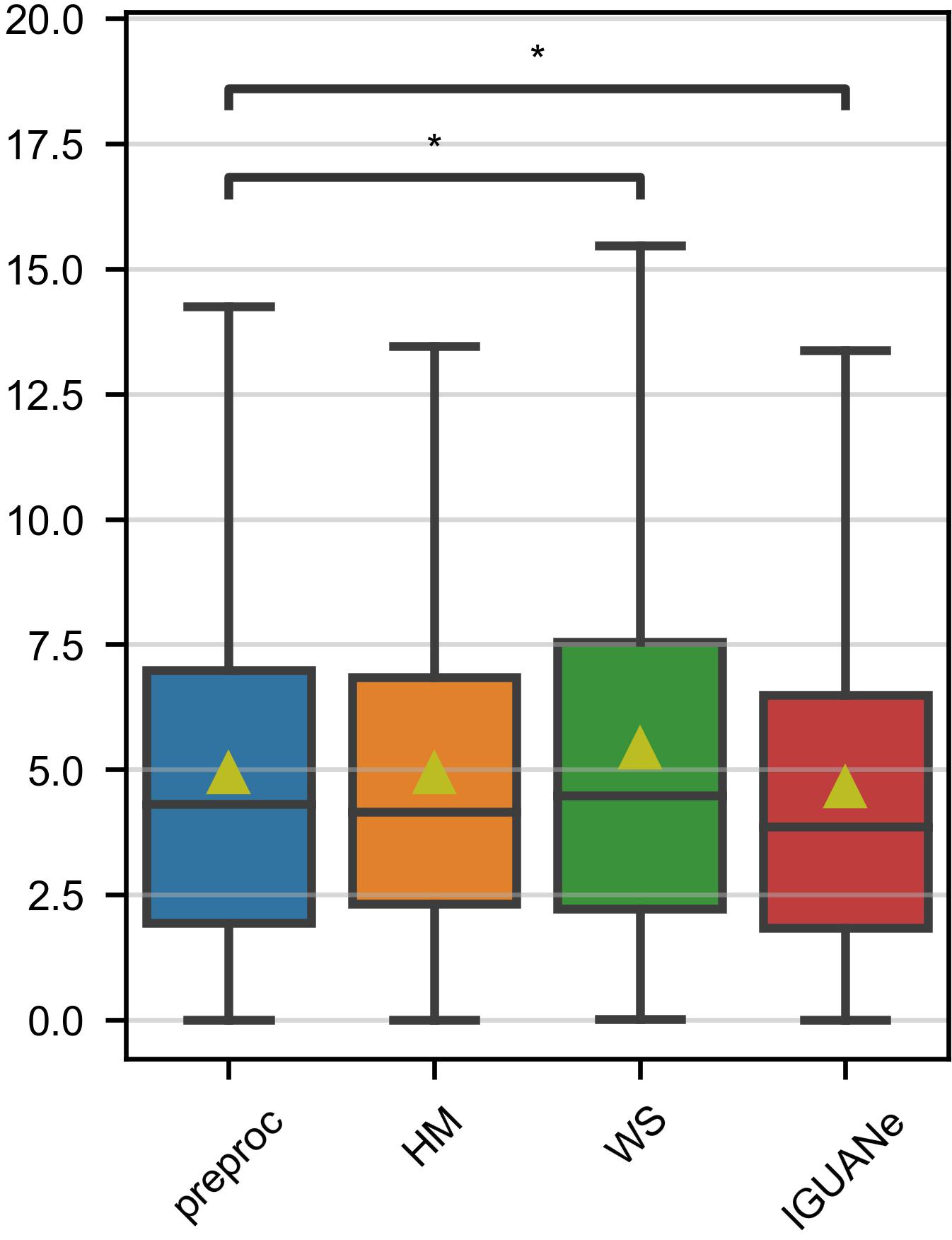
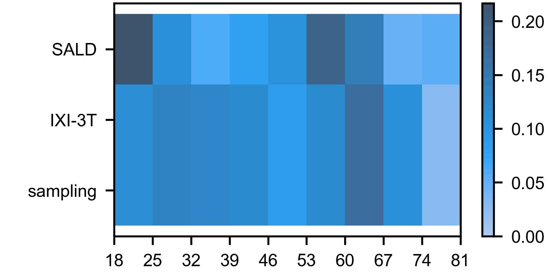
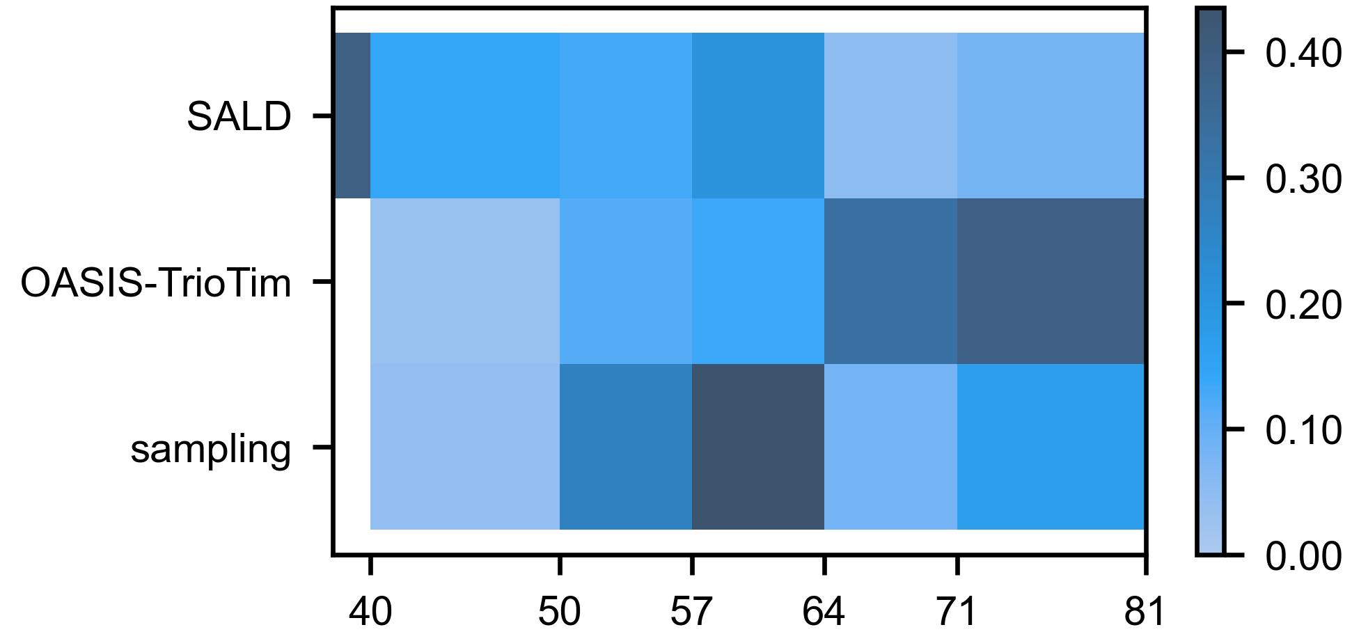
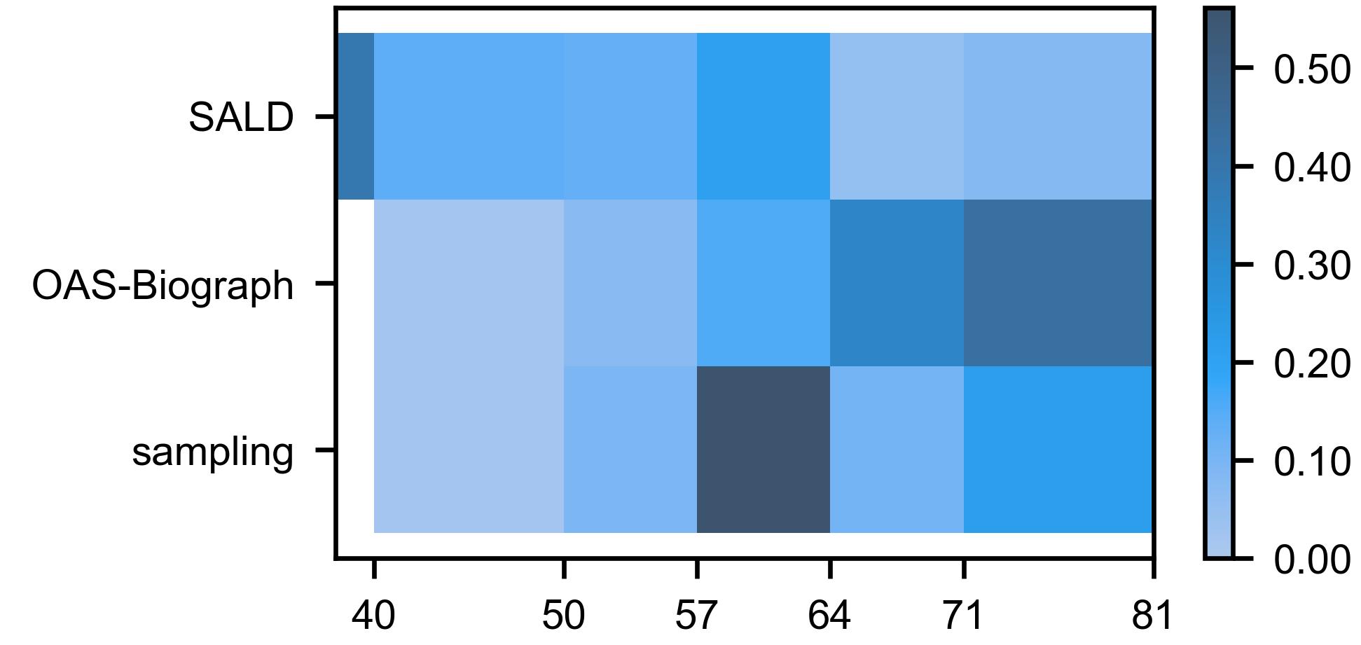
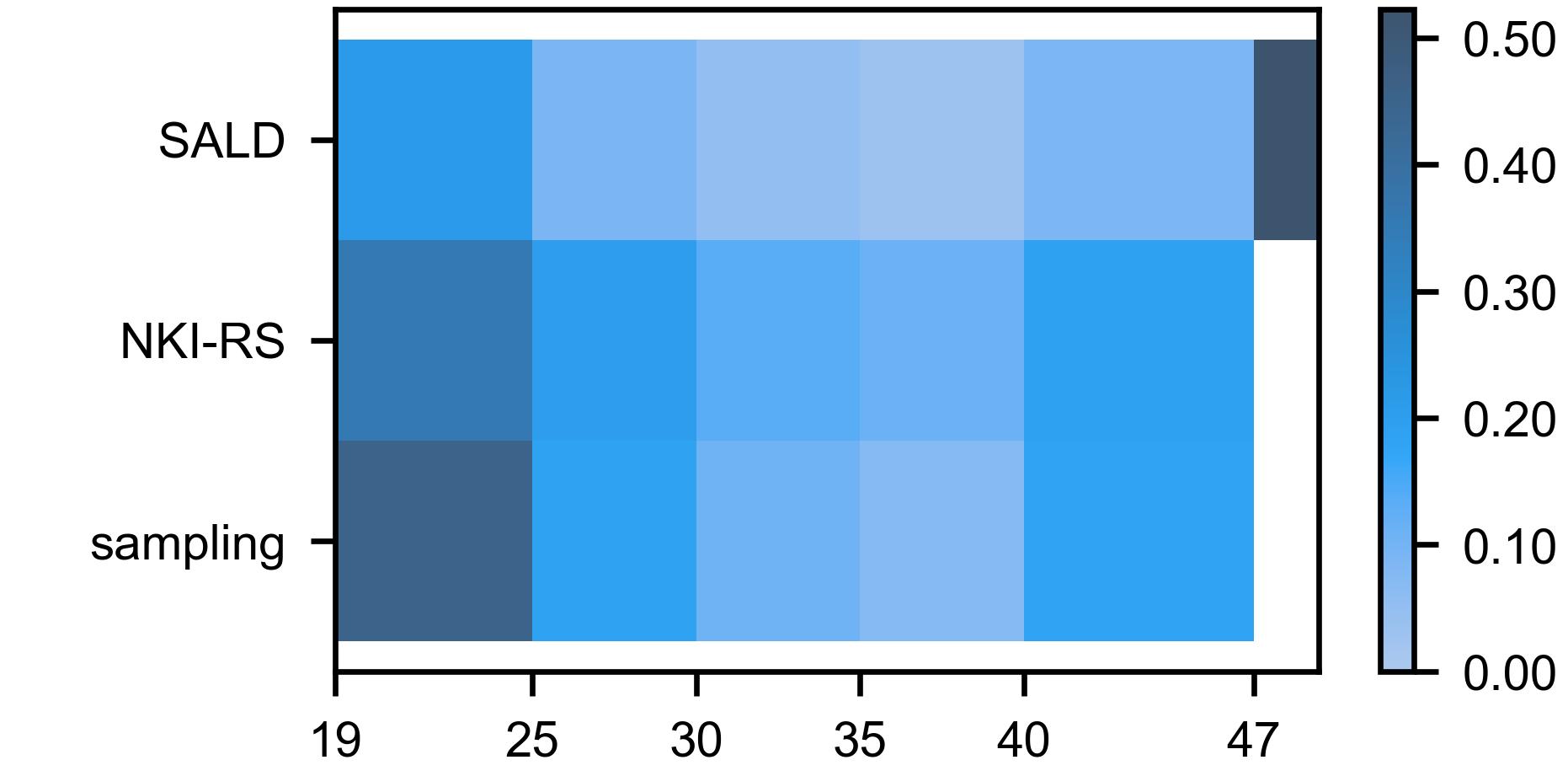
Fig. 1 illustrates the sampling strategy used for each source site of the Training dataset for IGUANe training.
Appendix C CALAMITI intensity normalization
The 3D T1w image provided in the online repository of CALAMITI did not seem to have been WS-normalized in a classical way as it contained no negative values. We therefore determined the normal appearing white-matter mask of the volume - using the code used in CALAMITI101010https://iacl.ece.jhu.edu/index.php?title=CALAMITI accessed 2023-07-27 - and computed the mean and the standard deviation inside this mask (mean = 1016.9; standard deviation = 7.8). Then, in order to implement the CALAMITI preprocessing, we matched these two values by scaling/shifting the MR intensities (after the three preprocessing steps explained online).
Appendix D Intensity shifts for structural similarity index
To avoid negative values that may hinder the SSIM computations, we added the following constant intensities to the images in our experiment on traveling subjects (section 3.4.1): WS +160; HM +10; CALAMITI +230 before harmonization, +10 after harmonization.
Appendix E Supplementary details for the predictive models
For both age regression and CN/AD classification, we trained predictive models for 400 epochs with a batch size of 16. We used an Adam optimizer [40] with a linear learning rate decay from 0.001 to 0.0001. A data-augmentation consisted in a random translation ( 5 voxels) along the three orthogonal axes and a random rotation ( 10 °) along a randomly selected orthogonal plane (rotation applied with a probability 1/2).
Each HM-normalized MR image was scaled/shifted with constants so that 1st and 99th percentiles of the brain intensities were -0.5 and 0.5, respectively.
Each WS-normalized MR image was scaled/shifted with constants so that the mean and the standard deviation of the normal appearing white-matter was 0.7 and 0.01, respectively.
The code we used in this study is available online (section 3.5).
References
- Aine et al. [2017] Aine, C.J., Bockholt, H.J., Bustillo, J.R., Cañive, J.M., Caprihan, A., Gasparovic, C., Hanlon, F.M., Houck, J.M., Jung, R.E., Lauriello, J., Liu, J., Mayer, A.R., Perrone-Bizzozero, N.I., Posse, S., Stephen, J.M., Turner, J.A., Clark, V.P., Calhoun, V.D., 2017. Multimodal neuroimaging in schizophrenia: Description and dissemination. Neuroinformatics 15, 343–364. doi:10.1007/s12021-017-9338-9.
- Basaia et al. [2019] Basaia, S., Agosta, F., Wagner, L., Canu, E., Magnani, G., Santangelo, R., Filippi, M., 2019. Automated classification of alzheimer’s disease and mild cognitive impairment using a single mri and deep neural networks. NeuroImage: Clinical 21, 101645. doi:10.1016/j.nicl.2018.101645.
- Bashyam et al. [2022] Bashyam, V.M., Doshi, J., Erus, G., Srinivasan, D., Abdulkadir, A., Singh, A., Habes, M., Fan, Y., Masters, C.L., Maruff, P., Zhuo, C., Völzke, H., Johnson, S.C., Fripp, J., Koutsouleris, N., Satterthwaite, T.D., Wolf, D.H., Gur, R.E., Gur, R.C., Morris, J.C., Albert, M.S., Grabe, H.J., Resnick, S.M., Bryan, N.R., Wittfeld, K., Bülow, R., Wolk, D.A., Shou, H., Nasrallah, I.M., Davatzikos, C., 2022. Deep generative medical image harmonization for improving cross‐site generalization in deep learning predictors. Journal of Magnetic Resonance Imaging 55, 908–916. doi:10.1002/jmri.27908.
- Bashyam et al. [2020] Bashyam, V.M., Erus, G., Doshi, J., Habes, M., Nasrallah, I.M., Truelove-Hill, M., Srinivasan, D., Mamourian, L., Pomponio, R., Fan, Y., Launer, L.J., Masters, C.L., Maruff, P., Zhuo, C., Völzke, H., Johnson, S.C., Fripp, J., Koutsouleris, N., Satterthwaite, T.D., Wolf, D., Gur, R.E., Gur, R.C., Morris, J., Albert, M.S., Grabe, H.J., Resnick, S., Bryan, R.N., Wolk, D.A., Shou, H., Davatzikos, C., 2020. Mri signatures of brain age and disease over the lifespan based on a deep brain network and 14 468 individuals worldwide. Brain 143, 2312–2324. doi:10.1093/brain/awaa160.
- de Bel et al. [2021] de Bel, T., Bokhorst, J.M., van der Laak, J., Litjens, G., 2021. Residual cyclegan for robust domain transformation of histopathological tissue slides. Medical Image Analysis 70, 102004. doi:10.1016/j.media.2021.102004.
- Billot et al. [2023] Billot, B., Greve, D.N., Puonti, O., Thielscher, A., Van Leemput, K., Fischl, B., Dalca, A.V., Iglesias, J.E., 2023. Synthseg: Segmentation of brain mri scans of any contrast and resolution without retraining. Medical Image Analysis 86, 102789. doi:10.1016/j.media.2023.102789.
- Cackowski et al. [2023] Cackowski, S., Barbier, E.L., Dojat, M., Christen, T., 2023. Imunity: A generalizable vae-gan solution for multicenter mr image harmonization. Medical Image Analysis 88, 102799. doi:10.1016/j.media.2023.102799.
- Chen et al. [2022] Chen, A.A., Srinivasan, D., Pomponio, R., Fan, Y., Nasrallah, I.M., Resnick, S.M., Beason-Held, L.L., Davatzikos, C., Satterthwaite, T.D., Bassett, D.S., Shinohara, R.T., Shou, H., 2022. Harmonizing functional connectivity reduces scanner effects in community detection. NeuroImage 256, 119198. doi:10.1016/j.neuroimage.2022.119198.
- Chen et al. [2014] Chen, J., Liu, J., Calhoun, V.D., Arias-Vasquez, A., Zwiers, M.P., Gupta, C.N., Franke, B., Turner, J.A., 2014. Exploration of scanning effects in multi-site structural mri studies. Journal of Neuroscience Methods 230, 37–50. doi:10.1016/j.jneumeth.2014.04.023.
- Chen et al. [2021] Chen, J., Sun, Y., Fang, Z., Lin, W., Li, G., Wang, L., 2021. Harmonized neonatal brain mr image segmentation model for cross-site datasets. Biomedical Signal Processing and Control 69, 102810. doi:10.1016/j.bspc.2021.102810.
- Choi et al. [2018] Choi, Y., Choi, M., Kim, M., Ha, J.W., Kim, S., Choo, J., 2018. Stargan: Unified generative adversarial networks for multi-domain image-to-image translation, in: 2018 IEEE/CVF Conference on Computer Vision and Pattern Recognition, IEEE. doi:10.1109/cvpr.2018.00916.
- Choi et al. [2020] Choi, Y., Uh, Y., Yoo, J., Ha, J.W., 2020. Stargan v2: Diverse image synthesis for multiple domains, in: 2020 IEEE/CVF Conference on Computer Vision and Pattern Recognition (CVPR), IEEE. doi:10.1109/cvpr42600.2020.00821.
- Cohen et al. [2018] Cohen, J.P., Luck, M., Honari, S., 2018. Distribution Matching Losses Can Hallucinate Features in Medical Image Translation. Springer International Publishing. p. 529–536. doi:10.1007/978-3-030-00928-1_60.
- Cole et al. [2017] Cole, J.H., Poudel, R.P., Tsagkrasoulis, D., Caan, M.W., Steves, C., Spector, T.D., Montana, G., 2017. Predicting brain age with deep learning from raw imaging data results in a reliable and heritable biomarker. NeuroImage 163, 115–124. doi:10.1016/j.neuroimage.2017.07.059.
- Cole et al. [2018] Cole, J.H., Ritchie, S.J., Bastin, M.E., Valdés Hernández, M.C., Muñoz Maniega, S., Royle, N., Corley, J., Pattie, A., Harris, S.E., Zhang, Q., Wray, N.R., Redmond, P., Marioni, R.E., Starr, J.M., Cox, S.R., Wardlaw, J.M., Sharp, D.J., Deary, I.J., 2018. Brain age predicts mortality. Molecular Psychiatry 23, 1385–1392. doi:10.1038/mp.2017.62.
- Costa [2011] Costa, J.F.P.d., 2011. Weighted Correlation. Springer Berlin Heidelberg. p. 1653–1655. doi:10.1007/978-3-642-04898-2_612.
- Dewey et al. [2019] Dewey, B.E., Zhao, C., Reinhold, J.C., Carass, A., Fitzgerald, K.C., Sotirchos, E.S., Saidha, S., Oh, J., Pham, D.L., Calabresi, P.A., van Zijl, P.C., Prince, J.L., 2019. Deepharmony: A deep learning approach to contrast harmonization across scanner changes. Magnetic Resonance Imaging 64, 160–170. doi:10.1016/j.mri.2019.05.041.
- Dewey et al. [2020] Dewey, B.E., Zuo, L., Carass, A., He, Y., Liu, Y., Mowry, E.M., Newsome, S., Oh, J., Calabresi, P.A., Prince, J.L., 2020. A Disentangled Latent Space for Cross-Site MRI Harmonization. Springer International Publishing. p. 720–729. doi:10.1007/978-3-030-59728-3_70.
- Dinsdale et al. [2021] Dinsdale, N.K., Jenkinson, M., Namburete, A.I., 2021. Deep learning-based unlearning of dataset bias for mri harmonisation and confound removal. NeuroImage 228, 117689. doi:10.1016/j.neuroimage.2020.117689.
- Ellis et al. [2009] Ellis, K.A., Bush, A.I., Darby, D., De Fazio, D., Foster, J., Hudson, P., Lautenschlager, N.T., Lenzo, N., Martins, R.N., Maruff, P., Masters, C., Milner, A., Pike, K., Rowe, C., Savage, G., Szoeke, C., Taddei, K., Villemagne, V., Woodward, M., Ames, D., 2009. The australian imaging, biomarkers and lifestyle (aibl) study of aging: methodology and baseline characteristics of 1112 individuals recruited for a longitudinal study of alzheimer’s disease. International Psychogeriatrics 21, 672–687. doi:10.1017/s1041610209009405.
- Enriquez Calzada [2021] Enriquez Calzada, P., 2021. Quantitative mr inter-scanner harmonization using image style transfer .
- Fatania et al. [2022] Fatania, K., Clark, A., Frood, R., Scarsbrook, A., Al-Qaisieh, B., Currie, S., Nix, M., 2022. Harmonisation of scanner-dependent contrast variations in magnetic resonance imaging for radiation oncology, using style-blind auto-encoders. Physics and Imaging in Radiation Oncology 22, 115–122. doi:10.1016/j.phro.2022.05.005.
- Fortin et al. [2018] Fortin, J.P., Cullen, N., Sheline, Y.I., Taylor, W.D., Aselcioglu, I., Cook, P.A., Adams, P., Cooper, C., Fava, M., McGrath, P.J., McInnis, M., Phillips, M.L., Trivedi, M.H., Weissman, M.M., Shinohara, R.T., 2018. Harmonization of cortical thickness measurements across scanners and sites. NeuroImage 167, 104–120. doi:10.1016/j.neuroimage.2017.11.024.
- Fortin et al. [2016] Fortin, J.P., Sweeney, E.M., Muschelli, J., Crainiceanu, C.M., Shinohara, R.T., 2016. Removing inter-subject technical variability in magnetic resonance imaging studies. NeuroImage 132, 198–212. doi:10.1016/j.neuroimage.2016.02.036.
- Gao et al. [2019] Gao, Y., Liu, Y., Wang, Y., Shi, Z., Yu, J., 2019. A universal intensity standardization method based on a many-to-one weak-paired cycle generative adversarial network for magnetic resonance images. IEEE Transactions on Medical Imaging 38, 2059–2069. doi:10.1109/tmi.2019.2894692.
- Gatys et al. [2016] Gatys, L.A., Ecker, A.S., Bethge, M., 2016. Image style transfer using convolutional neural networks, in: 2016 IEEE Conference on Computer Vision and Pattern Recognition (CVPR), IEEE. doi:10.1109/cvpr.2016.265.
- Gautherot et al. [2021] Gautherot, M., Kuchcinski, G., Bordier, C., Sillaire, A.R., Delbeuck, X., Leroy, M., Leclerc, X., Pruvo, J.P., Pasquier, F., Lopes, R., 2021. Longitudinal analysis of brain-predicted age in amnestic and non-amnestic sporadic early-onset alzheimer’s disease. Frontiers in Aging Neuroscience 13. doi:10.3389/fnagi.2021.729635.
- Ge et al. [2002] Ge, Y., Grossman, R.I., Babb, J.S., Rabin, M.L., Mannon, L.J., Kolson, D.L., 2002. Age-related total gray matter and white matter changes in normal adult brain. part i: volumetric mr imaging analysis. American journal of neuroradiology 23, 1327–1333.
- Gebre et al. [2023] Gebre, R.K., Senjem, M.L., Raghavan, S., Schwarz, C.G., Gunter, J.L., Hofrenning, E.I., Reid, R.I., Kantarci, K., Graff-Radford, J., Knopman, D.S., Petersen, R.C., Jack, C.R., Vemuri, P., 2023. Cross–scanner harmonization methods for structural mri may need further work: A comparison study. NeuroImage 269, 119912. doi:10.1016/j.neuroimage.2023.119912.
- Gollub et al. [2013] Gollub, R.L., Shoemaker, J.M., King, M.D., White, T., Ehrlich, S., Sponheim, S.R., Clark, V.P., Turner, J.A., Mueller, B.A., Magnotta, V., O’Leary, D., Ho, B.C., Brauns, S., Manoach, D.S., Seidman, L., Bustillo, J.R., Lauriello, J., Bockholt, J., Lim, K.O., Rosen, B.R., Schulz, S.C., Calhoun, V.D., Andreasen, N.C., 2013. The mcic collection: A shared repository of multi-modal, multi-site brain image data from a clinical investigation of schizophrenia. Neuroinformatics 11, 367–388. doi:10.1007/s12021-013-9184-3.
- Gourdeau et al. [2022] Gourdeau, D., Duchesne, S., Archambault, L., 2022. On the proper use of structural similarity for the robust evaluation of medical image synthesis models. Medical Physics 49, 2462–2474. doi:10.1002/mp.15514.
- Guan et al. [2021] Guan, H., Liu, Y., Yang, E., Yap, P.T., Shen, D., Liu, M., 2021. Multi-site mri harmonization via attention-guided deep domain adaptation for brain disorder identification. Medical Image Analysis 71, 102076. doi:10.1016/j.media.2021.102076.
- Han et al. [2006] Han, X., Jovicich, J., Salat, D., van der Kouwe, A., Quinn, B., Czanner, S., Busa, E., Pacheco, J., Albert, M., Killiany, R., Maguire, P., Rosas, D., Makris, N., Dale, A., Dickerson, B., Fischl, B., 2006. Reliability of mri-derived measurements of human cerebral cortical thickness: The effects of field strength, scanner upgrade and manufacturer. NeuroImage 32, 180–194. doi:10.1016/j.neuroimage.2006.02.051.
- Hedman et al. [2012] Hedman, A.M., van Haren, N.E., Schnack, H.G., Kahn, R.S., Hulshoff Pol, H.E., 2012. Human brain changes across the life span: A review of 56 longitudinal magnetic resonance imaging studies. Human Brain Mapping 33, 1987–2002. doi:10.1002/hbm.21334.
- Hu et al. [2023] Hu, F., Chen, A.A., Horng, H., Bashyam, V., Davatzikos, C., Alexander-Bloch, A., Li, M., Shou, H., Satterthwaite, T.D., Yu, M., Shinohara, R.T., 2023. Image harmonization: A review of statistical and deep learning methods for removing batch effects and evaluation metrics for effective harmonization. NeuroImage 274, 120125. doi:10.1016/j.neuroimage.2023.120125.
- Isensee et al. [2019] Isensee, F., Schell, M., Pflueger, I., Brugnara, G., Bonekamp, D., Neuberger, U., Wick, A., Schlemmer, H., Heiland, S., Wick, W., Bendszus, M., Maier‐Hein, K.H., Kickingereder, P., 2019. Automated brain extraction of multisequence mri using artificial neural networks. Human Brain Mapping 40, 4952–4964. doi:10.1002/hbm.24750.
- Isola et al. [2017] Isola, P., Zhu, J.Y., Zhou, T., Efros, A.A., 2017. Image-to-image translation with conditional adversarial networks, in: 2017 IEEE Conference on Computer Vision and Pattern Recognition (CVPR), IEEE. doi:10.1109/cvpr.2017.632.
- Jenkinson et al. [2002] Jenkinson, M., Bannister, P., Brady, M., Smith, S., 2002. Improved optimization for the robust and accurate linear registration and motion correction of brain images. NeuroImage 17, 825–841. doi:10.1006/nimg.2002.1132.
- Jonsson et al. [2019] Jonsson, B.A., Bjornsdottir, G., Thorgeirsson, T.E., Ellingsen, L.M., Walters, G.B., Gudbjartsson, D.F., Stefansson, H., Stefansson, K., Ulfarsson, M.O., 2019. Brain age prediction using deep learning uncovers associated sequence variants. Nature Communications 10. doi:10.1038/s41467-019-13163-9.
- Kingma and Ba [2017] Kingma, D.P., Ba, J., 2017. Adam: A method for stochastic optimization. arXiv:1412.6980.
- Kruggel et al. [2010] Kruggel, F., Turner, J., Muftuler, L.T., 2010. Impact of scanner hardware and imaging protocol on image quality and compartment volume precision in the adni cohort. NeuroImage 49, 2123–2133. doi:10.1016/j.neuroimage.2009.11.006.
- LaMontagne et al. [2019] LaMontagne, P.J., Benzinger, T.L., Morris, J.C., Keefe, S., Hornbeck, R., Xiong, C., Grant, E., Hassenstab, J., Moulder, K., Vlassenko, A.G., Raichle, M.E., Cruchaga, C., Marcus, D., 2019. Oasis-3: Longitudinal neuroimaging, clinical, and cognitive dataset for normal aging and alzheimer disease doi:10.1101/2019.12.13.19014902.
- Liu et al. [2023] Liu, M., Zhu, A.H., Maiti, P., Thomopoulos, S.I., Gadewar, S., Chai, Y., Kim, H., Jahanshad, N., 2023. Style transfer generative adversarial networks to harmonize multisite ¡scp¿mri¡/scp¿ to a single reference image to avoid overcorrection. Human Brain Mapping 44, 4875–4892. doi:10.1002/hbm.26422.
- Liu and Yap [2024] Liu, S., Yap, P.T., 2024. Learning multi-site harmonization of magnetic resonance images without traveling human phantoms. Communications Engineering 3. doi:10.1038/s44172-023-00140-w.
- Malone et al. [2013] Malone, I.B., Cash, D., Ridgway, G.R., MacManus, D.G., Ourselin, S., Fox, N.C., Schott, J.M., 2013. Miriad—public release of a multiple time point alzheimer’s mr imaging dataset. NeuroImage 70, 33–36. doi:10.1016/j.neuroimage.2012.12.044.
- Mao et al. [2017] Mao, X., Li, Q., Xie, H., Lau, R.Y., Wang, Z., Smolley, S.P., 2017. Least squares generative adversarial networks, in: 2017 IEEE International Conference on Computer Vision (ICCV), IEEE. doi:10.1109/iccv.2017.304.
- Marek et al. [2018] Marek, K., Chowdhury, S., Siderowf, A., Lasch, S., Coffey, C.S., Caspell‐Garcia, C., Simuni, T., Jennings, D., Tanner, C.M., Trojanowski, J.Q., Shaw, L.M., Seibyl, J., Schuff, N., Singleton, A., Kieburtz, K., Toga, A.W., Mollenhauer, B., Galasko, D., Chahine, L.M., Weintraub, D., Foroud, T., Tosun‐Turgut, D., Poston, K., Arnedo, V., Frasier, M., Sherer, T., 2018. The parkinson’s progression markers initiative (ppmi) – establishing a pd biomarker cohort. Annals of Clinical and Translational Neurology 5, 1460–1477. doi:10.1002/acn3.644.
- Micikevicius et al. [2018] Micikevicius, P., Narang, S., Alben, J., Diamos, G., Elsen, E., Garcia, D., Ginsburg, B., Houston, M., Kuchaiev, O., Venkatesh, G., Wu, H., 2018. Mixed precision training. arXiv:1710.03740.
- Nguyen et al. [2018] Nguyen, H., Morris, R.W., Harris, A.W., Korgoankar, M.S., Ramos, F., 2018. Correcting differences in multi-site neuroimaging data using generative adversarial networks. arXiv:1803.09375.
- Nooner et al. [2012] Nooner, K.B., Colcombe, S.J., Tobe, R.H., Mennes, M., Benedict, M.M., Moreno, A.L., Panek, L.J., Brown, S., Zavitz, S.T., Li, Q., Sikka, S., Gutman, D., Bangaru, S., Schlachter, R.T., Kamiel, S.M., Anwar, A.R., Hinz, C.M., Kaplan, M.S., Rachlin, A.B., Adelsberg, S., Cheung, B., Khanuja, R., Yan, C., Craddock, C.C., Calhoun, V., Courtney, W., King, M., Wood, D., Cox, C.L., Kelly, A.M.C., Di Martino, A., Petkova, E., Reiss, P.T., Duan, N., Thomsen, D., Biswal, B., Coffey, B., Hoptman, M.J., Javitt, D.C., Pomara, N., Sidtis, J.J., Koplewicz, H.S., Castellanos, F.X., Leventhal, B.L., Milham, M.P., 2012. The nki-rockland sample: A model for accelerating the pace of discovery science in psychiatry. Frontiers in Neuroscience 6. doi:10.3389/fnins.2012.00152.
- Palladino et al. [2020] Palladino, J.A., Fernandez Slezak, D., Ferrante, E., 2020. Unsupervised domain adaptation via cyclegan for white matter hyperintensity segmentation in multicenter mr images, in: Brieva, J., Lepore, N., Romero Castro, E., Linguraru, M.G. (Eds.), 16th International Symposium on Medical Information Processing and Analysis, SPIE. doi:10.1117/12.2579548.
- Pambrun and Noumeir [2015] Pambrun, J.F., Noumeir, R., 2015. Limitations of the ssim quality metric in the context of diagnostic imaging, in: 2015 IEEE International Conference on Image Processing (ICIP), IEEE. doi:10.1109/icip.2015.7351345.
- Pomponio et al. [2020] Pomponio, R., Erus, G., Habes, M., Doshi, J., Srinivasan, D., Mamourian, E., Bashyam, V., Nasrallah, I.M., Satterthwaite, T.D., Fan, Y., Launer, L.J., Masters, C.L., Maruff, P., Zhuo, C., Völzke, H., Johnson, S.C., Fripp, J., Koutsouleris, N., Wolf, D.H., Gur, R., Gur, R., Morris, J., Albert, M.S., Grabe, H.J., Resnick, S.M., Bryan, R.N., Wolk, D.A., Shinohara, R.T., Shou, H., Davatzikos, C., 2020. Harmonization of large mri datasets for the analysis of brain imaging patterns throughout the lifespan. NeuroImage 208, 116450. doi:10.1016/j.neuroimage.2019.116450.
- Ravano et al. [2022] Ravano, V., Démonet, J.F., Damian, D., Meuli, R., Piredda, G.F., Huelnhagen, T., Maréchal, B., Thiran, J.P., Kober, T., Richiardi, J., 2022. Neuroimaging Harmonization Using cGANs: Image Similarity Metrics Poorly Predict Cross-Protocol Volumetric Consistency. Springer Nature Switzerland. p. 83–92. doi:10.1007/978-3-031-17899-3_9.
- Robinson et al. [2020] Robinson, R., Dou, Q., Coelho de Castro, D., Kamnitsas, K., de Groot, M., Summers, R.M., Rueckert, D., Glocker, B., 2020. Image-Level Harmonization of Multi-site Data Using Image-and-Spatial Transformer Networks. Springer International Publishing. p. 710–719. doi:10.1007/978-3-030-59728-3_69.
- Robitaille et al. [2012] Robitaille, N., Mouiha, A., Crépeault, B., Valdivia, F., Duchesne, S., 2012. Tissue-based mri intensity standardization: Application to multicentric datasets. International Journal of Biomedical Imaging 2012, 1–11. doi:10.1155/2012/347120.
- Roca et al. [2023] Roca, V., Kuchcinski, G., Pruvo, J.P., Manouvriez, D., Leclerc, X., Lopes, R., 2023. A three-dimensional deep learning model for inter-site harmonization of structural mr images of the brain: Extensive validation with a multicenter dataset. Heliyon 9, e22647. doi:10.1016/j.heliyon.2023.e22647.
- Rosner et al. [2006] Rosner, B., Glynn, R.J., Lee, M.T., 2006. The wilcoxon signed rank test for paired comparisons of clustered data. Biometrics 62, 185–192. doi:10.1111/j.1541-0420.2005.00389.x.
- Schuff et al. [2009] Schuff, N., Woerner, N., Boreta, L., Kornfield, T., Shaw, L.M., Trojanowski, J.Q., Thompson, P.M., Jack, C.R., Weiner, M.W., 2009. Mri of hippocampal volume loss in early alzheimer’s disease in relation to apoe genotype and biomarkers. Brain 132, 1067–1077. doi:10.1093/brain/awp007.
- Shah et al. [2011] Shah, M., Xiao, Y., Subbanna, N., Francis, S., Arnold, D.L., Collins, D.L., Arbel, T., 2011. Evaluating intensity normalization on mris of human brain with multiple sclerosis. Medical Image Analysis 15, 267–282. doi:10.1016/j.media.2010.12.003.
- Shinohara et al. [2017] Shinohara, R., Oh, J., Nair, G., Calabresi, P., Davatzikos, C., Doshi, J., Henry, R., Kim, G., Linn, K., Papinutto, N., Pelletier, D., Pham, D., Reich, D., Rooney, W., Roy, S., Stern, W., Tummala, S., Yousuf, F., Zhu, A., Sicotte, N., Bakshi, R., 2017. Volumetric analysis from a harmonized multisite brain mri study of a single subject with multiple sclerosis. American Journal of Neuroradiology 38, 1501–1509. doi:10.3174/ajnr.a5254.
- Shinohara et al. [2014] Shinohara, R.T., Sweeney, E.M., Goldsmith, J., Shiee, N., Mateen, F.J., Calabresi, P.A., Jarso, S., Pham, D.L., Reich, D.S., Crainiceanu, C.M., 2014. Statistical normalization techniques for magnetic resonance imaging. NeuroImage: Clinical 6, 9–19. doi:10.1016/j.nicl.2014.08.008.
- Steiger [1980] Steiger, J.H., 1980. Tests for comparing elements of a correlation matrix. Psychological Bulletin 87, 245–251. doi:10.1037/0033-2909.87.2.245.
- Tanaka et al. [2021] Tanaka, S.C., Yamashita, A., Yahata, N., Itahashi, T., Lisi, G., Yamada, T., Ichikawa, N., Takamura, M., Yoshihara, Y., Kunimatsu, A., Okada, N., Hashimoto, R., Okada, G., Sakai, Y., Morimoto, J., Narumoto, J., Shimada, Y., Mano, H., Yoshida, W., Seymour, B., Shimizu, T., Hosomi, K., Saitoh, Y., Kasai, K., Kato, N., Takahashi, H., Okamoto, Y., Yamashita, O., Kawato, M., Imamizu, H., 2021. A multi-site, multi-disorder resting-state magnetic resonance image database. Scientific Data 8. doi:10.1038/s41597-021-01004-8.
- Tian et al. [2022] Tian, D., Zeng, Z., Sun, X., Tong, Q., Li, H., He, H., Gao, J.H., He, Y., Xia, M., 2022. A deep learning-based multisite neuroimage harmonization framework established with a traveling-subject dataset. NeuroImage 257, 119297. doi:10.1016/j.neuroimage.2022.119297.
- Torbati et al. [2023] Torbati, M.E., Minhas, D.S., Laymon, C.M., Maillard, P., Wilson, J.D., Chen, C.L., Crainiceanu, C.M., DeCarli, C.S., Hwang, S.J., Tudorascu, D.L., 2023. Mispel: A supervised deep learning harmonization method for multi-scanner neuroimaging data. Medical Image Analysis 89, 102926. doi:10.1016/j.media.2023.102926.
- Tustison et al. [2010] Tustison, N.J., Avants, B.B., Cook, P.A., Zheng, Y., Egan, A., Yushkevich, P.A., Gee, J.C., 2010. N4itk: Improved n3 bias correction. IEEE Transactions on Medical Imaging 29, 1310–1320. doi:10.1109/tmi.2010.2046908.
- Ulyanov et al. [2017] Ulyanov, D., Vedaldi, A., Lempitsky, V., 2017. Instance normalization: The missing ingredient for fast stylization. arXiv:1607.08022.
- Vieira et al. [2017] Vieira, S., Pinaya, W.H., Mechelli, A., 2017. Using deep learning to investigate the neuroimaging correlates of psychiatric and neurological disorders: Methods and applications. Neuroscience & Biobehavioral Reviews 74, 58–75. doi:10.1016/j.neubiorev.2017.01.002.
- Wang et al. [2022] Wang, R., Chaudhari, P., Davatzikos, C., 2022. Embracing the disharmony in medical imaging: A simple and effective framework for domain adaptation. Medical Image Analysis 76, 102309. doi:10.1016/j.media.2021.102309.
- Wang et al. [2004] Wang, Z., Bovik, A., Sheikh, H., Simoncelli, E., 2004. Image quality assessment: From error visibility to structural similarity. IEEE Transactions on Image Processing 13, 600–612. doi:10.1109/tip.2003.819861.
- Watanabe et al. [2013] Watanabe, M., Liao, J.H., Jara, H., Sakai, O., 2013. Multispectral quantitative mr imaging of the human brain: Lifetime age-related effects. RadioGraphics 33, 1305–1319. doi:10.1148/rg.335125212.
- Wei et al. [2018] Wei, D., Zhuang, K., Ai, L., Chen, Q., Yang, W., Liu, W., Wang, K., Sun, J., Qiu, J., 2018. Structural and functional brain scans from the cross-sectional southwest university adult lifespan dataset. Scientific Data 5. doi:10.1038/sdata.2018.134.
- Wrobel et al. [2020] Wrobel, J., Martin, M., Bakshi, R., Calabresi, P., Elliot, M., Roalf, D., Gur, R., Gur, R., Henry, R., Nair, G., Oh, J., Papinutto, N., Pelletier, D., Reich, D., Rooney, W., Satterthwaite, T., Stern, W., Prabhakaran, K., Sicotte, N., Shinohara, R., Goldsmith, J., 2020. Intensity warping for multisite mri harmonization. NeuroImage 223, 117242. doi:10.1016/j.neuroimage.2020.117242.
- Wu et al. [2019] Wu, S., Li, G., Deng, L., Liu, L., Wu, D., Xie, Y., Shi, L., 2019. -norm batch normalization for efficient training of deep neural networks. IEEE Transactions on Neural Networks and Learning Systems 30, 2043–2051. doi:10.1109/tnnls.2018.2876179.
- Zech et al. [2018] Zech, J.R., Badgeley, M.A., Liu, M., Costa, A.B., Titano, J.J., Oermann, E.K., 2018. Variable generalization performance of a deep learning model to detect pneumonia in chest radiographs: A cross-sectional study. PLOS Medicine 15, e1002683. doi:10.1371/journal.pmed.1002683.
- Zhu et al. [2017] Zhu, J.Y., Park, T., Isola, P., Efros, A.A., 2017. Unpaired image-to-image translation using cycle-consistent adversarial networks, in: 2017 IEEE International Conference on Computer Vision (ICCV), IEEE. doi:10.1109/iccv.2017.244.
- Zuo et al. [2021a] Zuo, L., Dewey, B.E., Carass, A., Liu, Y., He, Y., Calabresi, P.A., Prince, J.L., 2021a. Information-Based Disentangled Representation Learning for Unsupervised MR Harmonization. Springer International Publishing. p. 346–359. doi:10.1007/978-3-030-78191-0_27.
- Zuo et al. [2021b] Zuo, L., Dewey, B.E., Liu, Y., He, Y., Newsome, S.D., Mowry, E.M., Resnick, S.M., Prince, J.L., Carass, A., 2021b. Unsupervised mr harmonization by learning disentangled representations using information bottleneck theory. NeuroImage 243, 118569. doi:10.1016/j.neuroimage.2021.118569.
- Zuo et al. [2022] Zuo, L., Liu, Y., Xue, Y., Han, S., Bilgel, M., Resnick, S.M., Prince, J.L., Carass, A., 2022. Disentangling a Single MR Modality. Springer Nature Switzerland. p. 54–63. doi:10.1007/978-3-031-17027-0_6.