Autopoietic influence hierarchies in pancreatic cells
Abstract
cells are biologically essential for humans and other vertebrates. Because their functionality arises from cell-cell interactions, they are also a model system for collective organization among cells. There are currently two contradictory pictures of this organization: the hub-cell idea pointing at leaders who coordinate the others, and the electrophysiological theory describing all cells as equal. We use new data and computational modeling to reconcile these pictures. We find via a network representation of interacting cells that leaders emerge naturally (confirming the hub-cell idea), yet all cells can take the hub role following a perturbation (in line with electrophysiology).
The importance of the nutrient-sensing and insulin-secreting cells in vertebrates is hard to overstate. These cells reside in pancreatic islets, where they extensively communicate with each other and their environment [1]. The intercellular communication, in particular, serves to coordinate and synchronize cellular operations through which insulin is released in proportion to stimulation and metabolic requirements [2, 3]. The delicate nature of this task is reflected in the continual need to prevent oversecretion, and subsequent hypoglycemia [4], despite intracellular stores holding sufficient insulin to exceed the lethal dose by orders of magnitude if released at once.
On a microscopic scale, cells are electrically and metabolically coupled via gap junctions built from the protein connexin-36 (Cx36) [5]. The key role of such coupling is seen in the fact that the loss of Cx36 channels has a detrimental impact on -cell cooperation [6, 7], leading to uncoordinated plasma depolarizations, the desynchronization of calcium signals, and increases in basal insulin release. Sufficient Cx36 coupling, by contrast, curbs the -cell intrinsic heterogeneity in glucose sensitivity [8], and in mouse islets, limits the threshold for stimulatory glucose concentration [7] to a narrow band around 7 mM. Metaphorically, coupled cells are like individual soldiers who fall in line when the communication channels between them are open. A typical cell is coupled to between six and eight neighbors [9, 10]. According to electrophysiological theory [11], single cells lack mechanisms to become pacemakers beyond their immediate neighborhood. In terms of our metaphor, there are no generals in the army.
The above-mentioned lateral organization is diametrically opposed to the picture by functional studies of a large number of communicating cells (referred to as -cell collectives). For example, intrinsic cellular heterogeneity in glucose sensing [8], heterogeneous gap-junction coupling [12], and extensive paracrine signaling [13] all contribute to an islet-wide complicated cytoplasmic Ca2+ dynamics. This dynamics of the Ca2+ concentration in the cytosol [14, 15] is a key insulin-secretion driver [16]. It has recently become possible to record the Ca2+ dynamics with a great spatial and temporal precision using the functional multicellular confocal imaging [17, 18]. The high data content of such imaging enables mapping the functional organization of a -cell collective onto a complex network such that a pair of cells (i.e., network nodes) is linked if the cross-correlation between the corresponding Ca2+ signals is large enough [19, 20, 21, 22, 23, 24].
Complex-network representations of the functional organization of -cell collectives (i.e., functional networks) that arise from calcium cross-correlations in intact pancreatic islets reveal a modular structure [25] intertwined with small-world [26, 20] properties. Of particular interest is that some studies [22, 24] point to small subsets of highly active cells whose connectedness and impact on synchronization across islets make these cells candidate leaders. Because of their large degree, we call such candidate leaders hub cells. The proponents of the hub-cell idea list many reasons [27] why electrophysiological measurements may have missed detecting hubs. Overall, functional networks suggest that our army of cells is led by a few generals.
We will hereafter show that the hub-cell idea, rather than contradicting, actually complements electrophysiology. Based on a remarkable agreement between empirical and computational findings, we will argue in favor of an autopoietic influence hierarchy among cells. Leader cells emerge through cell-cell communication, and thus need not be genetically predisposed for leadership. In the language of the army metaphor, generals do lead, but not by birthright; they get promoted by their peers. Leader cells, furthermore, remain under the radar of electrophysiological measurements by communicating with immediate neighbors just as any other cell would. A direct implication is that a more-or-less arbitrary cell could emerge as a leader, which in turn makes for an ultra robust architecture of -cell collectives. Additional wide-reaching implications underpin some of the vital -cell features.
I Methods
We based our empirical analyses on a dataset obtained via Ca2+ imaging of an acutely prepared pancreatic tissue slice [28, 17] comprising a rodent oval-shaped islet (approx. dimensions: 370 µm200 µm). Ca2+ signals were recorded using a functional multi-cellular imaging technique at 10 Hz and 256256 pixel resolution in 8-bit grayscale color depth. The freely downloadable dataset [29] consisted of 65,536 Ca2+ signals, each with 23,873 data points. Our focus was on fast oscillations, which is why all signals were detrended and standardized before use [30].
For the purpose of constructing empirical functional networks, we randomly picked signals, denoted , from the dataset and computed cross-correlations , where is a time-averaging operator. The number of data points in each signal was , corresponding to 5-minute exposures of the pancreatic tissue slice to the stimulating glucose concentration of 8 mM. We link two signals and in the network if , where is a threshold selected to give a mean degree . We have previously shown [31] that the bulk cross-correlation spectrum of typical Ca2+ signals follows the predictions of random matrix theory, but the states outside the bulk-spectrum edges carry biological information. The delocalized states corresponding to the largest eigenvalues of the cross-correlation matrix, in particular, were found to harbor contributions from all Ca2+ signals, thus revealing an islet-wide collective mode [32].
The computational aspects of the study comprised constructing functional networks from simulated Ca2+ signals. Simulations were based on our model [30] that had been found to mimic empirical signals closely. Compared to typical electrophysiological modeling by means of differential-algebraic systems [21], the model’s structure is very simple. Each of the nodes represents a cell arranged in a random regular network with the degree distribution such that trivially the average node degree is . Nodes can change their binary state, indicating the calcium activity of individual cells, from active to inactive or vice versa in two ways. Internal activation is controlled by a forcing parameter, , that is interpretable as the inverse of the glucose concentration to which cells are exposed. External activation is controlled by the state of the nearest neighbors in conjunction with the coupling strength, , that is interpretable as the intensity of gap-junctional ion exchange. Functional networks were constructed from simulated Ca2+ signals in exactly the same way as from empirical ones.
II Results
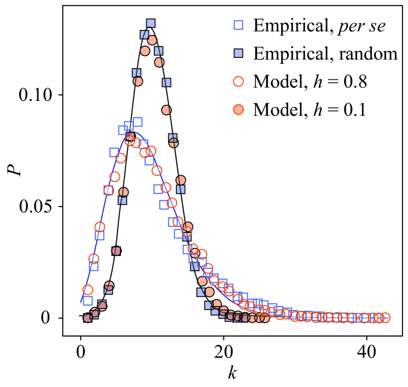
The degree distributions of empirical and simulated functional networks are practically the same (Fig. 1). We distinguish between two cases. Open symbols pertain to functional networks extracted from empirical data per se (squares), and from simulations with strong coupling, (circles). In this case, the obtained degree distributions closely follow a negative binomial distribution , where and is a numerical parameter. The opposite case, shown using filled symbols, consists of functional networks extracted upon randomizing empirical data (squares) and running simulations with weak coupling, (circles). Both data randomization and weak coupling should produce random functional networks, what indeed transpires given that the obtained degree distributions closely follow a binomial distribution with . The binomial distribution in turn converges to a Poisson distribution in the large network limit when is fixed.
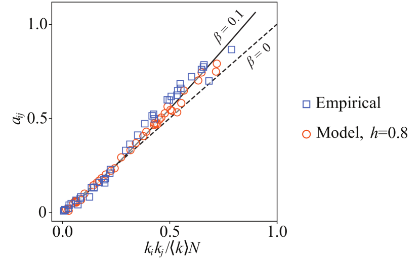
Aside from degree distributions, another informative way of characterizing functional networks is degree correlations, also known as degree assortativity. Ref. [33] explains that the elements of a configuration-ensemble adjacency matrix should satisfy the strictly linear relationship in zero-assortativity situations. Otherwise, , where () indicates negative (positive) assortative mixing. Plots of against (Fig. 2), while once again revealing an agreement between empirical and simulated functional networks, primarily show that . The degree assortativity of the functional networks is therefore positive, meaning that network nodes are preferentially linked to other nodes of similar degree.
The results so far allow us to estimate the assortativity coefficient of functional networks in an alternative way, which we can then compare with the definition [34, 35], that is, with the degree correlation coefficient between pairs of linked nodes , where is the average degree of the th node’s nearest neighbors. Using the exponent and the fact that the aforementioned negative binomial distribution converges to a gamma distribution for large , the assortativity coefficient is (see Ref. [33]). Following the definition of indeed yields a very similar value (Fig. 3; open symbols for ).
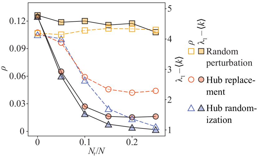
We furthermore tested how two different perturbations [36], signal randomization and replacement, affect the assortativity coefficient. Here, randomization and replacement respectively mean that a fraction of signals used in the construction of the original configuration ensemble were either randomly reshuffled or replaced with other random signals from the broader dataset. We found that assortativity of functional networks is robust to random perturbations, but not to perturbations targeting hubs (Fig. 3; open symbols for ). It is therefore hubs that preferentially link to other hubs, which emphasizes their importance in the overall structure of functional networks. Specifically, we are seeing the evidence of a purposeful correlating mechanisms at work among -cells because otherwise heterogeneous networks are expected to be disassortative [33, 35].
Another measure characterizing a network’s structure is the largest eigenvalue of the adjacency matrix, . Similarly as with the assortativity coefficient, the value of is robust to random perturbations, but not to perturbations targeting hubs (Fig. 3). As progressively more hubs get perturbed, the maximum adjacency-matrix eigenvalue falls to , which is the value characteristic of random networks with a Poisson degree distribution. This last result reaffirms the importance of hubs for the structure of functional networks and, in turn, hints at an interesting connection with recent experimental findings that generated much excitement.
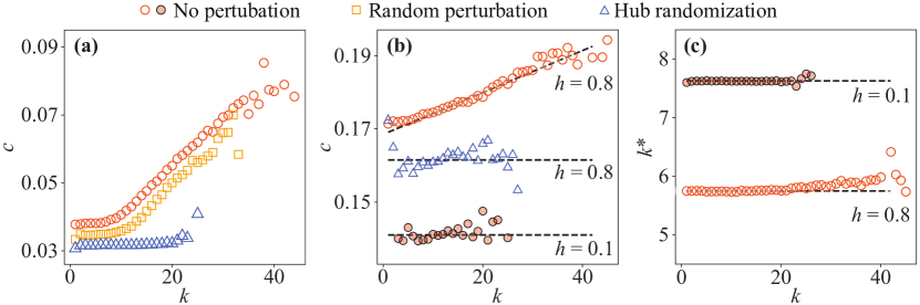
Experiments show [22, 24] that perturbing hub cells can quickly decorrelate a -cell collective. This implies that hubs carry most of the collective’s cross-correlation content, which could be tested using our empirical and simulated Ca2+ signals alike. To this end, we employed the pairwise cross-correlations to define , that is, the th node’s neighborhood-wide average cross-correlation. By pairing the values with the corresponding node degree , we obtained a function . If the -cell collective’s cross-correlation content were equally distributed among all nodes, then we should see , whereas if hubs carried most of the content, then should be an increasing function of . We find that the latter is true both for empirical (Fig. 4a) and simulated (Fig. 4b) Ca2+ signals. As with assortativity and the largest eigenvalue , random perturbations have little bearing on the function , but targeting high-degree nodes causes decorrelation. With the help of the dynamical network model, we see that perturbing about 10 % of hubs is very similar to running simulations with a weak coupling of (Fig. 4b).
The model additionally allows us to examine whether the node-degree dependence of the function could be due to hubs somehow communicating with more of their neighbors than lower-degree nodes. The number of active neighbors is, as expected, independent of the degree when the coupling is weak, (Fig. 4c). The same, however, approximately holds even when the coupling is strong, (Fig. 4c). Because, in the model, all nodes are equal and none of them communicate with an unusual number of neighbors, we are left with a conclusion that the emergence of hubs in functional networks is endogenous to cell-cell communication.
III Discussion
Herein, we used a combination of empirical and computational methods to shed new light on, among others, a fundamental tension between electrophysiological theory and the hub-cell idea as it pertains to pancreatic cells. We started by constructing functional -cell networks from empirical Ca2+ signals, and after that estimated the configuration ensemble [37, 38] of such networks. Meanwhile, we also simulated fast -cell activity using a dynamical network model [39, 40] whose outputs faithfully mimic the properties of said empirical Ca2+ signals [30]. Upon repeating the construction of functional networks, but now from simulated signals, we found remarkable quantitative agreement between the results.
Irrespective of the signal origin, empirical or simulated, the properties of functional networks—such as assortativity or the largest eigenvalue of the configuration-ensemble adjacency matrix—all critically depend on the presence of highly connected (i.e., hub) nodes. Our results thus support the hub-cell idea. A similar heterogeneous organization of functional networks has also been discovered in the collectives of chemosensing cells [41, 42] communicating via gap junctions.
The support for the hub-cell idea would be a strong blow to electrophysiological theory if nodes in the dynamical network model had any distinctive properties, for example, different degrees or an internal structure. In the model, however, all nodes are completely identical, upholding the ideas of electrophysiological theory that no cell is predisposed for leadership. Leader cells, in fact, emerge through cell-cell communication as evidenced by tracing the degree distributions of functional networks to the coupling strength in the dynamical network model. In the language of the army metaphor, generals do lead, but not by birthright; they get promoted by their peers. Our findings thus not only reconcile the concept of hubs with electrophysiological theory, but also point to an extremely robust architecture of collectives.
Before expanding on the last remark about the robustness of -cell collectives, let us examine a physiological advantage provided by the heterogeneity of functional networks. Cells have been shown to use cell-cell distance to optimize their sensing precision [43]. In compact microorgans such as pancreatic islets, cells cannot easily adjust mutual distances, but could instead use functional-network heterogeneity to sharpen their collective response. Let be the largest adjacency-matrix eigenvalue and the corresponding eigenvector [44, 32]. Because the components of the latter vector quantify the importance of network nodes, we assume that represents the signal that node integrates from the nearest neighbors , while all other signals are treated as constant-variance noise. Under this assumption, the signal-to-noise ratio of sensing cells is proportional to the variance , where is the cross-correlation threshold used during the construction of functional networks. The sensing precision is thus proportional to , which would equal if functional networks were random, but increases to because of heterogeneity. Accordingly, cell-cell communication imparts a sharp glucose-sensing acuity to -cell collectives.
The example on glucose-sensing acuity demonstrates a physiological advantage of heterogeneous functional networks over homogeneous ones. This advantage would, however, come at a cost of vulnerability to hub-node failures [36] if heterogeneity were imprinted in the underlying physical network of cells. Our results instead strongly favor an interpretation by which influential cells materialize endogenously within -cell collectives, giving rise to influence hierarchies that are autopoietic, both in the self-producing and self-maintaining sense of this term. All that is needed for hub cells to emerge among identical peers is cell-cell communication, and should a hub fail, there is no obstacle for another one to re-emerge as long as communication remains feasible. It would thus seem that in -cell collectives, biology has managed to create an ultra-robust architecture that is physiologically advantageous as well. This is an impressive feat that could perhaps inspire thinking about the design and engineering of next-generation critical infrastructure.
– – – – –
Acknowledgements. DK, AS, and MSR received financial support from the Slovenian Research Agency (research core funding program no. P3–0396 and projects no. N3-0048, no. N3-0133 and no. J3-9289). MSR was further supported by the Austrian Science Fund / Fonds zur Förderung der Wissenschaftlichen Forschung (bilateral grants I3562–B27 and I4319–B30). MJ received financial supported from the Japan Society for the Promotion of Science (JSPS) KAKENHI (grant no. JP 20H04288) as a co-investigator. BP received financial support from the Slovenian Research Agency (project no. J5-8236), the University of Zagreb’s project Advanced methods and technologies in Data Science and Cooperative Systems (DATACROSS; ref. KK.01.1.1.01.009), and the University of Rijeka. PH received financial support from JSPS KAKENHI (grant no. JP 18H01655).
Author contributions. All authors contributed substantially to all aspects of the study.
Conflict of interest. The authors declare no conflict of interest, financial or otherwise.
References
- Pipeleers [1987] D. Pipeleers, Diabetologia 30, 277 (1987).
- Pipeleers et al. [1982] D. Pipeleers, E. Maes, M. Van De Winkel, et al., Proc. Natl. Acad. Sci. USA 79, 7322 (1982).
- Korošak and Slak Rupnik [2018] D. Korošak and M. Slak Rupnik, Front. Physiol. 9, 31 (2018).
- Kolb et al. [2020] H. Kolb, K. Kempf, M. Röhling, and S. Martin, BMC Med. 18, 224 (2020).
- Meda [2018] P. Meda, Biochim. Biophys. Acta Biomembr. 1860, 124 (2018).
- Ravier et al. [2005] M. A. Ravier, M. Güldenagel, A. Charollais, A. Gjinovci, D. Caille, G. Söhl, C. B. Wollheim, K. Willecke, J.-C. Henquin, and P. Meda, Diabetes 54, 1798 (2005).
- Speier et al. [2007] S. Speier, A. Gjinovci, A. Charollais, P. Meda, and M. Rupnik, Diabetes 56, 1078 (2007).
- Benninger and Hodson [2018] R. K. Benninger and D. J. Hodson, Diabetes 67, 537 (2018).
- Zhang et al. [2008] Q. Zhang, J. Galvanovskis, F. Abdulkader, C. J. Partridge, S. O. Göpel, L. Eliasson, and P. Rorsman, Philos. Trans. R. Soc. A 366, 3503 (2008).
- Skelin Klemen et al. [2017] M. Skelin Klemen, J. Dolenšek, M. Slak Rupnik, and A. Stožer, Islets 9, 109 (2017).
- Satin et al. [2020] L. S. Satin, Q. Zhang, and P. Rorsman, Diabetes 69, 830 (2020).
- Farnsworth et al. [2014] N. L. Farnsworth, A. Hemmati, M. Pozzoli, and R. K. Benninger, J. Physiol. 592, 4431 (2014).
- Caicedo [2013] A. Caicedo, in Seminars in cell & developmental biology, Vol. 24(1), edited by S. D. Roper (Elsevier, 2013) pp. 11–21.
- Berridge et al. [2000] M. J. Berridge, P. Lipp, and M. D. Bootman, Nat. Rev. Mol. Cell Biol. 1, 11 (2000).
- Colecraft [2020] H. M. Colecraft, Biophys. J. 119, 1472 (2020).
- Idevall-Hagren and Tengholm [2020] O. Idevall-Hagren and A. Tengholm, in Seminars in cell & developmental biology, Vol. 103, edited by W. Han and D. Eizirik (Elsevier, 2020) pp. 20–30.
- Stožer et al. [2013a] A. Stožer, J. Dolenšek, and M. S. Rupnik, PLOS One 8, e54638 (2013a).
- Dolenšek et al. [2020] J. Dolenšek, M. S. Klemen, M. Gosak, L. Križančić-Bombek, V. Pohorec, M. S. Rupnik, and A. Stožer (2020), bioRxiv 2020.03.11.986893.
- Hodson et al. [2012] D. J. Hodson, M. Schaeffer, N. Romanò, P. Fontanaud, C. Lafont, J. Birkenstock, F. Molino, H. Christian, J. Lockey, D. Carmignac, et al., Nat. Commun. 3, 605 (2012).
- Stožer et al. [2013b] A. Stožer, M. Gosak, J. Dolenšek, M. Perc, M. Marhl, M. S. Rupnik, and D. Korošak, PLOS Comput. Biol. 9, e1002923 (2013b).
- Cherubini et al. [2015] C. Cherubini, S. Filippi, A. Gizzi, and A. Loppini, Phys. Rev. E 92, 042702 (2015).
- Johnston et al. [2016] N. R. Johnston, R. K. Mitchell, E. Haythorne, M. P. Pessoa, F. Semplici, J. Ferrer, L. Piemonti, P. Marchetti, M. Bugliani, D. Bosco, et al., Cell Metab. 24, 389 (2016).
- Gosak et al. [2018] M. Gosak, R. Markovič, J. Dolenšek, M. S. Rupnik, M. Marhl, A. Stožer, and M. Perc, Phys. Life Rev. 24, 118 (2018).
- Salem et al. [2019] V. Salem, L. D. Silva, K. Suba, E. Georgiadou, S. N. M. Gharavy, N. Akhtar, A. Martin-Alonso, D. C. Gaboriau, S. M. Rothery, T. Stylianides, et al., Nat. Metab. 1, 615 (2019).
- Markovič et al. [2015] R. Markovič, A. Stožer, M. Gosak, J. Dolenšek, M. Marhl, and M. S. Rupnik, Sci. Rep. 5, 7845 (2015).
- Rutter and Hodson [2013] G. A. Rutter and D. J. Hodson, Mol. Endocrinol. 27, 1984 (2013).
- Rutter et al. [2020] G. A. Rutter, N. Ninov, V. Salem, and D. J. Hodson, Diabetes 69, e10 (2020).
- Speier and Rupnik [2003] S. Speier and M. Rupnik, Pflügers Arch. 446, 553 (2003).
- Podobnik et al. [2020a] B. Podobnik, D. Korošak, M. S. Klemen, A. Stožer, J. Dolenšek, M. S. Rupnik, P. C. Ivanov, P. Holme, and M. Jusup, -cells operate collectively to help maintain glucose homeostasis: dataset (2020a), Archived at https://doi.org/10.17605/OSF.IO/NA5H3.
- Podobnik et al. [2020b] B. Podobnik, D. Korošak, M. S. Klemen, A. Stožer, J. Dolenšek, M. S. Rupnik, P. C. Ivanov, P. Holme, and M. Jusup, Biophys. J. 118, 2588 (2020b).
- Slak Rupnik and Korošak [2019] M. Slak Rupnik and D. Korošak, Front. Physiol. 10, 1194 (2019).
- Plerou et al. [2002] V. Plerou, P. Gopikrishnan, B. Rosenow, L. A. N. Amaral, T. Guhr, and H. E. Stanley, Phys. Rev. E 65, 066126 (2002).
- Johnson et al. [2010] S. Johnson, J. J. Torres, J. Marro, and M. A. Munoz, Phys. Rev. Lett 104, 108702 (2010).
- Newman [2002] M. E. Newman, Phys. Rev. Lett. 89, 208701 (2002).
- Qu et al. [2015] J. Qu, S.-J. Wang, M. Jusup, and Z. Wang, Sci. Rep. 5, 15450 (2015).
- Albert et al. [2000] R. Albert, H. Jeong, and A.-L. Barabási, Nature 406, 378 (2000).
- Molloy and Reed [1995] M. Molloy and B. Reed, Random Struct. Algorithms 6, 161 (1995).
- Nadakuditi and Newman [2013] R. R. Nadakuditi and M. E. Newman, Phys. Rev. E 87, 012803 (2013).
- Majdandzic et al. [2014] A. Majdandzic, B. Podobnik, S. V. Buldyrev, D. Y. Kenett, S. Havlin, and H. E. Stanley, Nat. Phys. 10, 34 (2014).
- Podobnik et al. [2017] B. Podobnik, M. Jusup, Z. Tiganj, W.-X. Wang, J. M. Buldú, and H. E. Stanley, Proc. Natl. Acad. Sci. USA 114, 11826 (2017).
- Sun et al. [2013] B. Sun, G. Duclos, and H. A. Stone, Phys. Rev. Lett. 110, 158103 (2013).
- Potter et al. [2016] G. D. Potter, T. A. Byrd, A. Mugler, and B. Sun, Proc. Natl. Acad. Sci. USA 113, 10334 (2016).
- Fancher and Mugler [2017] S. Fancher and A. Mugler, Phys. Rev. Lett. 118, 078101 (2017).
- Farkas et al. [2001] I. J. Farkas, I. Derényi, A.-L. Barabási, and T. Vicsek, Phys. Rev. E 64, 026704 (2001).