11email: {sourav,yamasaki}@ay-lab.org 22institutetext: exMedio Inc., Chiyoda-ku, Tokyo
22email: imaq@exmed.io
33institutetext: Microsoft Research Asia, Beijing
Supervised classification of dermatological diseases via Deep learning
Abstract
This paper introduces a deep learning based classifier for nine prevalent dermatological conditions. It is aimed at people without easy access to skin specialists. We report approximately 80% accuracy, in a situation where primary care doctors have attained 57% success rate. Our design rationale is centered on deploying it on hand-held devices in near future. With a shortage of dermatological expertise being observed in several countries and disease prevalence in every population sample, machine learning solutions can augment medical services. Our current attempt establishes that deep learning based techniques are viable avenues for preliminary information.
Keywords:
Dermatology Pattern detection Deep learning.1 Introduction
Access to quality health services is an established need today. Timely treatment can alleviate many medical issues. According to estimates by National Institutes of Health (NIH) in US, one out of five Americans could develop a serious dermatological anomaly such as skin cancer in their lifetimes. If a diagnosis is made early, the survival rate is close to 98% [1]. Skin diseases such as contact dermatitis and ringworm, although not life threatening, are communicable and spread virulently[2, 3]. At a time when demand for dermatological consultation has been rising, there has been a consistent under-supply of dermatologists in many countries. The number of practitioners in US has plateaued at 3.6 doctors per 100,000 people[4]. Japan is actively advocating use of telemedicine in areas which are not well serviced [5, 6, 7]. Because of shortage of specialists, immediate medical attention is often provided by general practitioners. Lowel et al. have argued that a general practitioner’s diagnosis is concurrent with a dermatologist’s opinion only 57% of the time [8]. It is difficult to diagnose a wide spectrum of diseases by classic rule based approaches. In such circumstances, machine learning aided techniques can be feasible means to apprise subjects of possible skin problems.
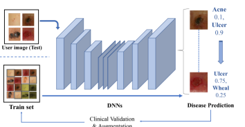
We attempt to provide such a solution which can indicate a subject if it is required to seek consultation urgently. It can also help doctors expedite consultancies based on the indicated detection. The mode of information exchange envisioned is via smartphone app(s) which can securely relay essential patient information. Our submission highlights the development of deep learning (DL) based method embedded at the core of this process.
Previously Esteva et al. used deep learning in detecting skin cancer [9]. Although their research was able to detect Melanoma with a dermatologist-level accuracy around 74%, it was limited to skin cancers, and distinguishing malignant from benign ones. Similar projects have been conducted by Shrivastava et al. in detecting Psoriasis [10]. In an attempt to detect multiple disease, Park et al. have introduced crowd-sourcing for common skin ailments [11]. Concurrently detecting multiple common skin diseases is unavailable along with lack of labeled data. Most of experiments focus on the accuracy, but not on training time or update schemes. We focus on detecting nine common skin diseases by training on curated data. We also explore the question of accuracy vis-a-vis time to make practical delivery schemes. With human-level accuracy in few classes, we hope such methods can gain traction towards affordability in health.
This paper is structured as follows: We discuss data preparation in Section 2. Our methodology is covered in Section 3. In Section 4, we elaborate on results and conclude with a brief discussion on shortcomings & future directions. The contribution of this paper is as follows:
-
•
We have curated an image database of nine common dermatological diseases. It comprises of about 4700 images per label. The process isolated only one disease per image. A significant amount of this database is being released to contribute to the machine learning community involved in healthcare.
-
•
We have evaluated classification strategies on popular DNN’s fine tuned to our requirements and subsequently attempted to understand the results.
-
•
We have compared network schemes and associated training times, which are indirectly related to cost of operation and drawn insights for future work.
2 Data Preparation
Since disease manifestation in Asian skin types could present differently from other races, we performed a systematic data collection via a smartphone application from volunteers. After anonymizing, 150,000 clinical images were labeled by trained medical professionals. Nine common conditions were chosen from this repository based on prevalence and relevancy. These diseases were: (i) Acne, (ii) Alopecia, (iii) Crust, (iv) Erythema, (v) Leukoderma, (vi) Pigmented Maculae, (vii) Pustule, (viii) Ulcers and (ix) Wheal. To avoid any skew in the process and balance the dataset, we performed augmentation on the dataset by standard techniques [12]. Making judgments from ablation studies we chose 4600 images per label approximately. The division of data between training and validation was done in a ratio of 90:10. A small corpus of test images, separate from the training and validation set, was kept at the outset to assess the classification as a blind experiment. Table 1 illustrates information about the various labels and their sizes.
| Disease | Training | Validation | Test |
|---|---|---|---|
| Acne | 4215 | 446 | 74 |
| Alopecia | 4119 | 441 | 65 |
| Crust | 4147 | 402 | 53 |
| Erythema | 4299 | 406 | 59 |
| Leukoderma | 4300 | 403 | 58 |
| P. Maculae | 4300 | 310 | 58 |
| Pustule | 4046 | 386 | 55 |
| Ulcer | 4514 | 395 | 58 |
| Wheal | 4120 | 385 | 50 |
3 METHODOLOGY
3.1 Statistical Basis
Our goal was to get the probabilistic predictions of the diseases as close as possible to ground truth. We chose to minimize cross-entropy loss as the basis of a good classification. Further information on them can be found in standard literature on statistical methods.
In addition to accuracy we paid attention to training time. Our long term objective requires us to frequently retrain models with new data. Training networks from scratch was found to be inefficient with best validation accuracy of less than 45%. We explored popular pre-trained DNNs such as ResNet18, ResNet50, ResNet152 and DenseNet161, initialized on ImageNet, as starting points for transfer learning[13, 14, 15]. Two strategies were evaluated. The first consisted of tuning the last fully-connected layer of these DNNs. The second approach was more rigorous by fine-tuning the entire network.
The classifier was built on PyTorch (v0.4) framework with Skorch library for scikit-learn modules. We chose a batch size of 16 and Stochastic gradient descent (SGD) with a learning rate of 0.001 along with appropriate decay for optimizer. The task was run on a system running NVIDIA Titan XP and CUDA v8. Five-fold cross-validation was adopted to deter over-fitting in addition. Best weights were recorded as soon as validation loss stabilized by Early-stopping.
3.2 Training the DNN’s
To test the first approach, we froze the network except for the final fully connected (FC) layer. Gradients were not computed in the backward direction, so as to not disturb the preceding layers. The results obtained were unsatisfactory in comparison to a full training, with maximum validation accuracy of 68% on any of the aforementioned model. We adopted training the full network for classification, although it was comparatively much slower. We highlight the results of this step in Table 2.
3.3 Test of classification
530 images, uniformly distributed across the nine labels, were left out of the training & validation corpus. Serving as unlabeled data, they were used to evaluate the quality of classification from our fine-tuned DNNs. To accomplish this step, a forward pass of the images on networks initialized with the corresponding best parameters was performed. The output score indicated the degree of match with each label. These outcomes were matched against the actual disease information tabulated by medical specialists. The average time to predict the class for a sample was approximately 0.4 seconds without needing a GPU. Test results have been elaborated in Table 3.
| Network | Validation | Time (min) |
|---|---|---|
| ResNet18 | 77.39% | 140.50 |
| ResNet50 | 78.19% | 374.11 |
| ResNet152 | 84.38% | 840.70 |
| DenseNet161 | 82.19% | 837.75 |
Noting that ResNet152 performs the best among candidate models, we have illustrated the class-wise prediction accuracy by a confusion matrix (Table 4).
| Tuned Network | Averaged Top-1 Accuracy |
|---|---|
| ResNet18 | 77.13% |
| ResNet50 | 78.81% |
| ResNet152 | 82.30% |
| DenseNet161 | 79.68% |
| Predicted (Rounded | |||||||||
| Actual | Acne | Alopecia | Crust | Erythema | Leukoderma | P. Macula | Pustule | Ulcer | Wheal |
| Acne | 83.8% | 0% | 0% | 10.8% | 0% | 2.7% | 0% | 2.7% | 0% |
| Alopecia | 0% | 90.8% | 6.2% | 3% | 0% | 0% | 0% | 0% | 0% |
| Crust | 0% | 0% | 60.4% | 3.8% | 0% | 30.2% | 0% | 5.7% | 0% |
| Erythema | 0% | 0% | 7% | 80% | 0% | 13% | 0% | 0% | 0% |
| Leukoderma | 0% | 0% | 0% | 0% | 93.0% | 3.5% | 0% | 0% | 3.5% |
| P. Macula | 3.6% | 0% | 0% | 0% | 0% | 96.4% | 0% | 0% | 0% |
| Pustule | 14.5% | 11.0% | 0% | 9.0% | 0% | 0% | 65.5% | 0% | 0% |
| Ulcer | 0% | 0% | 6.9% | 0% | 0% | 0% | 0% | 93.1% | 0% |
| Wheal | 0% | 0% | 0% | 28.0% | 0% | 0% | 0% | 0% | 72.0% |
4 DISCUSSION
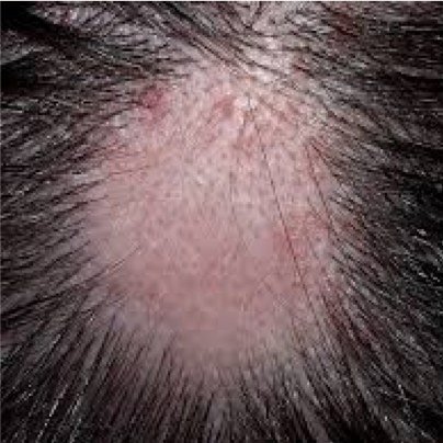
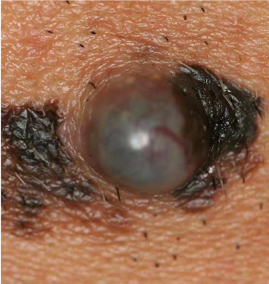
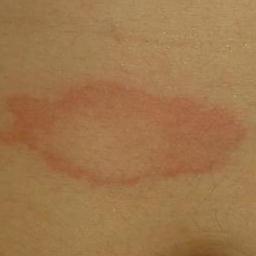
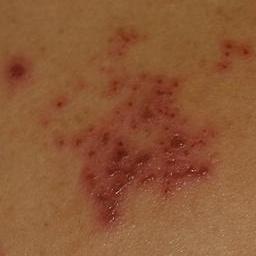
Despite some detection skew seen in Table 4, the model performed reasonably well. Five classes had test accuracy over 80%. Further, accuracy below 70% was observed only in two labels. Diseases such as Pigmented Macula, Ulcer and Alopecia are visually distinct in terms of contrast and structure. Hence, we hypothesize that extracted features are easy to distinguish in such cases. Labels such as Wheal or Crust, can present difficulty because of low amount of texture information in the images. This is consistent with our expectations. We illustrate our observation with samples in Fig. 2 and 3.
From our results, it is abundantly clear that common skin ailments are easy to detect. However, there are some caveats we would like to present. We concede that we assumed the existence of one of the disease types at the outset. We have not factored in separating normal skin from diseased conditions, which is a challenge by itself. Our task was easier than real world scenario, where several skin color and types could be involved. Our current results are limited to nine commonly seen conditions without any score of the severity. Also, observing the existence of two or more disease labels in a single sample is not uncommon. We hope to incorporate solutions to some of these situations in future works. In the absence of any network pre-trained on medical skin images strictly, our current insights advocate modestly large ResNet architectures when requirements deem rapid training and updating necessary.
5 CONCLUSION
This paper elucidates that several common skin problems can be successfully detected with deep learning techniques. In absence of dermatologists, this method can predict nine disease types, with accuracy surpassing that of general practitioners in many cases. We have also highlighted our choice of adopting a particular architecture for further development. Although there are some shortcomings, owing to the quantity and complexity of medical images, we anticipate overcoming some of these bottlenecks in future experiments. For reproducibility, dermatological image data, test codes, along with fine-tuned models are available at URL:http://bit.ly/2K76nwx.
References
- [1] Stern, R.S.: Prevalence of a history of skin cancer in 2007: results of an incidence-based model. Archives of dermatology 146(3) (2010) 279–282
- [2] Agbai, O.N., Buster, K., Sanchez, M., Hernandez, C., Kundu, R.V., Chiu, M., Roberts, W.E., Draelos, Z.D., Bhushan, R., Taylor, S.C., et al.: Skin cancer and photoprotection in people of color: a review and recommendations for physicians and the public. Journal of the American Academy of Dermatology 70(4) (2014) 748–762
- [3] Dawes, S.M., Tsai, S., Gittleman, H., Barnholtz-Sloan, J.S., Bordeaux, J.S.: Racial disparities in melanoma survival. Journal of the American Academy of Dermatology 75(5) (2016) 983–991
- [4] Kimball, A.B., Resneck, J.S.: The us dermatology workforce: a specialty remains in shortage. Journal of the American Academy of Dermatology 59(5) (2008) 741–745
- [5] Imaizumi, H., Watanabe, A., Hirano, H., Takemura, M., Kashiwagi, H., Monobe, S.: Hippocra: Doctor-to-doctor teledermatology consultation service towards future ai-based diagnosis system in japan. In: Consumer Electronics-Taiwan (ICCE-TW), 2017 IEEE International Conference on, IEEE (2017) 51–52
- [6] Dekio, I., Hanada, E., Chinuki, Y., Akaki, T., Kitani, M., Shiraishi, Y., Kaneko, S., Furumura, M., Morita, E.: Usefulness and economic evaluation of adsl-based live interactive teledermatology in areas with shortage of dermatologists. International journal of dermatology 49(11) (2010) 1272–1275
- [7] Lanzini, R.C., Fallen, R.S., Wismer, J., Lima, H.C.: Impact of the number of dermatologists on dermatology biomedical research: a canadian study. Journal of cutaneous medicine and surgery 16(3) (2012) 174–179
- [8] Lowell, B.A., Froelich, C.W., Federman, D.G., Kirsner, R.S.: Dermatology in primary care: prevalence and patient disposition. Journal of the American Academy of Dermatology 45(2) (2001) 250–255
- [9] Esteva, A., Kuprel, B., Novoa, R.A., Ko, J., Swetter, S.M., Blau, H.M., Thrun, S.: Dermatologist-level classification of skin cancer with deep neural networks. Nature 542(7639) (2017) 115
- [10] Shrivastava, V.K., Londhe, N.D., Sonawane, R.S., Suri, J.S.: Reliable and accurate psoriasis disease classification in dermatology images using comprehensive feature space in machine learning paradigm. Expert Systems with Applications 42(15-16) (2015) 6184–6195
- [11] Park, A.J., Ko, J.M., Swerlick, R.A.: Crowdsourcing dermatology: Dataderm, big data analytics, and machine learning technology (2017)
- [12] Russakovsky, O., Deng, J., Su, H., Krause, J., Satheesh, S., Ma, S., Huang, Z., Karpathy, A., Khosla, A., Bernstein, M., et al.: Imagenet large scale visual recognition challenge. International Journal of Computer Vision 115(3) (2015) 211–252
- [13] He, K., Zhang, X., Ren, S., Sun, J.: Deep residual learning for image recognition. In: Proceedings of the IEEE conference on computer vision and pattern recognition. (2016) 770–778
- [14] Iandola, F., Moskewicz, M., Karayev, S., Girshick, R., Darrell, T., Keutzer, K.: Densenet: Implementing efficient convnet descriptor pyramids. arXiv preprint arXiv:1404.1869 (2014)
- [15] Deng, J., Dong, W., Socher, R., Li, L.J., Li, K., Fei-Fei, L.: Imagenet: A large-scale hierarchical image database. In: Computer Vision and Pattern Recognition, 2009. CVPR 2009. IEEE Conference on, IEEE (2009) 248–255