\ul
SurvivalNet: Predicting patient survival from diffusion weighted magnetic resonance images using cascaded fully convolutional and 3D convolutional neural networks
Abstract
Automatic non-invasive assessment of hepatocellular carcinoma (HCC) malignancy has the potential to substantially enhance tumor treatment strategies for HCC patients. In this work we present a novel framework to automatically characterize the malignancy of HCC lesions from DWI images.
We predict HCC malignancy in two steps: As a first step we automatically segment HCC tumor lesions using cascaded fully convolutional neural networks (CFCN). A 3D neural network (SurvivalNet) then predicts the HCC lesions’ malignancy from the HCC tumor segmentation. We formulate this task as a classification problem with classes being “low risk” and “high risk” represented by longer or shorter survival times than the median survival. We evaluated our method on DWI of 31 HCC patients. Our proposed framework achieves an end-to-end accuracy of 65% with a Dice score for the automatic lesion segmentation of 69% and an accuracy of 68% for tumor malignancy classification based on expert annotations. We compared the SurvivalNet to classical handcrafted features such as Histogram and Haralick and show experimentally that SurvivalNet outperforms the handcrafted features in HCC malignancy classification. End-to-end assessment of tumor malignancy based on our proposed fully automatic framework corresponds to assessment based on expert annotations with high significance ().
Index Terms— Survival Prediction, 3D Neural Network, Fully Convolutional Neural Networks, MRI
1 Introduction
11footnotetext: Authors contributed equally22footnotetext: Corresponding authors: rbraren@tum.de and bjoern.menze@tum.de1.1 Motivation
Hepatocellular carcinoma (HCC) presents the sixth most common cancer and the third most common cause of cancer-related deaths worldwide [1]. HCC comprises a genetically and molecularly highly heterogeneous group of cancers that commonly arise in a chronically damaged liver. Importantly, HCC subtypes differ significantly in clinical outcome. The stepwise transformation to HCC is accompanied by major changes in tissue architecture including an increase in cellularity and a switch in vascular supply (i.e. arterialization). These differences provide the basis for the non-invasive detection of HCC [2]. In particular, diffusion weighted-magnetic resonance imaging (DW-MRI) detects differences in random Brownian motion, which is commonly reduced in highly cellular HCC due to an increase in cell membranes and macromolecules. The apparent diffusion coefficient (ADC) parameter value, which can be derived from two DW-MRI scans, quantifies this effect. DW-MRI imaging techniques provide a high level of sensitivity and specificity for tumor detection, the distinction of tumor subtypes requires the identification of more subtle differences. Computer aided analysis techniques allow medical image feature extraction far beyond the capabilities of the human eye and thus hold the potential for an imaging based differentiation of tumor subtypes. Non-invasive differentiation of tumor subtypes in HCC would enable pre-therapeutic patient stratification and the systematic testing of novel therapeutic strategies.
1.2 Related Works
Heid et al. (2016) have recently established a close relationship between the regional DW-MRI derived apparent diffusion coefficient (ADC) parameter value and distinct subtypes in pancreatic ductal adenocarcinoma (PDAC) [3]. Computer aided extraction of image features for tumor subtyping has previously been reported for several tumor entities. Prior work focused mostly on hand-crafted feature extraction such as histogram features [4], Gabor and Haralick features [5, 6], and grey level run length based features [7] to predict survival times for diverse tumor entities and image modalities. Recent works leveraged the discriminative power of the apparent diffusion coefficient (ADC) by extracting texture features for survival or malignancy characterization [8, 9]. Zhou et al. (2016) proposed a method to characterize malignancy of HCC in contrast enhanced MRI by extracting histogram and texture based features such as grey-level co-occurrence and run length (GLRL and GLCM) of HCC lesions [10]. However, their method required manual segmentation of the lesions beforehand.
1.3 Contribution
In comparison to prior work, we developed a method to predict HCC survival from DW-MRI volumes using automatic segmentations of tumors. Our contribution in this work is three-fold. First, we developed an automatic method to detect and segment HCC tumor lesions in DW-MRI data. Second, we found and analyzed quantitative biomarkers using handcrafted and CNN-based features to predict patient survival. Third, we experimentally demonstrated a fully automatic method to predict long/short survival of HCC patients from DW-MRI images.
2 Methods
Our proposed framework to fully automatically predict short/long survival from DW-MRI of HCC patients is depicted in figure 3.
2.1 Dataset
31 Patients underwent clinical assessment and MR imaging for the primary diagnosis of HCC. Barcelona Clinic Liver Cancer Classification was used to assess the clinical stage of the disease. Patients with a history of prior malignancy were excluded. No data with insufficient quality due to breathing artifacts, excessive banding or distortion, diffuse tumor growth or non-detectability of the lesions in the DW-MRI sequences was included in the dataset. Imaging was performed using a 1.5 T clinical MRI scanner (Avanto, Siemens) with a standard imaging protocol including axial and coronal T2w, axial T1w images before and after application of Gadolinium-DTPA contrast agent (Jenapharm Magnograf ® 0.5 mmol/ml per manufacturer’s instructions). Post-contrast T1w images were acquired in the early, mid and late arterial phases as well as in the portal venous phase. Diffusion weighted imaging was performed using a slice thickness of 5mm and a matrix size of 192 by 192. Institutional review board approval was obtained for this retrospective study.
2.2 Automatic Segmentation
To automatically detect and segment tumor lesions we applied a cascaded fully convolutional neural network to segment in step 1 the liver and in a step 2 the tumor lesions from a liver ROI volume [11]. We used the DW-MRI as input to the FCN architecture proposed by Ronneberger et al. (2015) [12]. We fine-tuned our networks using the liver and liver tumor model provided by Christ et al. (2016) [11] and applied a 5-fold cross-validation. Tumor margins were identified in the early arterial phase and in DWI images (b=600). Manual segmentation was performed by an experienced radiologist using the software TurtleSeg® and reviewed by two expert radiologists.
2.3 Survival Prediction
To predict the survival rate of HCC tumor patients we calculated different features using the detected and segmented tumor lesions applied in the ADC image sequences. We calculated handcrafted features and features trained end-to-end by a 3D Convolutional Neural Network (SurvivalNet).
2.3.1 Handcrafted Features
ADC value histograms were generated from the regions of the ADC map corresponding to the tumor ROI in the b=600 image. Histogram descriptors were obtained including mean, median, kurtosis and skewness. In addition, we extracted features representing ADC texture by calculating 3D Haralick statistics of the grey-level co-occurrence matrix [13]. We trained a k-nearest-neighbour classifier with k=4 and validated the results using 10-fold cross-validation.
2.3.2 SurvivalNet: 3D Convolutional Neural Network
Finally, we trained a 3D CNN to predict the survival rate in an end-to-end fashion. The SurvivalNet consists of two stacks of 3D convolution and max pooling layers, followed by 2 fully-connected layers. The 3D convolutions have 50 kernels with a kernel size of 3x3x3 pixels and a 3D spatial dropout with . The first fully-connected layer has 500 neurons. Figure 4 shows the SurvivalNet architecture. We trained the SurvivalNet from scratch using the Adadelta gradient-descent algorithm [14] at a learning rate of , and . We employed no data-augmentation.
Table 2 shows the performance of the handcrafted features and SurvivalNet.
3 Results
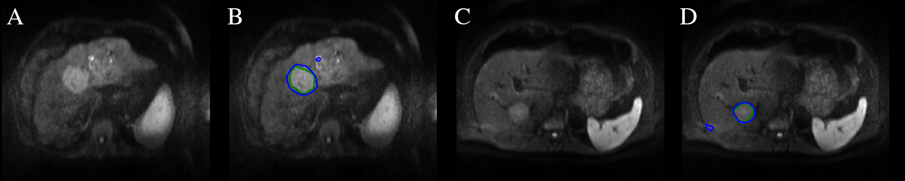
3.1 Qualitative
The qualitative results of the automatic segmentation are depicted in figure 1. The complex and heterogeneous shape of the tumor lesions was detected and segmented in both images using our automatic segmentation algorithm. The trained model achieves a Dice overlap score of 85% in both images. The segmentation reaches a high level of specificity by classifying all lesion pixels in the image as lesions. Small false positive outliers within the liver reduce the overall accuracy and Dice score.
3.2 Quantitative
| Method | Sensitivity | Precision | TNR | RVD | Dice |
| [%] | [%] | [%] | [%] | [%] | |
| Cascaded FCN on DWI | 91.1 | 70.0 | 99.6 | 52.1 | 69.7 |
| Features | ACC | Precision | Sensitivity | F1-Score | ||
| [%] | [%] | [%] | [%] | |||
| Manual Tumor Seg. | SurvivalNet CNN | 68 | 69 | 68 | 65 | |
| Histogram Features | 61 | 62 | 61 | 60 | ||
| Texture Features: 3D Haralick | 61 | 65 | 61 | 58 | ||
| Automatic Tumor Seg. | SurvivalNet CNN | 65 | 64 | 65 | 64 | |
| Histogram Features | 58 | 59 | 58 | 56 | ||
| Texture Features: 3D Haralick | 61 | 62 | 62 | 60 |
The quantitative results for our automatic segmentation method are shown in table 1. Our automatic HCC lesion segmentation algorithm achieves a Dice overlap score of 69.7% trained on DW-MRI images. The trained model is highly sensitive in recognizing HCC lesions with a Sensitivity of 91.1%, i.e. only few false negative errors occur.
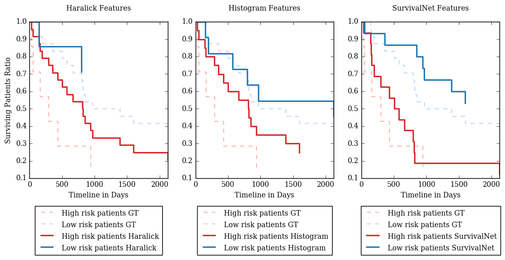
Table 2 shows the quantitative results of our proposed automatic survival prediction framework. Figure 2 shows a Kaplan-Meier plot of the survival prediction results. SurvivalNet achieves higher scores on both manual and automatic segmentation compared to handcrafted features. SurvivalNet trained on manual segmentations achieves an accuracy of 68% with a Precision and Sensitivity of 69% and 68% respectively. Furthermore, SurvivalNet accomplishes a classification accuracy of 65% at a Precision and Sensitivity of 64% and 65% when trained using our automatic tumor segmentation in a fully automatic fashion.
As a final experiment, we calculated a paired Wilcoxon signed-rank test with H0: the output posterior class probabilities of SurvivalNet with manual and automatic segmentation belong to the same distribution. At , we found H0 to be confirmed, i.e. SurvivalNet produces the same results with automatic segmentation or manual segmentation.
4 Conclusion and Discussion
The predictive value of various imaging parameters has previously been suggested in HCC. With the growing appreciation of tumor heterogeneity as a major obstacle to treatment response, more sophisticated image analysis algorithms are required. The complexity of such data analyses, especially considering multi-parametric multimodality imaging, requires computer aided techniques. We have presented a fully automatic framework to predict survival times of HCC patients. This approach based on fully convolutional and 3D convolutional neural networks outperformed state-of-the art handcrafted features, while still achieving the same diagnostic outcome as if human expert segmentations were provided. This work may have potential applications in HCC treatment planning.
5 Acknowledgement
This work was supported by the German Research Foundation (DFG) within the SFB-Initiative 824 (collaborative research center), “Imaging for Selection, Monitoring and Individualization of Cancer Therapies” (SFB824, project C6) and the BMBF project Softwarecampus. We thank NVIDIA and Amazon AWS for granting GPU and computation support.
References
- [1] Jacques Ferlay, Hai-Rim Shin, Freddie Bray, David Forman, Colin Mathers, and Donald Maxwell Parkin, “Estimates of worldwide burden of cancer in 2008: Globocan 2008,” International Journal of Cancer, vol. 127, no. 12, pp. 2893–2917, 2010.
- [2] European Association For The Study Of The Liver, “Easl–eortc clinical practice guidelines: management of hepatocellular carcinoma,” Journal of Hepatology, vol. 56, no. 4, pp. 908–943, 2012.
- [3] Irina Heid, Katja Steiger, Marija Trajkovic-Arsic, Marcus Settles, Manuela R Eßwein, Mert Erkan, Jorg Kleeff, Carsten Jäger, Helmut Friess, Bernhard Haller, Andreas Steingötter, Roland M Schmid, Markus Schwaiger, Ernst J Rummeny, Irene Esposito, Jens T Siveke, and Rickmer Braren, “Co-clinical assessment of tumor cellularity in pancreatic cancer,” Clinical Cancer Research, 2016.
- [4] Sang Ho Lee, Koichi Hayano, Dushyant V. Sahani, Andrew X. Zhu, and Hiroyuki Yoshida, “Kinetic textural biomarker for predicting survival of patients with advanced hepatocellular carcinoma after antiangiogenic therapy by use of baseline first-pass perfusion ct,” in Abdominal Imaging. Computational and Clinical Applications: 6th International Workshop, ABDI 2014, Held in Conjunction with MICCAI, pp. 48–61. 2014.
- [5] Jiawen Yao, Sheng Wang, Xinliang Zhu, and Junzhou Huang, “Imaging biomarker discovery for lung cancer survival prediction,” in MICCAI, pp. 649–657. 2016.
- [6] X. Zhu, J. Yao, X. Luo, G. Xiao, Y. Xie, A. Gazdar, and J. Huang, “Lung cancer survival prediction from pathological images and genetic data; an integration study,” in IEEE ISBI, 2016, pp. 1173–1176.
- [7] J. Song, D. Dong, Y. Huang, Z. Liu, and J. Tian, “Association between tumor heterogeneity and overall survival in patients with non-small cell lung cancer,” in IEEE ISBI, 2016, pp. 1249–1252.
- [8] Islam Reda, Ahmed Shalaby, Mohammed Elmogy, Ahmed Aboulfotouh, Fahmi Khalifa, Mohamed Abou El-Ghar, Georgy Gimelfarb, and Ayman El-Baz, “Image-based computer-aided diagnostic system for early diagnosis of prostate cancer,” in MICCAI, pp. 610–618. 2016.
- [9] M. Shehata, F. Khalifa, A. Soliman, M. Abou El-Ghar, A. Dwyer, G. Gimel’farb, R. Keynton, and A. El-Baz, “A promising non-invasive cad system for kidney function assessment,” in MICCAI, pp. 613–621. 2016.
- [10] Wu Zhou, Lijuan Zhang, Kaixin Wang, Shuting Chen, Guangyi Wang, Zaiyi Liu, and Changhong Liang, “Malignancy characterization of hepatocellular carcinomas based on texture analysis of contrast-enhanced mr images,” Journal of Magnetic Resonance Imaging, 2016.
- [11] Patrick Ferdinand Christ, Mohamed Ezzeldin A. Elshaer, Florian Ettlinger, Sunil Tatavarty, Marc Bickel, Patrick Bilic, Markus Rempfler, Marco Armbruster, Felix Hofmann, Melvin D’Anastasi, Wieland H. Sommer, Seyed-Ahmad Ahmadi, and Bjoern H. Menze, “Automatic liver and lesion segmentation in ct using cascaded fully convolutional neural networks and 3d conditional random fields,” in MICCAI, pp. 415–423. 2016.
- [12] O. Ronneberger, P. Fischer, and T. Brox, “U-net: Convolutional networks for biomedical image segmentation,” in MICCAI, vol. 9351, pp. 234–241. 2015.
- [13] R. M. Haralick, “Statistical and structural approaches to texture,” Proceedings of the IEEE, vol. 67, no. 5, pp. 786–804, May 1979.
- [14] Matthew D. Zeiler, “ADADELTA: an adaptive learning rate method,” CoRR, vol. abs/1212.5701, 2012.