Universality in the morphology and mechanics of coarsening amyloid fibril networks
Abstract
Above a critical concentration a wide variety of peptides and proteins self-assemble into amyloid fibrils which entangle to form percolating networks called hydrogels. Such hydrogels have important applications as biomaterials and in nanotechnology, but their applicability often depends on their mechanical properties for which we currently have no predictive capability. Here we use a peptide model to simulate the formation of amyloid fibril networks, and couple these to elastic network theory to determine their mechanical properties. The simulations reveal that the time-dependence of morphological quantities characterizing the network length scales can be collapsed onto master curves by using a time scaling function that depends on the interaction parameter between the peptides. The same scaling function is used to unveil a universal, non-monotonic dependence of the shear modulus with time. The obtained insight into the structure-function relationship between the peptide building blocks, network morphology and network mechanical properties can aid experimentalists to design amyloid fibril networks with tailored mechanical properties.
The formation of amyloid fibril networks proceeds in two stages. Firstly, peptides or proteins assemble into amyloid fibrils that share a common cross- structure formed of intertwined layers of -sheets extending in the direction parallel to the fibril axis 1; 2. The lengthening and thickening of the fibrils can be attributed to strongly directional backbone hydrogen bonding and weaker side-chain interactions, respectively. At later times, the amyloid fibrils can entangle into a percolating network 3 with a morphology characterised by various lengths including the distance between crosslinks. Although amyloid fibrils are often associated with devastating diseases including Alzheimer’s and Parkinson’s disease 4, amyloid networks are emerging as an important class of material with applications in biosensing, nanoelectronics, tissue engineering and drug delivery 5; 6; 7. However, many of these applications depend strongly on the network’s mechanical properties, for which we currently have no predictive structure-function relation between the bulk network stiffness and the properties of individual proteins without making assumptions regarding the fibril morphology, cross-linker dynamics, and the local deformation regime.
Theoretical and experimental studies characterizing the mechanical properties of single, isolated amyloid fibrils show that their elastic modulus and bending stiffness are comparable to semiflexible filaments like actin, keratin, collagen and spider silk 8; 9; 10; 11. Recent rheology experiments demonstrate that the shear modulus of amyloid fibril networks can be altered by changes in the amino acid sequence of the peptides 12; 13 and the ionic strength of the solution 14; 15; 16; 17, indicating that design principles for such networks might exist. The viscoelastic response of semiflexible polymer networks can be immediately related to the properties of individual filaments under the assumption of affinity, i.e. that the microscopic deformation field follows the applied macroscopic strain 18; 19; 20; 21; 22. Relaxing this assumption has thus far only been possible for the zero-frequency response of athermal networks, formally corresponding to the elastic plateau in rheological measurements where the crosslinks can be regarded as fixed 23; 24; 25; 26. Importantly, all of the mentioned theoretical approaches assume that the filaments are identical, and thus have the same bending rigidity, or equivalently persistence length, at every point in the network. This assumption is a good approximation for filaments such as actin, but is not valid for amyloid fibril networks, where the fibril thickness varies throughout the network 27; 28; 29. Furthermore, these previous studies considered procedurally-defined, static networks; the time evolution of the network morphology was not considered. The objective of this study is to model the formation of amyloid fibril networks to determine their mechanical properties from elastic network theory without making the aforementioned assumptions.
Modelling amyloid fibril network formation
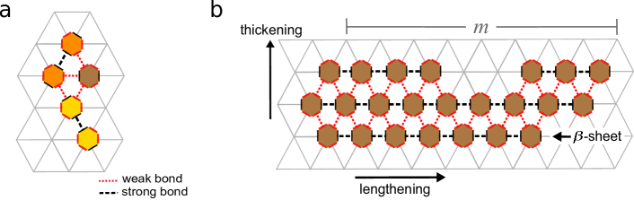
Our simulations use a peptide model similar to those used in recent studies on the aggregation of amyloid fibrils 30; 31; 32; 33, where the peptides in their virtually fully extended (-strand) conformation are described as hexagons positioned on a two-dimensional (2D) lattice with triangular symmetry (Supplementary Fig. 1a). The use of a triangular lattice ensures that the mechanical properties of the fibril network are isotropic at large length scales 34 as well as enabling the correct stacking of -sheets in fibrils 35 (Supplementary Fig. 1b). The model shares similarities with the anisotropic Potts model, where only nearest-neighbor interactions along the three symmetry axes of the triangular lattice are considered. The hexagon has two opposing strong bonding sides that allow the formation of directional backbone hydrogen bonds. This strong bond energy is denoted by the dimensionless parameter , where is Boltzmann’s constant and the temperature. The remaining four sides of the hexagon allow the formation of weaker bonds modelling the side-chain interactions between the peptides. This bond energy is denoted . The -strands can self-assembly laterally into -sheets by forming strong hydrogen bonds that are parallel to the fibril lengthening axis, and the fibril can thicken due to the formation of weaker bonds between side-chains (Supplementary Fig. 1b). Here we fix while varying , to study effect of the ratio of the hydrogen bond energy to the side-chain bond energy. A value of has been estimated for the A peptide 32.
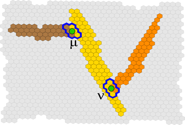
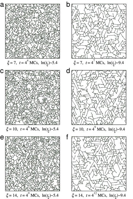
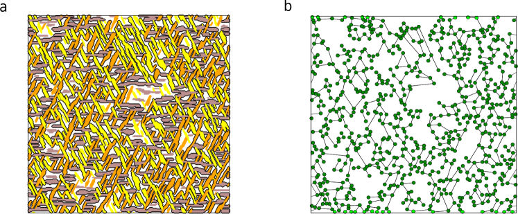
In order to model the formation of amyloid fibril networks we perform Monte Carlo (MC) simulations similar to those described in ref. 30. We perform displacement moves of peptides to nearest neighbor lattice sites, and rotation moves so that the peptide can change its orientation. At the beginning of a simulation, peptides are randomly placed and oriented on a periodic 2D triangular lattice of linear size . In all our simulations we set . As we only use physically plausible moves, there is a correspondence between the number of MC steps (MCs) and real time as suggested in refs. 30; 33; 36. A typical configuration is shown in Fig. 1a. The fibril network obtained is very similar to those observed in experiments 37; 38. We use the Hoshen-Kopelman algorithm 39 to identify individual amyloid fibrils, which enable us to characterized them by their thickness and length (see Supplementary Fig. 1b). The identification of fibrils also allows us to obtain a elastic network representation of the system as shown in Fig. 1b. The crosslink positions are taken to be the geometric centres of the overlap region between different fibrils (see Supplementary Fig. 2), with the distance between two connected crosslinks defined as the crosslink length . Note that not all fibrils identified in the system are part of the percolating network (Fig. 1a).
Morphology of coarsening amyloid fibril networks
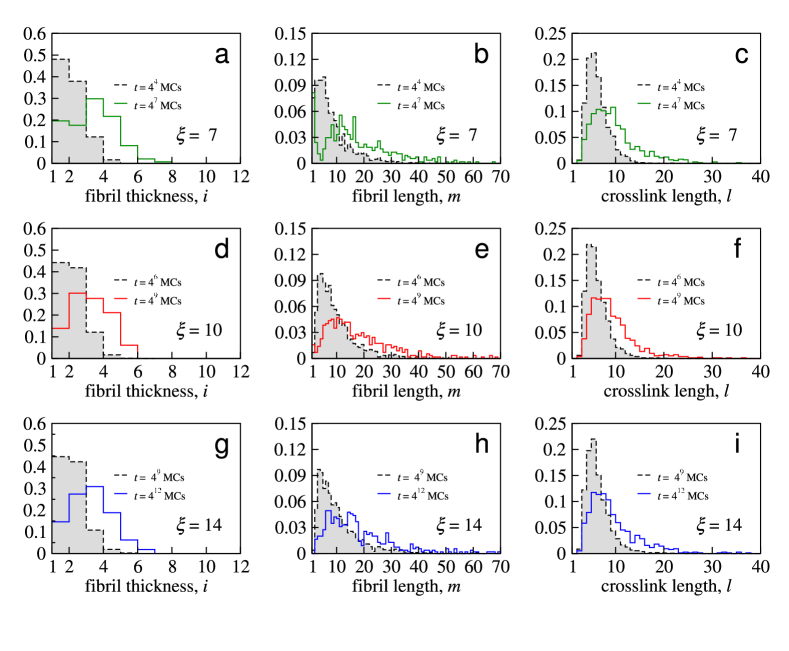
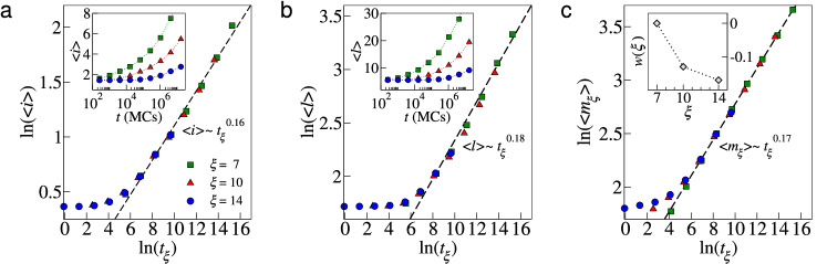
The time evolution of the mean fibril thickness , the mean crosslink distance , and the mean fibril length is shown in Fig. 2 for anisotropy ratios , , and , at a coverage . As can be seen in the insets to this figure, all lengths enter a coarsening regime where they monotonically increase, i.e. the fibrils become thicker and longer and the crosslink positions become further apart as the network evolves. Increasing the anisotropy ratio delays the onset of coarsening. A similar effect, where the dynamics of the system is slowed down due to stronger effective interparticle interactions, has been observed in colloidal gels and glasses 40; 41. As demonstrated in Fig. 2, the time dependence of each morphological quantity of the network obtained for different anisotropy ratios can be collapsed onto a single master curve by rescaling the time as , where with an arbitrary origin (here we take ). It is not only the mean of the morphological quantities that collapse, but also their distribution functions as shown in Supplementary Fig. 3. The coarsening exponents , , and of the morphological quantities can be obtained by fitting the data points for times to , , and , respectively. The mean fibril length was additionally scaled by an arbitrary function (inset of Fig. 2c), giving , but this does not affect the scaling with time. The measured exponents are consistent with a common value , demonstrating that all network lengths obey the same scaling during coarsening. Note that, if one identifies the fibril area as a domain size, the growth law is consistent with the 1/3 exponent derived analytically for the isotropic case, i.e. the 2D spin-exchange Ising model 42; 43, despite the fibrils in our networks forming anisotropic shapes.
Mechanics of amyloid fibril networks
We now turn our attention to understanding how the structural changes in the network affects its mechanical properties. Starting from the network representation as in Fig. 1b, one can mimic an applied strain by imposing horizontal displacements to the boundary crosslinks as shown in Supplementary Fig. 4. The internal crosslinks then relax to new positions, causing an increase in the total elastic energy of the network. For the linear response (), one can use to extract the shear modulus 26; 34,
| (1) |
where is the total system area. The shear modulus thus depends on the displacement vectors of all crosslinks, which contribute to as detailed in Methods. These displacement vectors are obtained by performing high-dimensional numerical optimization to minimize with respect to each displacement degree of freedom 24. For all measurements we consider to avoid non-linear responses due to high strain values. Our method generalises a previous lattice-based model 26 by permitting variations in fibril thickness and crosslink distances (see Methods).
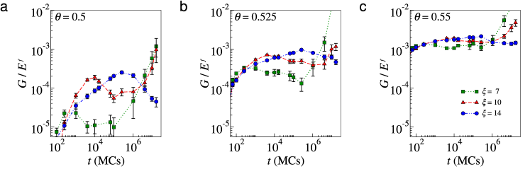
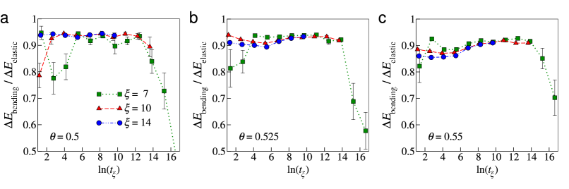
In Fig. 3 we present results for the time-dependent behaviour of the normalized shear modulus , where is the Young’s modulus of a fibril. It is evident that small changes in both the anisotropy and the coverage lead to large changes in spanning orders of magnitude, with an overall trend for higher values of with increasing . Moreover, the variation is non-monotonic in time, in contrast to the monotonic coarsening of the morphological quantities discussed above, and there are time periods when the thinner fibrils (formed for ) yield stronger networks than thicker fibrils. Most experiments on the formation of amyloid fibril networks are performed on a time scale of tens of minutes, where the shear modulus displays either an increasing or a constant behaviour 44; 14; 45; 16. However, one set of longer experiments suggested a slight decrease in after hours 46, and atomic force microscopy imaging has demonstrated significant changes to network morphology over a timescale of days 37; 47, for which mechanical properties are not usually measured. This suggests extending the data acquisition window may reveal a similar non-monotonicity to Fig. 3. In addition, the behaviour of resembles that of weakly-interacting colloidal aggregates which measurably weaken prior to visual collapse 48. We hypothesise this weakening shares a common mechanism to that observed in our results, although the final collapse under gravity (with its associated step change in the bulk symmetry) presumably has a different origin.
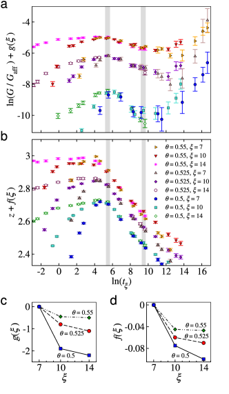
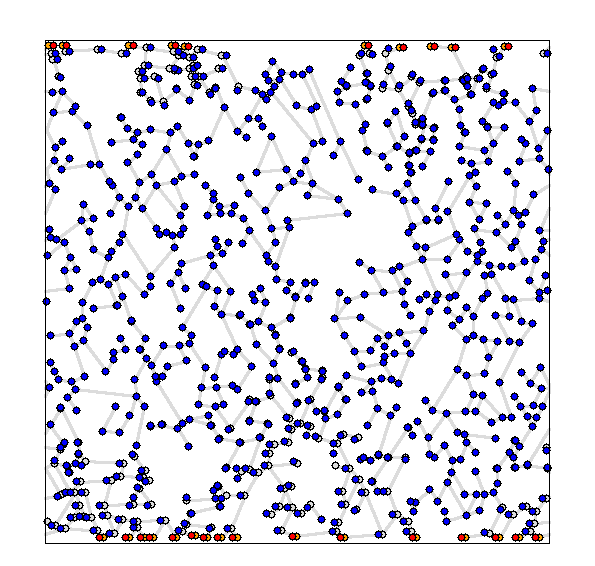
Insight into the mechanism underlying network weakening can be gained by measuring mechanical and morphological quantities simultaneously. We evaluated the shear modulus normalised to the affine prediction (computed from Eq. 1 for identical networks with affine displacements ) as in previous works 24; 26, and monitored the mean network connectivity given by the average value of the coordination numbers, , from all internal crosslinks. As depicted in Fig. 4a, we found that for different anisotropies can be collapsed onto a series of master curves, with one such curve for each coverage . The decreasing region of coincides with the onset of a decrease in as indicated in the diagram, and indeed snapshots reveal a reduction in network connectivity over these times (see Supplementary Fig. 5). Thus, the mechanical weakening of the network is due to its increased sparsity. As with the fibril length it was also necessary to scale the magnitude of by -dependent factors and as shown in Figs. 4c and 4d, but this does not alter the times corresponding to the local minimum and maximum in . These factors mean that, at fixed rescaled times , higher yields higher , and correspondingly higher values of . For all values of and , the values of are located below the central force threshold ( for 2D systems), and as evident in Fig. 4a. Thus all of our networks correspond to a non-affine deformation regime, and consistent with prior observations 26 most of the elastic energy takes the form of fibril bending, with fluctuating around 0.9 (see Supplementary Fig. 6).
Concluding remarks
Identifying universal behaviours, such as in time-cure superposition curves 46; 49; 50, can accelerate the development of novel materials by reducing the number of independent parameters that need to be assayed. Our simulations have revealed a simple time scaling function that depends on the anisotropy of interaction between peptides, which collapses data for both morphological and mechanical quantities. The proposed scaling function should benefit experimentalists in the design of amyloid-based materials, since it permits the extrapolation of the time-dependent mechanical response of the amyloid fibril networks from the behaviour of peptide systems with known interactions. Our findings indicate that features like the non-zero shear modulus for connectivities lower than and the non-affine response, which are commonly overlooked in the modelling of hydrogels 21; 22, should be included in further descriptions of amyloid fibril networks. Finally, we note that our hybrid approach to measure the elastic moduli of a fiber network as it forms and grows represents a new direction for fiber network modelling that can be extended to other fibrous and porous materials in general, including inorganic materials such as colloidal gels 40.
Methods
Elastic network model. Here we employ a modified version of a lattice-based elastic network model 26 to evaluate as the sum of the changes in stretching and bending energies. The change in the stretching energy is defined as
| (2) |
where the sum is over all unique pairs of adjacent connected crosslinks and , is the spring constant of the fibril segment connecting them, and is the segment’s extension due to the displacement vectors at each end, i.e. with and . The change in the bending energy is defined similarly,
| (3) |
where the sum is over adjacent crosslinks along the same fibril, is the bending rigidity of the corresponding fibril, is the mean crosslink length, and denotes the change in angle between the two consecutive fibril segments and . Note that the initial angle is generally not zero, but using the change in angle means that the unstrained network has zero elastic energy, i.e. there are no pre-stresses. We estimate the coupling constants and by treating the fibrils as slender, defect-free elastic bodies with a uniform circular cross-section 34. Thus the spring constant and bending rigidity are written, respectively, as and , where is the radius of the cross-section of the fibril, its Young’s modulus, and for connected crosslinks or otherwise. The radius is taken to be half of the fibril thickness of the fibril segment linking the crosslink pair and . We normalise both constants by , giving and . Note that although we have neglected the mechanical anisotropy of the fibrils, our key findings are expected to hold as long as the bending modulus scales with thickness as . This is predicted by theoretical calculations for protein fibre bundles with bundling springs times weaker than fibre springs, separated by one monomer, for which scaling is expected 51 up to , far in excess of our simulations. In addition, computational resources limited the network construction to 2D, but we employ 3D fibril properties so that our results can be experimentally realisable, for instance by confining the network to a Langmuir trough. Permitting out-of-plane displacements can significantly alter the measured moduli 52, but for actin networks the Langmuir trough was shown to suppress such modes 53 so we do not include these displacements in our calculations.
References
- (1) Adamcik, J. & Mezzenga, R. Proteins fibrils from a polymer physics perspective. Macromolecules 45, 1137 (2012).
- (2) Fitzpatrick, A. W. P. et al. Atomic structure and hierarchical assembly of a cross- amyloid fibril. P. Natl. Acad. Sci. USA 110, 5468 (2013).
- (3) Schleegert, M. et al. Amyloids: From molecular structure to mechanical properties. Polymer 54, 2473 (2013).
- (4) Chiti, F. & Dobson, C. M. Protein misfolding, functional amyloid, and human disease. Annu. Rev. Biochem. 75, 333 (2006).
- (5) Cherny, I. & Gazit, E. Amyloids: Not only pathological agents but also ordered nanomaterials. Angew. Chem. Int. Ed. 47, 4062 (2008).
- (6) Bowerman, C. J. & Nilsson, B. L. Self-assembly of amphipathic -sheet peptides: Insights and applications. Biopolymers 98, 169 (2012).
- (7) Reynolds, N. P., Charnley, M., Mezzenga, R. & Hartley, P. G. Engineered lysozyme amyloid fibril networks support cellular growth and spreading. Biomacromolecules 15, 599 (2014).
- (8) Smith, J. F., Knowles, T. P. J., Dobson, C. M., MacPhee, C. E. & Welland, M. Characterization of the nanoscale properties of individual amyloid fibrils. P. Natl. Acad. Sci. USA 43, 15806 (2006).
- (9) Keten, S., Xu, B., Ihle, B. & Buehler, M. J. Nanoconfinement controls stiffness, strength and mechanical toughness of beta-sheet crystals in silk. Nat. Mater. 9, 359 (2010).
- (10) Knowles, T. P. J. & Buehler, M. J. Nanomechanics of functional and pathological amyloid materials. Nat. Nanotechnol. 6, 469 (2011).
- (11) Volpatti, L. R. & Knowles, T. P. J. Polymer physics inspired approaches for the study of the mechanical properties. J. Polymer Sci. B: Pol. Phys 52, 281 (2014).
- (12) Tang, C., Ulijn, R. & Saiani, A. Effect of glycine substitution on Fmoc-diphenylalanine self-assembly and gelation properties. Langmuir 27, 14438 (2011).
- (13) Adler, J., Scheidt, H. A., Krüger, M., Thomas, L. & Huster, D. Local interactions influence the fibrillation kinetics, structure and dynamics of A(1-40) but leave the general fibril structure unchanged. Phys. Chem. Chem. Phys. 16, 7461 (2014).
- (14) Ozbas, B. B., Rajagopal, K. K., Schneider, J. P. J. & Pochan, D. J. Salt-triggered peptide folding and consequent self-assembly into hydrogels with tunable modulus. Macromolecules 37, 7331 (2004).
- (15) Riley, J. M., Aggeli, A., Koopmans, R. J. & McPherson, M. J. Bioproduction and characterization of a pH responsive self-assembling peptide. Biotech. Bioeng. 103, 241 (2009).
- (16) Bolisetty, S., Harnau, L., Jung, J.-M. & Mezzenga, R. Gelation, phase behaviour, and dynamics of -lactoglobulin amyloid fibrils at varying concentrations and ionic strengths. Biomacromolecules 13, 3241 (2012).
- (17) Boothroyd, S., Miller, A. F. & Saiani, A. From fibres to networks using self-assembling peptides. Farad. Discuss. 2013, 13 (2013).
- (18) Pritchard, R., Huang, Y. Y. S. & Terentjev, E. M. Mechanics of biological networks: From the cell cytoskeleton to connective tissue. Soft Matter 10, 1864 (2014).
- (19) Broedersz, C. P. et al. Cross-link governed dynamics of biopolymers networks. Phys. Rev. Lett. 105, 238101 (2010).
- (20) Wolff, L., Fernandez, P. & Kroy, K. Inelastic mechanics of sticky biopolymer networks. New J. Phys. 12, 053024 (2010).
- (21) Basu, A. et al. Nonaffine displacements in flexible polymer networks. Macromolecules 44, 1671 (2011).
- (22) Wen, Q., Basu, A., Janmey, P. A. & Yodhb, A. G. Non-affine deformations in polymer hydrogels. Soft Matter 8, 8039 (2012).
- (23) Head, D. A., Levine, A. & MacKintosh, F. Deformation of cross-linked semiflexible polymer networks. Phys. Rev. Lett. 91, 108102 (2003).
- (24) Head, D. A., Levine, A. & MacKintosh, F. Distinct regimes of elastic response and deformation modes of cross-linked cytoskeletal and semiflexible polymer networks. Phys. Rev. E 68, 061907 (2003).
- (25) Heussinger, C. & Frey, E. Floppy modes and nonaffine deformations in random fiber networks. Phys. Rev. Lett. 97, 105501 (2006).
- (26) Broedersz, C. P., Mao, X., Lubensky, T. C. & MacKintosh, F. C. Criticality and isostaticity in fibre networks. Nat. Phys. 7, 983 (2011).
- (27) Kolsofszki, M., Karsai, A., Soós, K., Penke, B. & Kellermayer, M. S. Z. Thermally-induced effects in oriented network of amyloid 25-35 fibrils. Prog. Colloid Polym. Sci. 135, 169 (2008).
- (28) Xue, W. F., Homans, S. W. & Radford, S. E. Amyloid fibril length distribution quantified by atomic force microscopy single-particle image analysis. Protein Eng. Des. Sel. 22, 489 (2009).
- (29) Morris, R. J. et al. Mechanistic and enviromental control of the prevalence and lifetime of amyloid oligomers. Nat. Comm. 4, 1891 (2013).
- (30) Zhang, J. & Muthukumar, M. Simulations of nucleation and elongation of amyloid fibrils. J. Chem. Phys. 130, 035102 (2009).
- (31) Cabriolu, R., Kashchiev, D. & Auer, S. Breakdown of nucleation theory for crystals with strongly anisotropic interactions between molecule. J. Chem. Phys. 137, 204903 (2012).
- (32) Kashchiev, D., Cabriolu, R. & Auer, S. Confounding the paradigm: Peculiarities of amyloid fibril nucleation. J. Am. Chem. Soc. 135, 1531 (2013).
- (33) Irback, A., Jonsson, S., Linnemann, N., Linse, B. & Wallin, S. Aggregate geometry in amyloid fibril nucleation. Phys. Rev. Lett. 110, 058101 (2013).
- (34) Landau, L. D. & Lifshitz, E. M. Theory of Elasticity (Butterworth-Heineman Oxford, 1986).
- (35) Nguyen, H. D. & Hall, C. K. Kinetics of fibril formation by polyalanine peptides. J. Biol. Chem. 280, 9074 (2005).
- (36) Linse, B. & Linse, S. Monte Carlo simulations of protein amyloid formation reveal origin of sigmoidal aggregation kinetics. Mol. Biosyst. 7, 2296 (2011).
- (37) Karsai, A. et al. Potassium-dependent oriented growth of amyloid 25-35 fibrils on mica. Nanotechnology 18, 345102 (2007).
- (38) Li, H. et al. Peptide diffusion and self-assembly in ambient water nanofilm on mica surfaces. J. Phys. Chem. B 113, 8795 (2009).
- (39) Hoshen, J. & Kopelman, R. Percolation and cluster distribution. I. Cluster multiple labeling technique and critical concentration algorithm. Phys. Rev. B 14, 3438 (1976).
- (40) Lu, P. J. & Weitz, D. A. Colloidal particles: crystals, glasses, and gels. Annu. Rev. Condens. Matter Phys. 4, 217 (2013).
- (41) Hunter, G. L. & Weeks, E. R. The physics of the colloidal glass transition. Rep. Prog. Phys. 75, 066501 (2012).
- (42) Amar, J. G., Sulivan, F. E. & Mountain, R. D. Monte Carlo study of growth in the two-dimensional spin-exchange kinetic Ising model. Phys. Rev. B 37, 196 (1988).
- (43) Mitchell, S. J. & Landau, D. P. Phase separation in a compressible 2D Ising model. Phys. Rev. Lett. 97, 025701 (2006).
- (44) Aggeli, A. et al. Responsive gels formed by the spontaneous self-assembly of peptides into polymeric -sheet tapes. Nature (1997).
- (45) Greenfield, M. A., Hoffman, J. R., de la Cruz, M. O. & Stupp, S. I. Tunable mechanics of peptide nanofiber gels. Langmuir 26, 3641 (2010).
- (46) Gosal, W. S., Clark, A. H. & Ross-Murphy, S. B. Fibrillar -lactoglobulin gels: part 2. dynamic mechanical characterization of heat-set systems. Biomacromolecules 5, 2420 (2004).
- (47) Usov, I., Adamcik, J. & Mezzenga, R. Polymorphism in bovine serum albumin fibrils: morphology and statistical analysis. Farad. Discuss. 166, 151 (2013).
- (48) Kamp, S. W. & Kilfoil, M. L. Universal behavior in the mechanical properties of weakly aggregated colloidal particles. Soft Matter 5, 2438 (2009).
- (49) Corrigan, A. M. & Donald, A. M. Particle tracking microrheology of gel-forming amyloid fibril networks. Eur. Phys. J. E 28, 457 (2009).
- (50) Corrigan, A. M. & Donald, A. M. Passive microrheology of solvent-induced fibrillar protein networks. Langmuir 25, 8599 (2009).
- (51) Bathe, M., Heussinger, C., Claessens, M. M. A. E., Bausch, A. R. & Frey, E. Cytoskeletal bundle mechanics. Biophys. J. 94, 2955 (2008).
- (52) Müller, P. & Kierfeld, J. Wrinkling of random and regular semiflexible polymer networks. Phys. Rev. Lett. 112, 094303 (2014).
- (53) Boatwright, T., Levine, A. J. & Dennin, M. Mechanical reorganization of cross-linked F-actin networks at the air-buffer interface. Soft Matter 7, 7851 (2011).
Acknowledgements
L.G.R. acknowledges support from the Brazilian agency CNPq (Grant N 245412/2012-3).
D.A.H. acknowledges support from the Biomedical Health Research Centre, University of Leeds, UK.
Author contributions
S.A., D.A.H., and L.G.R conceived and designed the experiments. L.G.R. performed the experiments. S.A., D.A.H., and L.G.R. analysed the data. All authors discussed the results, and wrote the manuscript.
Additional information
Supplementary information is attached in this arXiv version of the paper.
Competing financial interests
The authors declare no competing financial interests.
Supplementary Information: Universality in the morphology and mechanics of coarsening amyloid fibril networks.
L. G. Rizzi, D. A. Head, S. Auer