K-edge X-ray absorption spectra in transition metal oxides
beyond the single particle
approximation: shake-up many body
effects.
Abstract
The near edge structure (XANES) in K-edge X-ray absorption spectroscopy (XAS) is a widely used tool for studying electronic and local structure in materials. The precise interpretation of these spectra with the help of calculations is hence of prime importance, especially for the study of correlated materials which have a complicated electronic structure per se. The single particle approach, for example, has generally limited itself to the dominant dipolar cross-section. It has long been known however that effects beyond this approach should be taken into account, both due to the inadequacy of such calculations when compared to experiment and the presence of shake-up many-body satellites in core-level photoemission spectra of correlated materials. This effect should manifest itself in XANES spectra and the question is firstly how to account for it theoretically and secondly how to verify it experimentally. By using state-of-the-art first principles electronic structure calculations and 1s photoemission measurements we demonstrate that shake-up many-body effects are present in K-edge XAS dipolar spectra of NiO, CoO and CuO at all energy scales. We show that shake-up effects can be included in K-edge XAS spectra in a simple way by convoluting the single-particle first-principles calculations including core-hole effects with the 1s photoemission spectra. We thus describe all features appearing in the XAS dipolar cross-section of NiO and CoO and obtain a dramatic improvement with respect to the single-particle calculation in CuO. These materials being prototype correlated magnetic oxides, our work points to the presence of shake-up effects in K-edge XANES of most correlated transition metal compounds and shows how to account for them, paving the way to a precise understanding of their electronic structure.
I Introduction
Low-energy excitations of correlated materials are strongly influenced by electron-electron interaction effects beyond the single particle approximation. This is particularly evident in core-level photoemission spectra (XPS) of transition metal compounds where the occurrence of many-body satellites is well documented (for a review see Ref. Groot_Kotani_Book, ). As for x-ray absorption, interpretation of the dipolar K-edge XAS cross-section heavily relies on standard single-particle first principles calculations Gougoussis09a ; Taillefumier ; Hebert_Wien2k ; Joly that neglect shake-up excitations. Since dipolar L2,3 XAS mostly samples d-states of the absorbing atom which are more prone to effects of correlation than p-states, one normally assumes that shake-up effects are visible mostly in L2,3 XAS and not in K XAS. However a recent work Gougoussis09b shows that in NiO the single particle dipolar K-edge spectrum misses some near-edge and far-edge features present in the experimental measured one.
We concentrate on shake-up many body excitations arising from a valence electron excitation following the creation of a core hole by the incident x-ray Agren92 . In the past, the description of shake-up effects in core-hole photoemission spectra has been investigated in the framework of quantum-chemical calculations Carravetta-12 , by using approaches based on model hamiltonians Veenendal93 ; Groot_Kotani_Book ; Taguchi or first-principles modified approaches Takahashi-12 . The occurrence of these effects in XPS is well established but they have also been shown to occur in M4,5 edges of mixed-valent compounds Gunnarsson and in L2,3 X-ray absorption spectra (XAS) of transition metals and rare earths compounds Hammoud ; Malterre and were proposed as a possible explanation of the double peak structure in dipolar K-edge XAS of high Tc cuprates tolentino_PhysRevB.45.8091 and copper compounds in general Bair_PhysRevB.22.2767 . However this attribution was questioned in Ref. Kosugi_PhysRevB.41.131 ; Gougoussis09a and the double peak structure was suggested to be single particle in origin.
Nailing down the importance of these effects has been difficult due to complications related to many-body calculations but also to the paucity of experimental 1s photoemission spectra. In this work, following earlier suggestions, we demonstrate that shake-up manybody effects can be included in a simple way in K-edge XAS spectra by convoluting the single particle first principles calculations with experimental 1s photoemission spectra, some of which we have freshly measured. We show that this procedure explains all features in K-edge XAS spectra of NiO and CoO and strongly improves the agreement with experimental data in CuO. Our work points out the relevance of these effects in K-edge dipolar XAS of all compounds displaying multiple structures in photoemission spectra.
The structure of the paper is the following. In section II.1, following Ref OhtakaRMP, , we briefly sketch the demonstration of the convolution formula relating the many body XAS cross section to the single particle one via the photoemission spectrum. We then present experimental details concerning our measured photoemission spectra in sec. III. The experimental results and the theoretical undestanding are presented in sec. IV.
II Theory
II.1 Shake-up theory
Shake up satellites are many-body peaks present in core-electron spectra. They originate from a valence electron excitation following the creation of a core hole by the incident x-ray Agren92 . Quantum chemical calculations of shake-up satellites have been recently reviewed by Carravetta and Ågren Carravetta-12 .
An electric dipole transition between two Slater determinants built from the same set of orbitals do not allow for shake-up satellites. Indeed, the orthogonality of orbitals allows for only one transition from the core level to the empty one. Therefore, a shake-up can only be obtained by describing the (initial) state with a linear combination of Slater determinants or by using different orbitals for the initial and final determinants Martin-76 . The first approach was extensively used by Sawatzky and collaborators Veenendal93 . Here we use dipole transitions between single Slater determinants using non-orthogonal orbitals, the orbitals of the final state being relaxed in the presence of the core hole.
The possibility of describing the electronic state of NiO by a single Slater determinant was suggested in Refs Brandow-77 ; Brandow-92 ; Harrison-98 . Moreover, relaxed Slater determinants can sometimes describe a state much better than the sum of a small number of unrelaxed Slater determinants Brandow-77 .
A single Slater determinant is also the non-interacting ground state of the Kohn-Sham version of density funtional theory (DFT). The corresponding Kohn-Sham orbitals are usually considered to have no physical meaning. This would be a problem for our approach that calculates electric dipole transitions between these orbitals. The success of DFT calculations of XAS seems to indicate that Kohn-Sham orbitals are physically meaningful and indeed Gidopoulos Gidopoulos-11 discovered that the non-interacting Kohn-Sham ground state is the best approximation of the true ground state in a subtle way. To describe his finding, let be a one-body potential and be the corresponding non-interacting many-body Hamiltonian. Denote by the (Slater determinant) ground state of and by the ground state of the interacting Hamiltonian . By the Rayleigh-Ritz minimum principle we have . Gidopoulos proved that the potential that minimizes this difference is precisely the Kohn-Sham potential. In that sense, the Kohn-Sham determinant and the Kohn-Sham potential provide the best single-particle description of the ground state of an interacting system.
Therefore, it is relevant to describe shake-up processes with non-orthogonal Slater determinants. Other calculations were carried out within this framework by Tyson TysonPhD , who could calculate double-electron excitations in XAS Chaboy-94 for LN4,5-edges. Similar calculations for x-ray photoemission spectroscopy are more common Takahashi-12 .
II.2 Cross section
The manybody X-ray photoemission cross section can be written as almbladh
| (1) | |||||
where is the photoelectron kinetic energy, is the energy of the electrons ground state , and characterize the excited energy and state of the electron system with a core hole and is the energy of the incident X-ray beam. The electric-dipole transition operator is labeled T. The transform of the XPS cross-section is defined as,
| (2) |
where and Eq. 2 has to be understood as the limit for .
The manybody X-ray absorption cross section in the dipolar approximation can be written as
| (3) | |||||
where now is the energy of the incident beam and is proportional to the dipole transition operator , namely .
Similarly to what was done for the case of XPS and using a similar notation, we can define the the transform of the XAS cross-section as
| (4) |
Under the assumption that both and are single determinant states, Ohtaka and TanakeOhtaka83 ; OhtakaRMP demonstrated that
| (5) |
where both the terms and (see Eq. 4.43 in Ref. OhtakaRMP ) includes manybody shake-up processes at all orders. Eq. 5 holds for a generic static core-hole potential. A similar relation was found for the less-general case of a contact core-hole potential by Nozières and DeDominicis Nozieres69 using the linked cluster theorem.
If shake-up processes are neglected only in the term then reduces to , namely the transform of the single-particle XAS cross section calculated in the presence of a static core-hole potential. Thus, it is possible to include to some extent many body effects in the XAS cross section by performing the convolution between the measured XPS cross section that fully includes manybody shake-up processes and the single-particle calculated XAS cross section , namely
| (6) |
In our work the single-particle cross section is calculated in the framework of density functional theory with inclusion of a static core-hole and is measured.
II.3 Technical details
The single particle XAS cross section is calculated in the framework of density functional theory using the implementation of Ref. Gougoussis09a distributed with the Quantum-Espresso QE distribution. The technical details for the NiO calculation are the same as in ref. Gougoussis09b . We used norm-conserving pseudopotentials with inclusion of semicore states. The energy cutoffs used in the calculations were 140 Ryd and 160 Ryd for CuO and CoO, respectively. In the case of CoO, we neglect the tetragonal structural distortion below the 290K Néel temperature and adopt magnetic and crystal structures similar to those of NiO. The electron-momentum grids for the Brillouin zone integration and the choice of the supercell for the XAS calculation are the same as for the NiO case in ref. Gougoussis09b . The CuO XAS cross-section was calculated in the supercell obtained by doubling the antiferromegnetic cell along the shortest direction. The antiferromagnetic cell is obtained from the non-magnetic one by defining as new lattice vectors , , and where ,, and are the direct lattice vectors. We then use a electron-momentum grid in the supercell to obtain the self consistent charge density and a electron-momentum grid in the supercell to calculate the XAS cross-section.
Finally we employ the DFT+U approximation in all case with eV and eV for CoO and CuO respectively. These values of the Hubbard repulsion are calculated from first principles using the method of ref. Cococcioni05 .
III Experiment
The Ni-1s photoemission spectrum in NiO were measured at the HIKE station of the KMC-1 beamline at BESSY Gorgoi2009 ; Schaefers2007 . The spectra were recorded with a SCIENTA R4000 photoelectron analyzer placed at 90∘ from the x-ray beam. The incident x-ray beam (9 keV) was monochromatized by a pair of Si(422) crystals providing 500 meV energy bandwidth. To avoid charging effects, a 25 nm thick NiO thin film was grown on a Ag substrate in the presence of oxygen, and capped by 3 nm of MgO. The growth of NiO was found fully epitaxial with the NiO(001) direction parallel to Ag(001) as confirmed by the LEED patterns. The sample was positioned at a grazing angle of 89.99∘ from the incident x-rays in order to reduce the penetration depth of photons and enhance the photoelectron yield.
The XAS spectra of NiO and CoO were borrowed from Refs. Vedrinskii and Modrow respectively.
IV Results
IV.1 Nickel Oxide
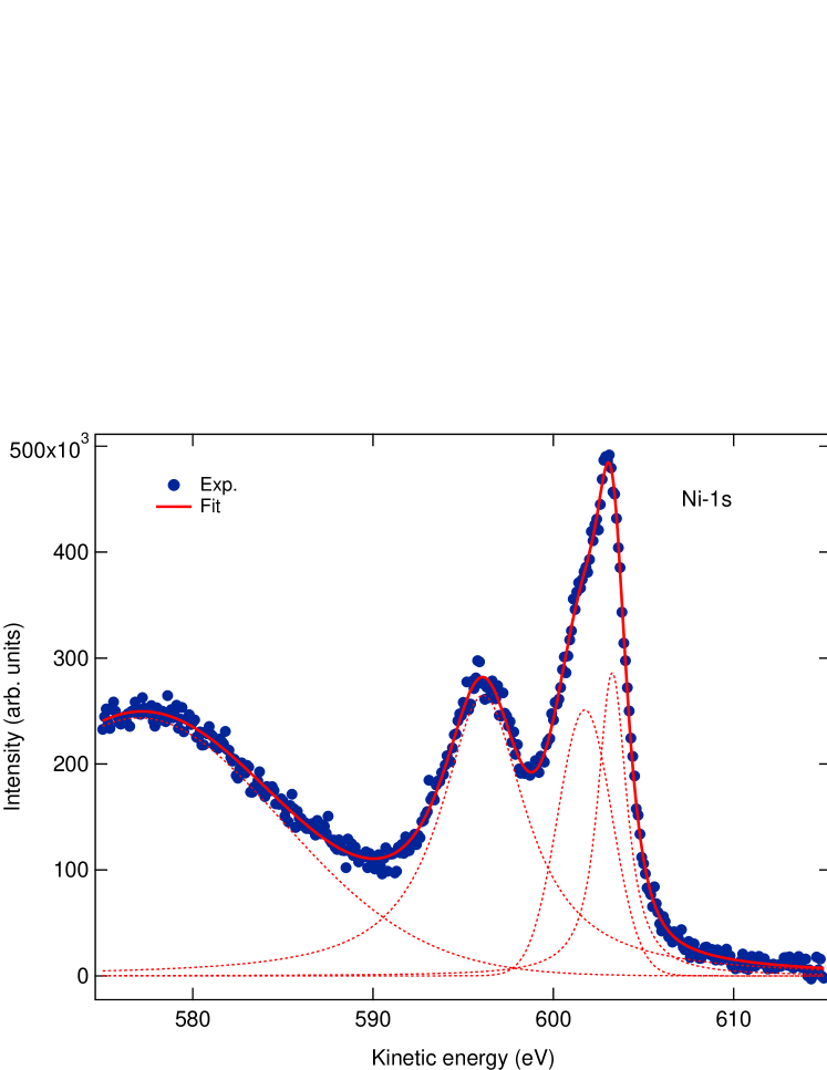
The measured 1s photoemission spectra of NiO are shown in Fig. 1. The fit to the data is consistent with a three peak structure in the 590-610 eV energy region. The results closely resemble 2p3/2 Ni photoemission in NiO Taguchi ; Parmigiani99 in this energy region. In literature the attribution of the different features in 2p3/2 Ni XPS is very controversial and was subject to several reinterpretations. Van Veenendaal and Sawatzky Veenendal93 attributed the main feature at high energy to a 2p53d9L state, where L means a hole in the ligand state. The satellite of the main peak (shoulder) was attributed to non-local screening coming from the nearest neighbours Ni atoms, while the lower energy satellite at eV was attributed to a 2p63d10L. Recently this was reconsidered in ref. Taguchi where the main feature was attributed to a 2p53d9Z, where Z is a Zhang-Rice k-dispersing bound stateKunes . The shoulder of the main peak is attributed to a 2p53d9L state and the lowest energy feature to a 2p53d8 state. Here we show that, regardless of their attribution, the features measured in 1s Ni NiO XPS are present also in the dipolar Ni K-edge XAS spectrum of NiO.
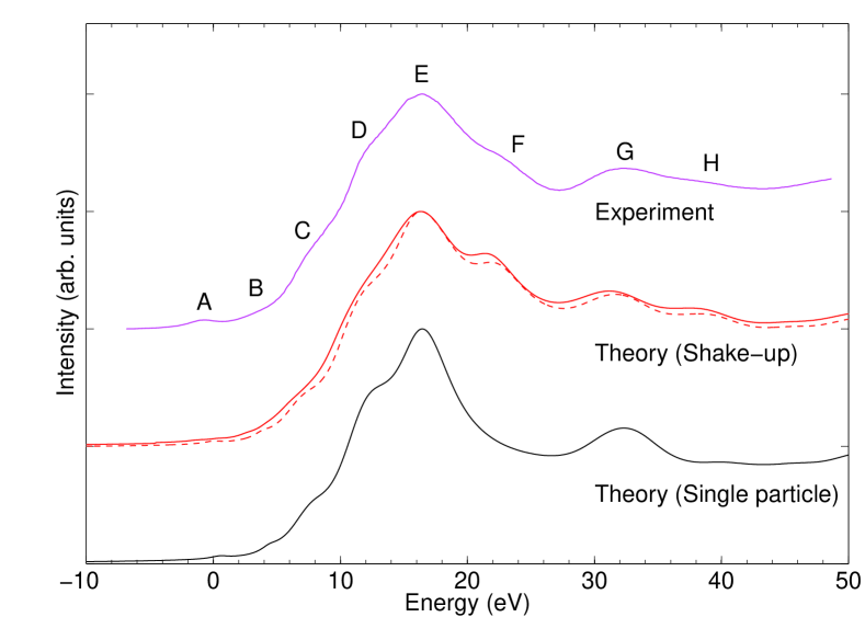
In Fig. 2 we show the measured and calculated XAS cross sections. The single particle cross section is generally in good agreement with the dipolar part of the measured spectrum except for the two peaks indicated by the letters F and H that are missing in the single particle calculation. In order to determine if the missing excitations are manybody in nature, and eventually due to multi-determinant or shake-up processes, we then proceed by using Eq. 6 and obtain new XAS spectra. We first perform the convolution using the complete three-peaks structure of the XPS spectra. We find that the convolution of the DFT calculated XAS cross-section with the photoemission spectra greatly improves the agreement with experiments. In particular the missing peaks are now present in the spectrum and a better agreement occurs at all energy scales. The F and H peaks are then replicas of the single particle E and G peaks respectively and are manybody in nature. We can further test to what extent this interpretation is robust by altering the XPS spectrum before convolution with the theoretical single-particle XAS calculation. We do this for NiO by artificially supressing the main peak of the XPS spectrum, leaving the shoulder and the satellite. We find that the resulting absorption spectrum (dashed line Fig xxx)is less in agreement with the experimental spectrum supporting our interpretation.
IV.2 Cobalt Oxide
Co 1s photoemission data of CoO are not available in literature. However 2p3/2and 3s Co XPS data ShenCoO ; Parmigiani99 are extremely similar and composed by two main peaks and a small shoulder at low energy visible only in the 3s data. We then consider 3s photoemission data of Ref. Parmigiani99 and fit them with a two-peak structure and neglect the very small shoulder, invisible in 2p photoemission data. The results are shown in Fig. 3.
The situation is very similar to NiO, namely the F peak is missing missing from the single-particle spectrum and the H peak is weak. Convolution with photoemission improves substantially the agreement although the main edge peak is narrower than the experimental data.
IV.3 Copper Oxide
The copper oxide CuO has a monoclinic crystal structure with symmetry group C/2c . The dipolar part in CuO was measured in ref. Bocharov01 ; BocharovTh . We follow the notation of refs. Bocharov01 ; BocharovTh and label the configuration of the crystal with respect to the incident beam by the three angles . These angles correspond to the three rotation angles of the goniometer. In particular, when the three angles are zero, then the polarization is parallel to the axis of the goniometer and to the c-axis of the crystal. At zero angles, the plate holding the sample is orthogonal to the incident beam and parallel to one of the plaquette chains in the crystal (see Ref. BocharovTh Fig. 10 for more details).
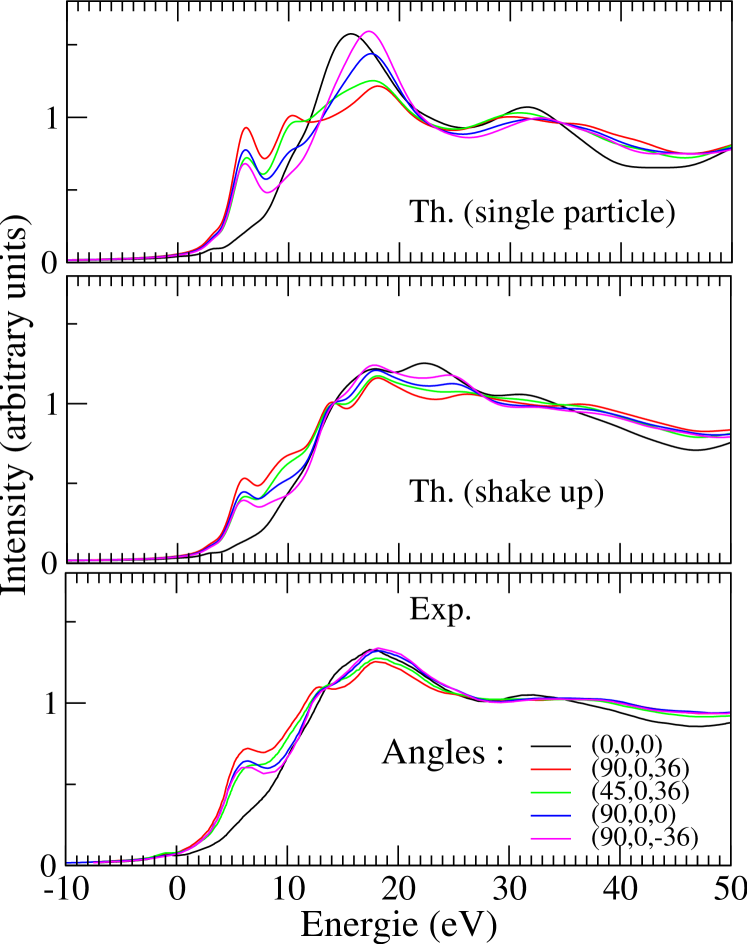
Given the low symmetry of the crystal, the polarization dependence of CuO K-edge XAS spectra is very complicated, as can be seen in Fig. 4. The calculated single particle spectra are in strong disagreement with experiments. Both the peak positions and the polarization dependence of the intensities disagree with the measured data. In order to see if the disagreement is due to the lack of manybody effects in the XAS cross-section, we consider the convolution with Cu 1s photoemission spectra HiroshimaXPS . Cu 1s photoemission spectra of CuO are composed of two peaks, usually attributed to 3d9 and 3d10L. Performing the convolution with the calculated single particle Cu K-edge XAS leads to an impressive improvement. The convoluted spectrum is in much better agreement with experimental data, demonstrating that the Cu K-edge XAS spectrum in CuO is dominated by shake-up manybody processes.
V Conclusions
We have demonstrated that shake-up processes occur in dipolar K-edge XAS spectra of NiO, CoO, CuO. As these are prototype correlated transition metal oxides, we expect these excitations to be present in all XAS data of correlated materials. To be more precise, whenever charge transfer satellites occur in XPS core-hole spectra, then shake-up satellites must also occur in the corresponding X-ray absorption edge, at all energy scales.
We have also shown that a practical way to include these effects in first principle calculations is to perform the convolution with the XPS spectrum at the same edge, as suggested by Eq. 6. Despite the fact that Eq. 6 was obtained many years ago Nozieres69 ; Ohtaka83 ; OhtakaRMP , we are currently unaware of other works trying to explicitly apply this equation to K-edge XAS by using state of the art calculations. Our work that full includes core-hole attraction and Hubbard U at the DFT+U level demonstrates that this approach is feasible and allows, for the first time, the attribution of all dipolar peaks in NiO and CoO dipolar K-edge XAS spectra.
VI Acknowledgement
M. C. acknowledges fruitful discussion with F. Mauri. Calculations were carried out at the IDRIS supercomputing center (proposal number: 091202).
References
- (1) F. de Groot and A. Kotani, Core Level Spectroscopy of Solids, Taylor and Francis 2008.
- (2) C. Gougoussis, M. Calandra, A. P. Seitsonen, and F. Mauri, Phys. Rev. B 80, 075102 (2009)
- (3) Mathieu Taillefumier, Delphine Cabaret, Anne-Marie Flank, and Francesco Mauri Phys. Rev. B 66, 195107 (2002)
- (4) C.Hebert, Micron 38 12 (2007)
- (5) Y. Joly, Phys. Rev. B 63, 125120 (2001)
- (6) C. Gougoussis, M. Calandra, A. Seitsonen, Ch. Brouder, A. Shukla, and F. Mauri, Phys. Rev. B 79, 045118 (2009)
- (7) H. Ågren and V. Carravetta. Inter. J. Quant. Chem., 42, 685 (1992)
- (8) V. Carravetta and H. Ågren. Computational x-ray spectroscopy. In V. Barone, editor, Computational Strategies for Spectroscopy: From Small Molecules to Nano Systems, pages 137–205, Hoboken, 2012. Wiley.
- (9) M. A. van Veenendaal and G. A. Sawatzky, Phys. Rev. Lett. 70, 2459 (1993)
- (10) M. Taguchi, M. Matsunami, Y. Ishida, R. Eguchi, A. Chainani, Y. Takata, M. Yabashi, K. Tamasaku, Y. Nishino, T. Ishikawa, Y. Senba, H. Ohashi and S. Shin, Phys. Rev. Lett. 100, 206401 (2008)
- (11) M. Takahashi and J. I. Igarashi. Phys. Rev. B, 85, 085128 (2012)
- (12) Fuggle, J. C., Hillebrecht, F. U., Esteva, J.-M. , Karnatak, R. C. , Gunnarsson, O. , Schönhammer, K., Phys. Rev. B 27, 4637 (1983)
- (13) Y. Hammoud, J. C. Parlebas, F. Gauthier, J. Phys. F, 17, 503 (1987)
- (14) D. Malterre, Phys. Rev. B 43, 1391 (1991)
- (15) H. Tolentino, M. Medarde, A. Fontaine, F. Baudelet, E. Dartyge, D. Guay and G. Tourillon, Phys. Rev. B 45, 8091 (1992)
- (16) R. Bair and W. Goddard, Phys. Rev. B 22 2767 (1980)
- (17) N. Kosugi, Y. Tokura, H. Takagi and S. Uchida, Phys. Rev. B 41, 131–137 (1990)
- (18) R. L. Martin and D. A. Shirley. J. Chem. Phys., 64, 3685 (1976)
- (19) B. H. Brandow. Adv. Phys., 26, 651 (1977)
- (20) B. H. Brandow. J. Alloys Compounds, 181, 377 (1992)
- (21) N. M. Harrison, V. R. Saunders, R. Dovesi, and W. C. Mackrodt. Phil. Trans. R. Soc. Lond. A, 356, 75 (1998)
- (22) N. I. Gidopoulos. Phys. Rev. A, 83, 040502 (2011)
- (23) T. A. Tyson. X-ray Absorption Spectroscopy: Experimental K, L and KL Spectra with Quantitative Models. Ph.D. thesis, Stanford University, 1991
- (24) J. Chaboy and T. A. Tyson. Phys. Rev. B, 49, 5869 (1994)
- (25) C. O. Almbladh and L. Hedin, Handbook of Synchrotron radiation, Vol. 1b, 607, 1983, North-Holland Amsterdam
- (26) K. Ohtaka and Y. Tanabe, Phys. Rev. B 28, 6833 (1983)
- (27) K. Ohtaka and Y. Tanabe, Rev. Mod. Phys. 62, 929 (1990)
- (28) P. Nozières and C. T. DeDominicis, Phys. Rev. 178, 1097 (1969)
- (29) P. Gianozzi et al., J. Phys.: Condens. Matter 21, 395502 (2009)
- (30) M. Cococcioni and S. de Gironcoli, Phys. Rev. B 71, 035105 (2005)
- (31) M. Gorgoi, S. Svensson, F. Schäfers, G. Öhrwall, M. Mertin, P. Bressler, O. Karis, H. Siegbahn, A. Sandell, H. Rensmo, W. Doherty, C. Jung, W. Braun, and W. Eberhardt, Nucl. Instrum. Meth. A 601, 48 (2009)
- (32) F. Schaefers, M. Mertin, and M. Gorgoi, Rev. Sci. Instrum. 78, 123102 (2007)
- (33) R. V. Vedrinskii, V. L. Kraizman, A. A. Novakovich, Sh. M. Elyafi, S. Bocharov, Th. Kirchner, and G. Dräger, Phys. Status Solidi B 226, 203 (2001)
- (34) H. Modrow, S. Bucher, J. J. Rehr and A. L. Ankudinov Phys. Rev. B 67, 035123 (2003)
- (35) F. Parmigiani and L. Sangaletti, Journal of Electron Spectroscopy and Related Phenomena, 98-99, 287, 1999.
- (36) J. Kunes, V. I. Anisimov, S. L. Skornyakov, A. V. Lukoyanov, and D. Vollhardt, Phys. Rev. Lett. 99, 156404 (2007)
- (37) Shen, Z.-X., J. W. Allen, P. A. P. Lindberg, D. S. Dessau, B. O. Wells, A. Borg, W. Ellis, J. S. Kang, S.-J. Oh, I. Lindau, and W. E. Spicer, Phys. Rev. B 42, 1817 (1990)
- (38) S. Bocharov, Th. Kirchner, G. Dräger, O. Sipr, and A. Simunek, Phys. Rev. B 63, 045104 (2001).
- (39) S. Bocharov. Winkelabhn̈gige K-Rn̈tgenabsorption und Elektronenstruktur von 3d-Metallverbindungen. PhD thesis, VanMartin-Luther-Universitẗ Halle-Wittenberg, 2001.
- (40) See for example experimental Cu 1s hard X-ray photoemission collected at the Hiroshima Synchrotron radiation center at http://www.hsrc.hiroshima-u.ac.jp/cuprates.htm