Elastic Correlations in Nucleosomal DNA Structure
Abstract
The structure of DNA in the nucleosome core particle is studied using an elastic model that incorporates anisotropy in the bending energetics and twist-bend coupling. Using the experimentally determined structure of nucleosomal DNA [T.J. Richmond and C.A. Davey, Nature 423, 145 (2003)], it is shown that elastic correlations exist between twist, roll, tilt, and stretching of DNA, as well as the distance between phosphate groups. The twist-bend coupling term is shown to be able to capture these correlations to a large extent, and a fit to the experimental data yields a new estimate of nm for the value of the twist-bend coupling constant.
pacs:
87.15.-v, 87.15.La, 87.14.Gg, 82.39.PjThe DNA in eukaryotes is tightly bound to an equal mass of histone proteins, forming a repeating array of DNA-protein complexes called nucleosomes Cell . A stretch of 147 base pair (bp) DNA is wrapped in 1.84 left-handed superhelical turns around the histone octamer that forms the nucleosome core particle, which is connected via a linker DNA to the next core particle. Each nucleosome core is a tiny sized spool with a radius of 5 nm and a height of 6 nm Luger . The wrapped DNA-histone octamer complex is essentially ubiquitous in nature and has a major role in many cell life processes such as gene expression and transcription Seacker .
In a recent high precision measurement, Richmond and Davey have determined the structure of the 147 base pair DNA in the nucleosome core particle with resolution Richmond . They have observed that the structure of the bent DNA segment is modulated in the curvature, roll, and tilt, and that the twist structure appears to be most affected by the specific interactions with the protein substrate. This experiment provides a wealth of information about the conformational structure of such highly bent and strongly interacting DNA, among which we can highlight a number of quantitative observations: (1) the period of modulation in curvature is set by half of the DNA pitch bp, where either the major or the minor grooves face the histone octamer, (2) roll appears to have the main contribution to the curvature, as it is favored over tilt by , and (3) the DNA segment is stretched by about 1-2 bp as compared to its unbent conformation Richmond .
The conformational properties of relatively long DNA segments, as well as their elastic response to mechanical stresses such as pulling forces and torques, have been successfully studied using a coarse-grained elastic description Benham ; Tanaka ; Marko ; Kamien1 ; Fain1 that could take into account thermal fluctuations Marko-PRE ; Bouchiat1 ; Panyukov1 . Other approaches for studying DNA structure include first-principle computer simulations Schlick ; Olson2 and phenomenological modelings using base-stacking interactions Kamien2 ; Mergel . In light of the recent experimental determination of the nucleosomal DNA structure, a corresponding theoretical analysis is called for, and one naturally wonders which of the above approaches could more easily accommodate the additional complications due to the high degree of bending and the specific DNA-protein interactions.
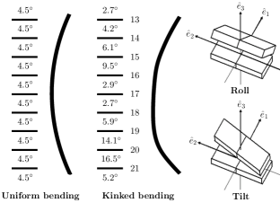
Here, we attempt to use an augmented elastic description to account for a number of observations made by Richmond and Davey. We consider an elastic energy expression that includes anisotropic bending rigidities and twist-bend coupling. We show that the anisotropic bending elasticity is responsible for the modulations in curvature with the period of 5 bp Farshid1 , and calculate the shape of DNA, an example of which is shown in Fig. 1. Using an analysis of the experimental data of Ref. Richmond , we show that the specific features in twist and bend are correlated to a large extent through the twist-bend coupling, and extract an estimate of nm for the twist-bend coupling constant that best describes this correlation. We calculate the components of curvature as well as the axial strain using the experimental values for twist as input, and find that roll is favored over tilt by , and that the overall stretching of DNA is about 1 bp, both in encouraging quantitative agreement with the experiment. We have also studied another parameter called , which shows the super-helix-component of the difference of the lengths of the two phosphodiester chains Richmond , and found good agreement with the experimental data.
To study the structure of the nucleosomal DNA, we consider a simple model in which the molecule is represented as an elastic rod Farshid1 . The rod is parameterized by the arclength and at each point, an orthonormal basis is defined with the unit vectors , , and , where shows the direction from minor groove to major groove, and is the unit tangent to the axis (see Fig. 1). Note that due to the helical structure of DNA, and rotate with the helix. The deformation of the double helix is characterized by the angular strains corresponding to bending in the plain perpendicular to and corresponding to twist and torsion. We should mention that each slice of the rod is labeled by , which is corresponding to arc length along the unstressed helix axis and so always changes from to . The actual arc length along the deformed axis is given by , which in terms of axial strain one have note1 . The elastic energy for the deformation of DNA in units of thermal energy is written as Marko ; Kamien1
| (1) | |||||
where and are the bending rigidities for the “hard” and “easy” axes of DNA cross section, is the twist rigidity, is the stretch modulus, is the twist-stretch coupling, and is the twist-bend coupling. The spontaneous twist of the helix is defined via its pitch as , and for B-DNA we have . Note that the twist-bend coupling can be ruled out by symmetry ( to ) for a non-chiral rod , and thus its presence is a direct consequence of a spontaneous twist structure, as manifested by the form .
The local strains can be written in terms of the curvature , the torsion , and the twist angle as , , and Marko . It can be shown that the mean curvature and torsion imposed by the nucleosomal structure are and , where is the radius of the nucleosome and denotes the pitch of the wrapped DNA around the histone octamer. For the nucleosome case, and Richmond , which yields . Since typical values of is of the order of , we can estimate the relevance of torsion with respect to twist by the ratio . These estimates justify neglecting torsion with respect to twist and curvature. After defining , and , we can write Eq. (1) as
| (2) | |||||
For the coupling parameters, we use nm Marko-PRE , nm Olson , nm Wang , nm Vologodskii , and nm Marko3 . Since the only estimate available for the value of the twist-bend coupling constant has been rather indirect Marko , we treat as a tuning parameter and find its value by fitting to the experimental data of Ref. Richmond .
To find the shape of the DNA, we need to incorporate the interactions with the binding substrate. The wrapping of DNA by an overall angle of around the protein octamer can be imposed as a global constraint, which reads . To take account of the specific and local interactions, we assume that it is mostly the twist degree of freedom that is affected by those interactions Schiessel-Review ; a view that is supported by the experimental observations of Ref. Richmond . This allows us to write the interaction energy term, which is the final addition to the energy expression for the DNA conformation Eq. (2), as . We can now minimize the sum of with respect to , , and , subject to the wrapping constraint above. This yields expressions for the curvature and the axial strain as functions of the twist angle as
| (3) | |||||
where is the Lagrange multiplier for the constraint, as well as an equation for the twist that contains unknown interaction terms arising from . Instead of elaborating on the possible forms of this interaction, we simplify the procedure by directly reading off the twist angle from the experimental results of Richmond and Davey. This allows us to make a direct comparison between the calculated values for the curvature and stretching and the corresponding experimentally measured values for them, and hence put the elastic model to a stringent test.
The twist angle is obtained from the twist strain by integration, and the corresponding integration constant is chosen by noting that at the dyad axes of the nucleosome the major groove-minor groove direction is perpendicular to the surface of the nucleosome core particle. This means that the first base pair in either the left or the right half of the nucleosomal DNA should have an offset twist angle of . Finally, we note that following Ref. Richmond the calculations are performed for half of the DNA length corresponding to 73 base pairs, to make a direct comparison possible.
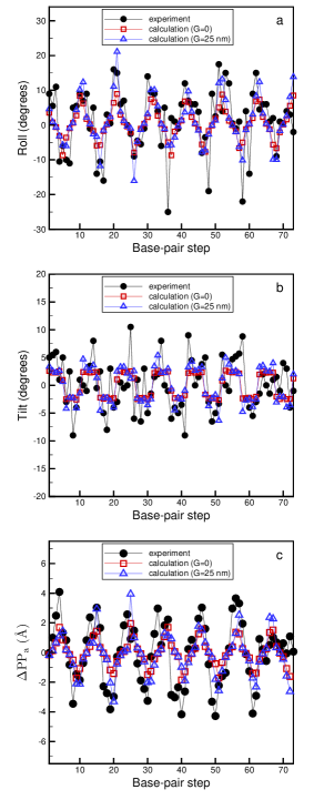
In Figs. 2a and 2b, we have plotted the calculated roll and tilt Dickerson as functions of the position of the base pairs, together with the experimental data of Ref. Richmond , where is the base-pair step for B-DNA. The two quantities appear to be modulated with a near periodicity of 10 bp, which is imposed by the near periodicity in . The best fit to the experimental data yields , which results in the roll being favored over tilt by , to be compared with the experimental value of . This shows that in such a highly bent structure, the DNA prefers by a ratio of nearly 2 to 1 to use the bending over the easy axis as opposed to the hard one.
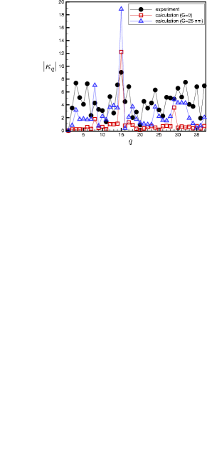
To make a more refined quantitative comparison with the experiment, we consider the net curvature (as opposed to its components) and take its Fourier transform defined as for a list of length ( here), to better resolve its features. A plot of the absolute value of the curvature Fourier transform is shown in Fig. 3, comparing the experimental data with the calculated ones. Note that the absolute value of the Fourier transform is symmetric with respect to the transformation , hence only the first half of the plot is shown. The Fourier transform of the curvature shows a distinct peak at corresponding to a periodicity of 5 bp, which is a result of the anisotropic bending elasticity Farshid1 . Moreover, Fig. 3 shows that while the simple elastic description fails to account for the detailed features of the curvature without a twist-bend coupling, once equipped with such a term it can give a considerably improved account, with the best fit corresponding to nm. We also note that using the values for the curvature the shape of the bent DNA can be determined upon integration. We have provided such an example in Fig. 1, where the presence of two kinks at the distance of 5 base pairs from each other can be visibly noted.
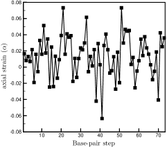
Using Eq. (LABEL:alpha), one can also determine the stretching of the nucleosomal DNA. In Fig. 4, the axial strain is plotted as a function of the base-pair position. A positive (negative) value of shows a stretching (compression) for the corresponding base-pair. The overall length of the DNA in the nucleosome can be found as bp, which suggests that the DNA in the nucleosome is stretched by about 1 bp, in agreement with the observations of Richmond and Davey Richmond .
We have also calculated the difference between the components of the phosphate-phosphate distances lying parallel to the path of the superhelix , where gives the phosphate-phosphate distances on the th strand. Using the geometrical definitions, we find , where , and nm is the diameter of the undeformed DNA. The calculated values of are shown in Fig. 2c, which are in good agreement with the experimental values taken from Ref. Richmond .
We have also examined the effect of other elastic couplings—such as the bend-stretch coupling—by trying to fit to the experimental data, and have found no significant effect. Higher order elastic terms such as the cubic terms in curvature etc. Marko , are expected to add corrections of the order of , which become (marginally) important in the highly bent (kink) regions. While the addition of such terms would definitely help improve the results quantitatively, the fact that it will introduce more unknown coupling constants make such a direction not particularly appealing. Moreover, there are also other structural properties of the nucleosomal DNA such as shift and slide Dickerson , which appear to be beyond such a simple elastic description. This suggests that a more promising direction for an improved theory that can better describe the conformation of nucleosomal DNA is a generalization of the base-stacking model of O’Hern et al. Kamien2 ; ToBePub .
In conclusion, we have shown that an elastic theory that takes into account anisotropic bending and twist-bend coupling can account to a considerable degree for the observed structure of the nucleosomal DNA. Since a full microscopic computer simulation of such a large DNA-protein complex appears to be out of reach with the computational power at hand, such simplified phenomenological approaches could be helpful in understanding the structural properties of biomolecules.
We thank R. Bruinsma, H. Flyvbjerg, T.B. Liverpool, P. D. Olmsted, Z.-C. Ou-Yang, W.C.K. Poon, M. Rao, H. Schiessel, and A. Travers for very helpful discussions.
References
- (1) B. Alberts et al., Molecular Biology of the cell (Garland, New York, 2002).
- (2) K. Luger, A.W.Mäder, R.K. Richmond, D.F. Sargent, T.J. Richmond, Nature 389, 251 (1997).
- (3) R.M. Saecker, M.T. Record, Curr. Opin. Struct. Biol. 12, 311 (2002).
- (4) T.J. Richmond, C.A. Davey, Nature 423, 145 (2003).
- (5) C.J. Benham, Biopolymers 22, 2477 (1983).
- (6) F. Tanaka, and H. Takahashi, J. Chem. Phys. 83, 6017 (1985).
- (7) J.F. Marko and E.D. Siggia, Macromolecules 27, 981 (1994); Erratum in: Macromolecules 29, 4820 (1996).
- (8) R.D. Kamien, T.C. Lubensky, P. Nelson, C.S. O’Hern, Europhys. Lett. 38, 237 (1997).
- (9) B. Fain, J. Rudnick, and S. Östlund, Phys. Rev. E 55, 7364 (1997); B. Fain and J. Rudnick, Phys. Rev. E 60, 7239 (1999).
- (10) J.F. Marko and E.D. Siggia, Phys. Rev. E 52, 2912 (1995).
- (11) C. Bouchiat and M. Mézard, Phys. Rev. Lett. 80, 1556 (1998).
- (12) S. Panyukov and Y. Rabin, Phys. Rev. Lett. 85, 2404 (2000); Europhys. Lett. 57, 512 (2002); A. De Col and T.B. Liverpool, Phys. Rev. E 69, 061907 (2004).
- (13) T. Schlick and W.K. Olson, Science 257, 1110 (1992).
- (14) W.K. Olson, Curr. Opin. Struct. Biol. 6, 242 (1996).
- (15) C.S. O’Hern, R.D. Kamien, T.C. Lubensky, and P. Nelson, Eur. J. Phys. B 1 95 (1998).
- (16) B. Mergell, M.R. Ejtehadi, and R. Everaers, Phys Rev E 68 021911 (2003).
- (17) F. Mohammad-Rafiee and R. Golestanian, Eur. Phys. J. E 12, 599 (2003); J. Phys.: Condens. Matter, 17, S1165 (2005).
- (18) The fact that the backbone is considered as inextensible does not contradict with the introduction of the axial strain, which as a field defined on the inextensible coordinate system of the backbone describes the extension of the rod.
- (19) W.K. Olson, N.L. Marky, R.L. Jernigan, and V.B. Zhurkin, J. Mol. Biol. 232, 530 (1993).
- (20) M.D. Wang, H. Yin, R. Landick, J. Gelles, S.M. Block, Biophys. J. 72, 1335 (1997).
- (21) A.V. Vologodskii, S.D. Levene, K.V. Klenin, M. Frank-Kamenetskii, N.R. Cozzarelli, J. Mol. Biol. 227, 1224 (1992).
- (22) J.F. Marko, Europhys. Lett. 38, 183 (1997).
- (23) H. Schiessel, J. Phys.: Condens. Matter 15, R699 (2003).
- (24) R.E. Dickerson et al., EMBO J. 8, 1 (1989).
- (25) F. Mohammad-Rafiee and R. Golestanian, work in progress.