Topological classification of RNA structures
Abstract
We present a novel topological classification of RNA secondary structures with pseudoknots. It is based on the topological genus of the circular diagram associated to the RNA base-pair structure. The genus is a positive integer number, whose value quantifies the topological complexity of the folded RNA structure. In such a representation, planar diagrams correspond to pure RNA secondary structures and have zero genus, whereas non planar diagrams correspond to pseudoknotted structures and have higher genus. We analyze real RNA structures from the databases wwPDB and Pseudobase, and classify them according to their topological genus. We compare the results of our statistical survey with existing theoretical and numerical models. We also discuss possible applications of this classification and show how it can be used for identifying new RNA structural motifs.
Keywords: Secondary structure, pseudoknot, RNA structure
classification.
PACS: 82.39.Pj, 87.14.Gg
Introduction
In their biologically active form, RNA molecules are folded in fairly well defined three dimensional structures [1]. These structures are strongly constrained by the pairing of conjugate bases along the sequence, but depend also on the ionic strength of the solution [2]. It has proved very useful to describe the pairing of RNA in terms of secondary structures and pseudoknots [3]. These structural elements can be viewed as motifs which appear repeatedly in the folds. The main structural motifs of secondary structures are helical duplexes, single stranded regions, hairpin stems, hairpin loops, bulges and internal loops, junctions and multiloops (see table 1). It is convenient at this stage to introduce some standard graphical representations of RNA structures. In the linear representation, one writes the base sequence on an oriented straight line, starting from the 5’ to the 3’ end. By replacing the straight line by a closed circle one obtains the circular representation. The pairing of two bases is represented by a dotted line, or colored line, joining the two bases in the upper side of the straight 5’-to-3’ line. In the case of a circular representation, pairings are drawn inside the circle. This representation associates a unique diagram to any set of base pairings of RNA.
![[Uncaptioned image]](/html/q-bio/0607032/assets/x1.png)
In the circular and linear representation, a diagram represents a secondary structure if it involves only pairings which do not cross [4]. In table 2 (top row), we show a secondary structure, together with its two representations (linear and circular in the fourth and fifth column, respectively). Similarly, a diagram contains a pseudoknot if it contains pairings which do cross ( see, e.g., the bottom row in table 2).
![[Uncaptioned image]](/html/q-bio/0607032/assets/x2.png)
There are quite a few methods to predict secondary structures. Energy-based methods have proven to be the most reliable (as, e.g., [16, 17]). They assign some energy to the base pairings and some entropy to the loops and bulges. In addition, they take into account stacking energies, and assign precise weights to specific patterns (tetraloops, multiloops, etc.) [18]. The lowest free energy folds are obtained either by dynamic programming algorithms [19], or by computing the partition function of the RNA molecule [20]. The main drawback of these energy-based methods is that they deal solely with secondary structures and cannot take into account pseudoknots in a systematic way.
There are several computer programs that attempt to predict RNA-folding with pseudoknots, but the problem is still mostly unsolved (see, e.g. [21, 22, 23, 24, 25, 26, 27, 28, 29]; the list is not exhaustive) . There exists however a novel approach: in order to include the pseudoknots, the RNA folding problem has been formulated in terms of a sophisticated mathematical theory, namely a quantum matrix field theory [30]. These types of field theories were first introduced in particle physics, more precisely in Quantum Chromodynamics, in order to model the theory of strong interactions [31]. Since then, these field theories have been used in many mathematical problems, such as combinatorics, number theory, etc. (for a recent review see [32]). They involve a parameter , the linear dimension of the matrices, which can be used as an expansion parameter for the theory (large expansion) [31]. In the RNA folding problem, the matrix field theory can be expanded diagrammatically in various parameters. The simplest development is in terms of the number of pairings and can easily be represented in terms of diagrams. These diagrams, which are the usual Feynman diagrams of quantum field theory, can be viewed as the set of all the possible pairings of the RNA, with the correct corresponding Boltzmann weights [30, 33]. Another possible expansion is in powers of . As was shown in a previous paper [30, 34], this expansion relies on a topological number called the genus which characterizes the pairing. As we shall see, the genus of a diagram is defined by its embedding on a two-dimensional surface. It is the minimal number of handles that the surface should have so that the diagram can be drawn on the surface without crossing.
Secondary structures correspond to zero genus, that is planar structures: They can be drawn on a sphere without crossing. The simplest pseudoknots, such as the “H-pseudoknot” (see table 2) or the kissing hairpin, correspond to genus 1: they can be drawn on a torus without crossing. This classification of RNA structures allows us to completely grasp the topological complexity of a pseudoknot with a single integer number, the genus. It can be viewed as a kind of “quantum” number. It is reminiscent of the superfold families, such as CATH or SCOP [35], which have proven so useful in protein structure classification. In the literature other possible classifications of RNA structures with pseudoknots have been proposed, such as the ones in, e.g. , [36]. However, the one we propose in this paper is the only one that is purely topological, i.e. independent of any three-dimensional embedding and which is based only on the classical topological expansion of closed bounded surfaces. This is also the reason why this expansion can be derived mathematically with standard tools of combinatorial topology. We believe that such a mathematical framework can be exploited far beyond the simple classification of RNA pseudoknots, and could be applied also for RNA-folding predictions [34]. In this work however we restrict only to the problem of classifying known RNA-structures.
In the following, we shall define more precisely the genus for a given diagram, and show how it can be simply calculated. We then present an analysis of the genii of two main databases which contain RNA structures, namely PSEUDOBASE [37] and the wwPDB (the Worldwide Protein Data Bank which contains some RNAs). The RNA structures in the latter are also listed in the RNABase database [38], that we also used as a reference database. We find that RNAs of sizes up to about 250 have a genus smaller than 2, whereas long RNAs, such as ribozomal RNA may have a genus up to 18.
Materials and Methods
The genus. The topological classification of RNA secondary structures with pseudoknots that we propose is based on the concept of topological genus. We first review the definition of genus of a given diagram. Consider a diagram representing a pairing in the linear representation. The matrix field theory representation of the problem suggests representing a pairing not by a single dotted line, but rather by a double line (which should never be twisted) [30, 31]. Therefore, a unique diagram in the double line representation corresponds to each dotted-line diagram. Some examples are shown in fig.1.

Each double line diagram is characterized by its number of double lines (i.e. the number of pairings of the diagram) which we denote by , and by its number of loops denoted , which is the total number of closed loops made with the (single) lines of the diagram. For instance, in fig.1 (bottom) and in fig.2, the diagram has double lines and loop. The genus of the diagram is the integer defined by

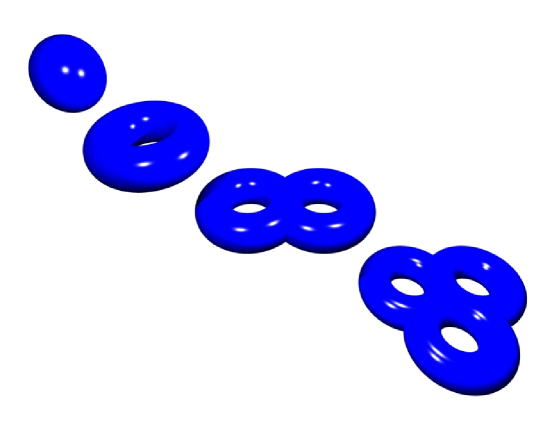
It is related to the Euler characteristics of the diagram, and is a topological invariant of the diagram. Its geometrical interpretation is quite simple. Consider a sphere with handles: a sphere with 0 handles is a sphere, a sphere with one handle is topologically equivalent to a torus, a sphere with 2 handles is topologically equivalent to a double-torus, etc. (see fig.3).
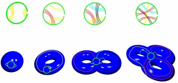
The genus of a diagram is the minimum number of handles a sphere must have in order to be able to draw the diagram on it without any crossing. The precise way to do so, is unambiguously defined only when the diagram does not have open dangling lines, such as the or ends. Therefore it is important to connect the ends, as is done in the circular representation. However, it is more convenient to close the two ends below the backbone-line, which results in drawing the pairing arcs all at the exterior of the backbone-circle. In that way it is simple to see how the embedding of a pseudoknotted RNA structure on a high-genus surface works. Mathematically speaking, the circle of the RNA-backbone (when the are connected) becomes the boundary of a hole or puncture on the surface, and the arcs corresponding to the RNA base-pairs are drawn on the surface without that hole. In fig.4, we show explicit examples of diagrams having different genus. As can be seen, a diagram with genus 0 is planar, in that it can be drawn on the sphere without crossing, and corresponds to a secondary structure. More generally, it was shown in [30] that the secondary structure diagrams are all the planar diagrams with . Likewise, in fig.4 one sees also how diagrams with non-zero genus can be drawn without any crossing on a surface with handles.
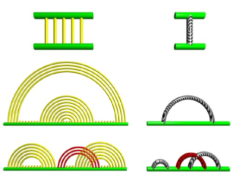
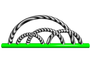
Clearly, different diagrams can have the same genus. Thus, in order to further simplify the classification, we first note that adding a line of pairing parallel to an existing one does not change the genus of the diagram, since it increases by one the number of pairing lines, and increases by one the number of loops of the diagram. Therefore, all diagrams with parallel pairings are equivalent topologically. We will thus use a reduced representation of the diagrams, where each pairing line can be replaced by any number of parallel pairings as in fig.5.
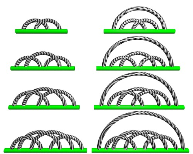
With this convention, it has been shown in [39] that there are exactly 8 topologies of pseudoknots of genus 1, see fig.6. Those topologies can be uniquely identified also as a) ABAB, b) ABACBC, c) ABCABC, d) ABCADBCD, where each letter A,B, etc. indicates a specific helix (or set of helices) along the RNA-backbone from the end to the end. Note that one recognizes the standard H-pseudoknot (ABAB) and the kissing hairpin (ABACBC) (diagrams a) and b) on the left of fig.6, respectively). Among the 8 pseudoknots of genus 1, four are quite common in the databases (the rows a) and b) of fig.6), two are very rare (the row c) fig.6), and the remaining two have not been reported as of yet. We will discuss these pseudoknots in more details in the next section.
Let us insist again that the genus captures the topological complexity of the pseudoknots. It is not simply related to the number of crossings, or of pairings. It depends on the intrinsic complexity of the pseudoknot. This complexity itself depends on what kind of pairings are considered. This is of course conventional. Before discussing the statistics of the genus of pseudoknots from the databases, let us address this question. As discussed in [40], there are many possible non-canonical bonds between base-pairs. We emphasize that our classification of RNA structures according to their genus is well defined and possible even when including non-canonical bonds, or more general definitions of RNA-binding interactions (as far as such interactions are binary). The larger the number of pairings, the higher the genus of the structure might be. However, the weaker bonds, such as the Hoogsteen bonds, or even the wobble pairs, do not form the structure, they merely stabilize a structure already formed by canonical pairings. Therefore, in the following, we shall consider only Watson-Crick pairs between conjugate bases and G-U wobble pairs.
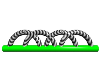
Irreducibility and nesting. In many cases, the genus of a diagram is an additive quantity. For instance, if we consider a succession of two H-pseudoknots (see fig.7, left), each one has genus 1, and the total genus of the diagram is 2. In order to characterize the intrinsic complexity of a pseudoknot, it is thus desirable to define the notion of irreducibility. A diagram is said to be irreducible if it can not be broken into two disconnected pieces by cutting a single line. The diagram on the left of fig.7 is reducible, whereas the one on the right of fig.7 is irreducible. Any diagram can thus be decomposed in a unique way into irreducible parts. It is obvious that the genus of a non-irreducible diagram is the sum of the genii of its irreducible components.
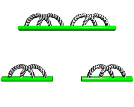
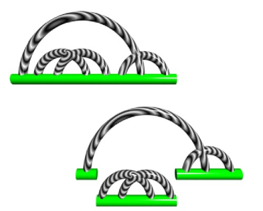
Similarly, if one considers the diagram of fig.8 (left), its genus is equal to 2. It is composed of an H pseudoknot, embedded inside another H pseudoknot. A diagram is said to be embedded or nested in another, if it can be removed by cutting two lines while the rest of the diagram stays connected in a single component. The diagram on the left of fig.8 is nested, whereas the one on the right is not. It is clear that the genus of a nested diagram is the sum of the genii of its nested components. As a result, to any non-nested diagram of genus there corresponds a nested diagram of same genus, obtained by adding a pairing line between the first base and the last base of the diagram. For instance, the 8 diagrams of genus 1 in fig.6 can be decomposed in 4 non-nested diagrams (left column) and 4 nested diagrams (right column). Therefore, there are only 4 irreducible non-nested diagrams (a,b,c,d) of genus 1. As we shall see in the next section, pseudoknots (a) and (b) are quite common, pseudoknot (c) has been seen but is rare, and pseudoknot (d) has not yet been seen. In the following, a pseudoknot which is irreducible and non nested is said to be primitive. Clearly, all RNA structures can be constructed from primitive pseudoknots. The primitive diagram for secondary structures is obviously a single pairing.
Results and Discussion
Analysis of databases. There are several databases containing RNA structures. We have analyzed two of them, namely Pseudobase [37] and the wwPDB [1] (modulo the RNAbase database [38]).
Pseudobase
Pseudobase is a database, containing 246 pseudoknots, at the time of writing this work. These pseudoknots have been deposited and validated by several research groups. They are subsegments of larger RNA sequences, and are displayed in bracket form using several symbols (see fig.9). As an example, we show below one of the pseudoknots from Pseudobase (accession number PKB210)
CGCUGCACUGAUCUGUCCUUGGGUCAGGCGGGGGAAGGCAACUUCCCAGGGGGCAACCCCGAACCGCAGCAGCGAC
((((((::(((:::[[[[[[[::))):((((((((((::::)))))):((((::::)))):::)))):))))))::

AUUCACAAGGAA
:::::]]]]]]]

A simple analysis shows that this is an H pseudoknot, of the type ABAB. Likewise, we analyzed all the 246 pseudoknots of Pseudobase and found that:
-
•
there are 238 H pseudoknots (or nested H pseudoknots) of the ABAB type with genus 1
-
•
there are 6 kissing hairpin pseudoknots (or nested) of the ABACBC type with genus 1
-
•
there is 1 pseudoknot of the type ABCABC (number PKB71) with genus 1
-
•
there is 1 pseudoknot of the type ABCDCADB (number PKB75) with genus 2
Note that the pseudoknot PKB71, from the regulatory region of the alpha ribosomal protein operon (E.coli organism) is the unique example of the ABCABC pseudoknot in Pseudobase. Its structure is [37, 41]:
UGUGCGUUUCCAUUUGAGUAUCCUGAAAACGGGCUUUUCAGCAUGGAACGUACAUAUUAAAUAGUAGGAGUGC
(((((((:(((((::::::::[[[[::::[[[[::::{{{{:)))))))))))):::::::::::::::::::

AUAGUGGCCCGUAUAGCAGGCAUUAACAUUCCUGA
:::::::]]]]:::::]]]]:::::::::::}}}}
Its irreducible structure is given in figure 6 (third from the top, on the left). However, looking at sequence alignment, it is very likely that in fact at least more than 20 other RNA sequences in the EMBL database [42] contain pseudoknots of this kind (A. Mondragón, A. Torres-Larios and K.K. Swinger, Department of Biochemistry, Molecular Biology and Cell Biology, Northwestern University, Evanston, IL: private communication).
The wwPDB databank
The world wide Protein Data Bank (wwPDB) is a collection of databases comprising mostly crystallographic and NMR structures of proteins [1]. In addition, as of today, it contains approximately 850 structures containing at least one RNA molecule. Among these structures, there are about 300 single RNA structures, 200 containing several RNA fragments, 30 RNA/DNA complexes, 250 RNA/protein complexes and 60 transfer RNA.
Among these 850 structures, there are about 650 structures which have obviously genus 0 (very short sequences, or single or double stranded RNA helices). The number of bases ranges from 22 (2g1w.pdb) which is an H pseudoknot, to 2999 (chain 3 of 1s1i.pdb) which has genus 15.
We have analyzed the remaining 200 structures according to the following scheme:
-
•
removal of non RNA molecules and extraction of the molecule of interest
-
•
search for all pairings using the program RNAview
-
•
selection of relevant pairings (Watson-Crick and G-U wobble)
-
•
computation of the genus of the corresponding diagram
Our results can be summarized in the following way
-
•
Transfer RNAs, which are among the smallest RNAs (length of 78), are made of a single primitive pseudoknot (irreducible and non-nested) of genus 1 (a kissing-hairpin) nested inside an arch (see fig.10)
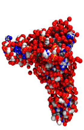
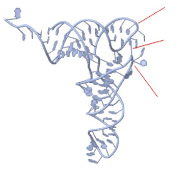
Figure 10: A typical tRNA (PDB 1evv, [45]). It has the genus 1 of a kissing hairpin pseudoknot. -
•
Larger RNAs, such as RNA ribosomal 50s subunits (length larger than 2000), have total genii less than 18. For an RNA with a non designed random sequence of length and without steric constraints, the typical genus should be [43], which in the present case would be around 500. Even by including steric constraints [44], the genus would be around . In addition, if we analyze these sequences in terms of primitive pseudoknots, we find that most of the structures are built from very simple primitive blocks, with genii 1 or 2, nested inside a more complex pseudoknot, of genus smaller than 8. In fig.11, we show an RNA of genus 7 and of length 2825 (the B chain of 1vou.pdb [46]) made of 3 H-pseudoknots, 3 kissing hairpins, nested inside a large kissing hairpin. In fig.12, we display an RNA of genus 9 and of length 2825 (the B chain of 1vp0.pdb, of the 50s subunit of E.Coli [46]), which is made of 3 H-pseudoknots and 2 kissing hairpins, nested in a primitive pseudoknot of genus 4.
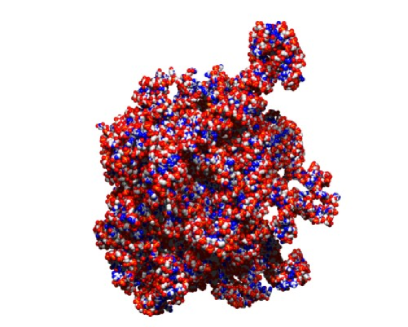

Figure 11: The B chain of PDB 1vou is an RNA of genus 7 and of length 2825 bases. On the right, the outermost primitive arc structure is the pseudoknot type b) of the second column in fig.6, which has genus 1. Such a primitive structure is decorated by 6 additional simple pseudoknots of type and (type a) and b) in the first column of fig.6, respectively). 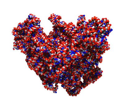
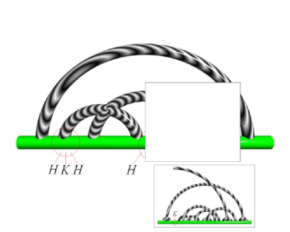
Figure 12: The B chain of PDB 1vp0 is an RNA of genus 9 and of length 2825 bases. The outermost primitive structure is similar to the one of fig.11, with a more complex decoration on the right-hand part. There, a complex pseudoknot with genus 4 is included. Five simple and pseudoknots complete the full decoration. -
•
There is no hierarchical nesting of the pseudoknots: The general structure observed in all RNAs of the PDB is that of several low genus primitive pseudoknots in serie, nested inside a possibly higher genus “scaffold pseudoknot". We show in fig.11 one example of decomposition of a structure (1vou.pdb, which is a 30s subunit of E. Coli).
-
•
In fig.13 (left), we plot the distribution of genii as a function of the length of the RNA. As mentioned before, the genii are much lower than what is expected for random sequences, and this is a manifestation of the specific design of RNA.
-
•
In fig.13 (right), we plot a histogram of the statistics of primitive pseudoknots in the PDB. We see that the genus of primitive pseudoknots is small, typically one or 2, and that the probability to observe large genii is very small. This reflects the fact that complex pseudoknots are built from many small primitive pseudoknots with low genii.
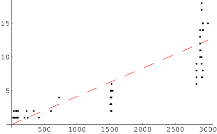
Figure 13: On the left: total genus as a function of the number of bases in the RNA molecule. The interpolating dashed line emphasizes an overall linear behavior. On the right: histogram distribution of the number of primitive pseudoknots as a function of their genus for all RNA molecules in the wwPDB database.
We conclude by reporting in table 3 the sorted list of all the PDB files with non-zero genus, according to our classification. Note that our statistical analysis is biased by the inherent bias of the PDB: the PDB sometimes contains many structures of the same molecules, and thus those utilized for the statistical analysis are not independent.
Conclusion
We have shown that RNA structures can be characterized by a topological number, namely their genus. This genus is 0 for secondary structures (planar structures), and non zero for pseudoknots. We have shown how the complexity of the RNA structure can be analyzed in terms of so-called “primitive pseudoknots". Any complex RNA structure can be uniquely decomposed as a sequence of primitive pseudoknots concatenated sequentially and nested. A survey of the existing RNA structures shows that even for large RNA ( 3 kb), the genus remains small (smaller than 18), and natural RNA have a genus which is much smaller than that of paired structures obtained from random sequences. By capturing the intrinsic complexity of the structure, the genus provides a natural and powerful classification of RNA. Finally, a statistical study shows that complex RNA structures are built from low genii primitive pseudoknots (genii 0, 1 or 2), and that the most complex primitive pseudoknots have genus 13. In a forthcoming work, we will show how this concept of genus can be utilized to actually predict the folded structure of RNA molecules.
| total genus | PDB file accession number |
|---|---|
| 1 | 1b23, 1c0a, 1e8O, 1ehz, 1eiy, 1euq, 1euy, 1f7u, 1f7v, 1fcw, 1ffy, 1fir, 1g59, 1gix-B, 1gix-C, 1grz, |
| 1gtr, 1i9v, 1il2, 1j1u, 1jgo-D, 1jgp-D, 1jgq-D, 1kpd, 1kpy, 1kpz, 1l2x, 1l3d, 1mj1, 1mzp, 1n77, | |
| 1o0b, 1o0c, 1qf6, 1qrs, 1qrt, 1qru, 1qtq, 1qu2, 1qu3, 1ser, 1sz1, 1tn2, 1tra 1ttt 1u6b-B, 1x8w, | |
| 1yfg, 1yg3, 1ymo, 1zzn-B, 2a43, 2a64, 2csx, 2fk6, 2g1w, 2tpk, 2tra, 437d, 4tna, 4tra, 6tna, | |
| 1asy-R, 1asy-S, 1asz-R, 1asz-S | |
| 2 | 1cx0, 1ddy, 1drz, 1et4, 1exd, 1ffz, 1fg0, 1fka, 1pnx, 1sj3, 1sj4, 1sjf, 1u8d, 1vbx, 1vby, 1vbz, |
| 1vc0, 1vc5, 1vc6, 1vc7, 1y0q, 1y26, 1y27, 1yoq, 2a2e | |
| 3 | 1i97, 1n34, 1s1h, 1voz, 1yl4-A |
| 4 | 1ibm, 1fjg, 1hnw, 1hnx, 1hnz, 1hr0, 1i95, 1ibk, 1ibl, 1n32, 1n33, 1q86 1vov, 1vox, 1xmo, |
| 1xmq-A, 1xnr, 1j5e | |
| 5 | 1i94, 1i96, 1n36, 1voq, 1vos, 1xnq, 2avy, 2aw7, 2aw7-A |
| 6 | 1pns, 1voy-B |
| 7 | 1c2w, 1vou-B, 1yl3-A |
| 8 | 1ffk-0, 1vow-B |
| 9 | 1vp0-B, 2aw4-B |
| 10 | 1njm, 1njn, 1njo, 1njp, 2awb-B |
| 11 | 1k01, 1p9x, 1pnu, 1pny |
| 12 | 1j5a, 1jzx, 1jzy, 1jzz, 1nwx, 1nwy-0, 1sm1-0, 1xbp-0, 1y69-0 |
| 13 | 1nkw-0, 1ond, 2d3o |
| 14 | 1jj2, 1k73, 1k8a-A, 1k9m-A, 1kc8, 1kd1, 1kqs-0, 1m1k, 1m90, 1n8r, 1nji, 1q7y, 1q82, 1qvf-0, |
| 1s72, 1vq4-0, 1vq5-0, 1vq7-0, 1vq8-0, 1vq9-0, 1vqk, 1vql, 1vql-0, 1vqm, 1vqn, 1vqo-0, 1vqp-0, | |
| 1yhq-0, 1yi2-0, 1yij-0, 1yit-0, 1yj9-0, 1yjn-0, 1yjw-0, 2aar | |
| 15 | 1q81, 1qvg, 1s1i-3, 1vq6-0 |
| 16 | - |
| 17 | 2aw4-B |
| 18 | 2awb-B |
Acknowledgements
This work was supported in part by the National Science Foundation under Grant No. PHY 99-07949 and Grant No. DMR 04-14446, and by the European program MEIF-CT-2003-501547. G.V. acknowledges Professor Monica Olvera de la Cruz (Northwestern University) for support and stimulating discussions.
References
- [1] Berman,H.M. , Henrick,K. and Nakamura,H. (2003) “Announcing the worldwide Protein Data Bank.” Nature Structural Biology 10, 980–980.
- [2] Misra,V.K. and Draper,D.E. (1998) “On the role of magnesium ions in RNA stability.” Biopolymers 48, 113–135.
- [3] Pleij,C.W. , Rietveld,K. and Bosch,L. (1985) “A new principle of RNA folding based on pseudoknotting." Nucleic Acids Research 13, 1717–1731.
- [4] Waterman,M.S. and Smith,T.F. (1978) “RNA secondary structure: a complete mathematical analysis." Mathematical Biosciences 42,257–266.
- [5] Baeyens,K.J. , De Bondt,H.L. , Pardi,A. and Holbrook,S.R. (1996) “A curved RNA helix incorporating an internal loop with G.A and A.A non-Watson-Crick base pairing." Proc. Natl. Acad. Sci. USA 93, 12851–12855.
- [6] Pan,B. , Mitra,S.N. and Sundaralingam,M. (1998) “Structure of a 16-mer RNA duplex r(GCAGACUUAAAUCUGC)2 with wobble C.A+ mismatches." J.Mol.Biol. 283, 977–984.
- [7] Michiels,P.J. , Schouten,C.H. , Hilbers,C.W. and Heus,H.A. (2000) “Structure of the ribozyme substrate hairpin of Neurospora VS RNA: a close look at the cleavage site." RNA 6, 1821–1832.
- [8] Du,Z. , Ulyanov,N.B. , Yu,J. , Andino,R. and James,T.L. (2004) “NMR Structures of Loop B RNAs from the Stem-Loop IV Domain of the Enterovirus Internal Ribosome Entry Site: A Single C to U Substitution Drastically Changes the Shape and Flexibility of RNA." Biochemistry 43, 5757–5771.
- [9] Kieft,J.S. , Zhou,K. , Grech,A. , Jubin,R. and Doudna,J.A. (2002) “Crystal structure of an RNA tertiary domain essential to HCV IRES-mediated translation initiation." Nat.Struct.Biol. 9, 370–374.
- [10] MolPov 2.0 by D. Richardson, Department of Chemistry, University of Florida, available http://www.chem.ufl.edu/ der/der_pov2.htm
- [11] Available at www.jmol.org
- [12] Available at www.povray.org
- [13] Dallas,A. and Moore,P.B. (1997) “The loop E-loop D region of Escherichia coli 5S rRNA: the solution structure reveals an unusual loop that may be important for binding ribosomal proteins." Structure 5, 1639–1653.
- [14] Kolk,M.H. , van der Graaf,M. , Wijmenga,S.S. , Pleij,C.W. , Heus,H.A. and Hilbers,C.W. (1998) “NMR structure of a classical pseudoknot: interplay of single- and double-stranded RNA." Science 280, 434–438.
- [15] Yang,H. , Jossinet,F. , Leontis,N. , Chen,L. , Westbrook,J. , Berman,H.M. , Westhof,E. (2003) “Tools for the automatic identification and classification of RNA base pairs." Nucleic Acids Research 31, 3450–3460.
- [16] Zuker,M. (2003) “Mfold web server for nucleic acid folding and hybridization prediction." Nucleic Acids Res 31, 3406–3415.
- [17] Hofacker,I.L. (2003) “Vienna RNA secondary structure server." Nucleic Acids Research 31, 3429–3431.
- [18] Mathews,D.H. , Sabina,J. , Zuker,M. and Turner,D.H. (1999) “Expanded Sequence Dependence of Thermodynamic Parameters Improves Prediction of RNA Secondary Structure." J. Mol. Biol. 288, 911–940.
- [19] Zuker,M. and Stiegler,P. (1981) “Optimal computer folding of larger RNA sequences using thermodynamics and auxiliary information." Nucleic Acids Res. 9, 133–148.
- [20] McCaskill,J.S. (1990) “The equilibrium partition function and base pair binding probabilities for RNA secondary structure." Biopolymers 29, 1105–1119.
- [21] Rivas,E. and Eddy,S.R. (1999) “A dynamic programming algorithm for RNA structure prediction including pseudoknots." Journal of Molecular Biology 285, 2053–2068.
- [22] Liu,H.J. , Xu,D. , Shao,J.L. and Wang,Y.F. (2006) “An RNA folding algorithm including pseudoknots based on dynamic weighted matching." Computational Biology and chemistry 30, 72–76.
- [23] Li,H.W. and Zhu,D.M. (2005) “A new pseudoknots folding algorithm for RNA structure prediction." Computing and combinatorics, proceedings lecture notes in computer science 3595, 94–103.
- [24] Ren,J.H. , Rastegari,B. , Condon,A. and Hoos,H.H. (2005) “HotKnots: Heuristic prediction of RNA secondary structures including pseudoknots." RNA 11, 1494–1504.
- [25] Xayaphoummine,A. , Bucher,T. and Isambert,H. (2005) “Kinefold web server for RNA/DNA folding path and structure prediction including pseudoknots and knots." Nucleic Acids Research 33, W605-W610.
- [26] Xayaphoummine,A. , Bucher,T. , Thalmann,F. and Isambert,H. (2003) “Prediction and statistics of pseudoknots in RNA structures using exactly clustered stochastic simulations." PNAS 100, 15310–15315.
- [27] Reeder,J. and Giegerich,R. (2004) “Design, implementation and evaluation of a practical pseudoknot folding algorithm based on thermodynamics." Bmc Bioinformatics 5: Art. No. 104.
- [28] Ruan,J.H. , Stormo,G.D. and Zhang,W.X. (2004) “ILM: a web server for predicting RNA secondary structures with pseudoknots." Nucleic Acids Research 32, W146-W149.
- [29] Dirks,R.M. and Pierce,N.A. (2004) “An algorithm for computing nucleic acid base-pairing probabilities including pseudoknots." Journal of computational Chemistry 25, 1295–1304; also (2003) “A partition function algorithm for nucleic acid secondary structure including pseudoknots." Journal of computational Chemistry 24, 1664–1677.
- [30] Orland,H. and Zee,A. (2002) “RNA folding and large N matrix theory." Nucl. Phys. B620, 456–476.
- [31] ’t Hooft,G. (1974) “A planar diagram theory for strong interactions.", Nucl. Phys. B72, 461–473.
- [32] Series: Mathematical Sciences Research Institute Publications (No. 40), (2001) Random Matrix Models and their Applications. Bleher,P. and Its,A. (Eds.).
- [33] Zee,A. (2005) “Random Matrix Theory and RNA Folding." Acta Physica Polonica B 36, 2829–2836.
- [34] Vernizzi,G. and Orland,H. (2005) “Large-N Random Matrices for RNA Folding." Acta Physica Polonica B 36, 2821–2828.
- [35] Orengo,C.A. , Michie,A.D. , Jones,S. , Jones,D.T. , Swindells,M.B. and Thornton,J.M. (1997) “CATH - a hierarchic classification of protein domain structures." Structure 5, 1093–1108. Murzin,A.G. , Brenner,S.E. , Hubbard,T. and Chothia,C. (1995) “SCOP - a structural classification of proteins database for the investigation of sequences and structures." Journal of Molecular Biology 247, 536–540.
- [36] Condon,A. , Davy,B. , Rastegari,B. , Zhao, S. and Tarrant,F. (2004) “Classifying RNA pseudoknotted structures." Theoretical Computer Science 320, 35–50. Gan,H.H. , Pasquali,S. and Schlick,T. (2003) “Exploring the repertoire of RNA secondary motifs using graph theory; implications for RNA design." Nucleic Acids Research 31, 2926–2943. Kim,N. , Shiffeldrim,N. , Gan,H.H. and Schlick,T. (2004) “Candidates for novel RNA topologies." Journal of Molecular Biology 341, 1129–1144.
- [37] Batenburg,F.H.D.van , Gultyaev,A.P., Pleij,C.W.A. , Ng,J. and Oliehoek,J. (2000) “Pseudobase: a database with RNA pseudoknots." Nucleic Acids Research 28, 201–204.
- [38] Murthy,V.L. and Rose,G.D. (2003) “RNABase: an annotated database of RNA structures." Nucleic Acids Research 31, 502–504.
- [39] Pillsbury, M., Orland,H. and Zee,A. (2005) “Steepest descent calculation of RNA pseudoknots." Physical Review E 72, Art. No. 011911.
- [40] Leontis,N.B. and Westhof,E. (2001) “Geometric nomenclature and classification of RNA base pairs." RNA 7, 499–512.
- [41] Tang,C.K. and Draper,D.E. (1989) “Unusual messenger-RNA pseudoknot structure is recognized by a protein translational repressor." Cell 57, 531–536; also Gluick,T.C. and Draper,D.E. (1994) “Thermodynamics of folding a pseudoknotted messanger-RNA fragment." J. Mol. Biol. 241, 246–262; Gluick,T.C. , Gerstner,R.B. and Draper,D.E. (1997) “Effects of Mg2+, K+, and H+ on an equilibrium between alternative conformations of an RNA pseudoknot." J. Mol. Biol. 270, 451–463.
- [42] Cochrane G., et al. (2006) “EMBL Nucleotide Sequence Database: developments in 2005." Nucleic Acids Research 34, D10–D15.
- [43] Vernizzi,G. , Orland,H. and Zee,A. (2005) “Enumeration of RNA structures by matrix models.", Physical Review Letters 94, Art. No. 168103.
- [44] Vernizzi,G. , Ribeca,P. , Orland,H. and Zee,A. (2006) “Topology of pseudoknotted homopolymers.", Physical Review E 73: Art. No. 031902.
- [45] Jovine,L., Djordjevic,S. and Rhodes,D. (2000) “The crystal structure of yeast phe nylalanine tRNA at 2.0 A resolution: cleavage by Mg(2+) in 15-year old crystals." J.Mol.Biol. 301, 401–414.
- [46] Vila-Sanjurjo,A. , Schuwirth,B.S. , Hau,C.W. and Cate,J.H. (2004) “Structural basis for the control of translation initiation during stress.” Nat.Struct.Mol.Biol. 11, 1054–1059.