Mass Fractal Dimension of the Ribosome and Implication of its Dynamic Characteristics
Abstract
Self-similar properties of the ribosome in terms of the mass fractal dimension are investigated. We find that both the 30S subunit and the 16S rRNA have fractal dimensions of 2.58 and 2.82, respectively; while the 50S subunit as well as the 23S rRNA has the mass fractal dimension close to 3, implying a compact three dimensional macromolecule. This finding supports the dynamic and active role of the 30S subunit in the protein synthesis, in contrast to the pass role of the 50S subunit.
pacs:
87.14.Gg, 87.15.-v, 61.43.HvThe structure of biomolecules is important because not only the structure dictates its biological function, but it is the target of antibiotics. In this sense, finding characteristics of the three-dimensional shape of biomolecules is important for a better understanding of their biological functions and associated applications in medicine. In the case of the ribosome ribosome , as a large protein-RNA complex, in contrast to most cellular machines, it has been known that the ribosomal function heavily rely on the ribosomal RNA (rNRA), as a ribozyme, rather than protein components function . In particular, the protein synthesis is closely related to the dynamic structure of the ribosome, which is too complicated for direct studies by physical methods. However, careful study on the static structure from a quantitative perspective may reveal an important aspect of its dynamic properties.
The quantitative investigation on the structure of the ribosome is relatively less studied than that of protein, mainly due to the difficulty of the highly resolved structure conformation. In contrast to the ribosome, the geometric and self-similar properties of the proteins have been studied extensively. It was reported that the relation between the average radius and the mass of the protein chains can be described by a fractal dimension moret . The mass fractal dimension of the protein was shown to lie near 3, suggesting a compact three-dimensional object elber . However, more recent and extensive studies, including a statistical analysis in estimating the dimension, argue smaller values that are consistent with the result of the vibrational analysis of proteins enright . These results suggest that the fractal dimension of less than 3 may be intrinsic and universal characteristics of the protein chain.
Since the ribosome, as a tightly packed macromolecule, was considered too large for a high-resolution structural analysis, quantitative studies toward an understanding of the structure proved difficult until recent progress in the high resolution crystallography has been made. In fact, the ribosome and its subunits are the largest asymmetric molecules that have been resolved at the atomic level so far by the crystallography. The 2.4 high-resolution of the 50S subunit from the Haloarcula marismortuii ban and the 3.05 structure of the 30S subunit from the Thermus thermophilus wimberly provided the first detailed views of the structure of both ribosomal subunits; the intact 70S ribosome from a Escherichia coli of 3.5 resolution schuwirth revealed the features of the inter-subunit bridges. In addition to these, there are other X-ray crystal structures available for the ribosome as well as its subunits harms ; schluenzen ; cate ; yusupov . With these considerable advances in the ribosome structures at the atomic level, we are now able to investigate quantitative characteristics of the ribosome from the statistical physics perspective.
In bacteria, the ribosome is a particle of size about 250 in diameter and consists mainly of two subunits: 30S and 50S subunits, together forming the 70S. The unit “S” stands for Svedberg, which is a measure of the sedimentation rate. The 30S subunit plays a crucial role in decoding mRNA by monitoring base pairing between codon and anticodon; whereas the 50S subunit catalyzes peptide bond formation between the incoming amino acid and the nascent peptide chain ramakrishnan . The 30S subunit, in turn, contains the 16S rRNA molecule in addition to about 20 different proteins, and the 50S subunit consists of the 5S and the 23S rRNAs besides about 30 different proteins. The 16S and 23S rRNAs are composed of approximately 1500 and 3000 nucleotides respectively, each of which is composed of one of four different bases (denoted as A, C, G, and U), and sugar-phosphate backbones.
With this structural information of the ribosome at the atomic level, we investigate the self-similar property of the ribosome structure and its biological implication. We especially focus on the scale invariance, by estimating the mass fractal dimension, for structures of Thermus thermophilus 30S subunit including the 16S rRNA, and Haloarcula marismortui 50S subunit including the 23S rRNA data . It has been known that the protein synthesis occurs in the context of the intact ribosome and the moving part of the ribosome enables the dynamic process of the translation. In this sense, the ribosome function is closely related to its spatial conformation in the physiological medium. Thus, the mass fractal dimension analysis may help to reveal any characteristics of the ribosome, especially the dynamics of the ribosome.
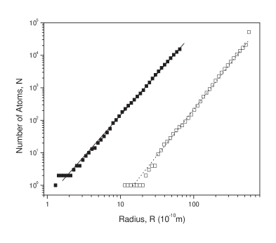
The mass fractal dimension, which can be used as a measure of the compactness, is defined as the number of monomers (atoms in our case), , enclosed in a sphere of radius . When a molecule has a fractal structure, it is expected that
| (1) |
where is the mass fractal dimension. It can be estimated by plotting the number of all atoms contained inside concentric spheres of varying radius on a log-log scale.
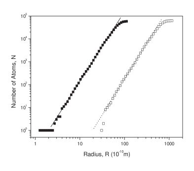
To test the sensitivity of the result to the choice of the origin, we set the origin at the geometric center of each molecule, and vary the origin by in the XYZ directions (thus 27 different origins) with respect to the coordinate system adopted from the PDB data. We ignore hydrogen atoms since the X-ray crystal structures do not contain the geometric information of hydrogen atoms, which cannot be seen but in very high resolution. Incidentally, this is also true for the protein case. Thus, most of descriptions of the ribosome focus on the position of the heavy atoms, such as C, N, O, and P. Note also that the PDB for the 30S subunit does not contain water molecules; where as that for the 50S does. For a fair comparison, we exclude water molecules from the mass fractal calculation. Due to the finite-size effect, there are both upper and lower size limits beyond which a macromolecule is no longer fractal.
| molecule | number of atoms | |
|---|---|---|
| 16S rRNA | 32514 | 2.58 (0.06) |
| 30S subunit | 51742 | 2.82 (0.07) |
| 23S rRNA | 59017 | 3.11 (0.07) |
| 50S subunit | 62673 | 3.07 (0.08) |
We estimate the mass fractal dimension for the 16S rRNA, the 30S subunit, the 23S rRNA, and the 50S subunit with 27 different origins. The result is summarized in Table 1, and Fig. 1 and 2 present a typical log-log plot of the enclosed “mass” as a function of radius . Note that the geometric origin and the center of mass for all molecules are almost identical, and evaluating the corresponding physical mass rather than the number of atoms does not affect the result. For the 23S rRNA and the 30S subunit, we are able to estimate the fractal dimensions for all 27 cases; while for the 23S rRNA and the 50S subunit, respectively 17 and 19 cases out of 27 shows the scaling behavior.
The mass fractal dimension exhibits the molecule’s space-filling ability: the larger is , the more atoms are in the sphere. When fractal dimension is less than 3, the structure has “empty” or “void” space. From the above result, we see that the mass fractal dimensions for both the 23S rRNA and the 50S subunit are close to 3, implying that these are compact three-dimensional collapsed objects. On the other hand, the average for the 16S rRNA and the 30S subunit are found to be 2.58 and 2.82, respectively, smaller than that of a completely compact three-dimensional collapsed polymer. This indicates that the mass inside a radius does not increase with the Euclidean dimension as an exponent but with some lesser power. Thus, we find that both the 23S rRNA and 50S subunit are more compact than either the 16S rRNA or the 30S subunit.
The fact that the 16S rRNA and the 30S subunit have the mass fractal dimension less than 3 leaves a room for the 16S rRNA to make any movement during the protein synthesis, and can be related to the rigid body motion of domains within the subunit. It has been found from many studies that it is the 30S subunit that makes a motion in translocation. It was reported that the 30S subunit makes a ratchet-like rotations relative to the large 50S subunit schuwirth ; frank , particularly, the rotational rigid body motions of the “head” domain domain within the 30S subunit schuwirth . This reveals a high degree of flexibility between the head and the rest of the 30S subunit. Furthermore, not only domains for the entrance channel such as the “shoulder” and the head domains in the 30S subunit are dynamic during decoding, but the exit channel formed between the “platform” and the head domains are known to be variable schuwirth ; frank ; spirin ; serdyuk . Thus, it is the 30S subunit that is either dynamic or variable, playing an active role, which is possible due to the fractal characteristics of the 30S subunit.
The 50S subunit, on the other hand, cannot make movements due to a highly compact structure except peripheral regions. As an example, the L1 stalk, located in a peripheral region of the 50S subunit, makes a bifurcation frank and moves toward the inter-subunit space playing a pivotal role in the transcription process valle .
In this paper, we investigated a symmetry embedded in the ribosome structure under a change of the length scale via the mass fractal dimension. We found the 30S and the 50S subunit (also the 16S and the 23S rRNAs) differs in their fractal dimensions: the 30S subunit and the 16S rRNA have fractal dimensions, while the 50S subunit and the 23S rRNA can be regarded as three-dimensional compact molecules. The fractality of both the 16S rRNA and the 30S subunit supports the dynamic nature of the ribosome in the protein synthesis.
Although the power of the self-similarity approach to the ribosome structure is in its simplicity and generality, it also true that their detail dynamic properties and realization are not obvious because detailed properties of the ribosome which determine their function are averaged out. Nevertheless, the fractal property of the 30S subunit (also the 16S rRNA) provides a partial, if not whole, evidence of its movement during the transcription.
This work was supported by the Korea Research Foundation Grant funded by the Korean Government (MOEHRD) (KRF-2005-041-H00052).
References
- (1) For a general reference of the ribosome and its function, see, for example, Protein Synthesis and Ribosome Structure: Translating the Genome, edited by Knud H. Nierhaus and Daniel N. Wilson (Wiley-VCH, Weinheim, 2004); The Ribosome: Structure, Function, Antibiotics, and Cellular Interactions, edited by R. Garrett, S. Douthwaite, A. Liljas, A. Matheson, P. Moore, and H. Noller (ASM Press, Washington, DC, 2000).
- (2) H. Noller, Science 309, 1508 (2005); P. Nissen, J. Hansen, N. Ban, P. B. Moore, T. A. Steitz, Science 289, 920 (2000).
- (3) M. A. Moret, J. G. V. Miranda, E. Nogueira, Jr., M. C. Santana, and G. F. Zebende, Phys. Rev. E 71, 012901 (2005).
- (4) R. Elber, in Fractal analysis of protein in The Fractal Approach to Heterogeneous Chemistry, edited by D. Avnir (John Wiley Sons, New York, 1989), p. 407.
- (5) Matthew B. Enright and David M. Leitner, Phys. Rev. E 71, 011912 (2005); X. Yu and D. M. Leitner, J. Chem. Phys. 119, 12673 (2003).
- (6) N. Ban, P. Nissen, J. Hansen, P. Moore, T. Steitz, Science 289, 905 (2000).
- (7) B. Wimberly, D. Brodersen, W. Claemons, R. Morgan-Warren, A. Carter, C. Vonhein, T. Hartsch, and V. Ramakrishnan, Nature 407, 327 (2000).
- (8) B. Schuwirth, M. Borovinskaya, C. Hau, W. Zhang, A. Vila-Sanjurjo, J. Holton, J. Cate, Science 310, 827 (2005).
- (9) J. Harms, F. Schluenzen, R. Zarivach, A. Bashan, S. Gat, I. Agmon, H. Bartels, F. Franceschi, and A. Yonath, Cell 107, 679 (2001).
- (10) F. Schluenzen, A. Tocilj, R. Zarivach, J. Harms, M. Gluehmann, D. Janell, A. Bashan, H. Bartels, I. Agmon, F. Franceschi, and A. Yonath, Cell 102, 615 (2000).
- (11) J. H. Cate, M. M. Yusupov, G. Z. Yusupova, T. N. Earnest, H. F. Noller, Science 285, 2095 (1999).
- (12) M. Yusupov et al, Science 292, 883 (2001).
- (13) V. Ramakrishnan, Cell 108, 557 (2002).
- (14) These are the highest resolution results for each subunit. Anything worse than about 3.5 would normally not be possible to construct an accurate model of a macromolecule. We also exclude the 5S subunit from the analysis because it is too small to perform any statistical analysis. The structure information for the subunits can be found in the Protein Data Bank (PDB) at http://www.rcsb.org/pdb/. The access numbers are 1J5E for Thermus thermophilus 30S subunit including the 16S rRNA, and 1JJ2 for Haloarcula marismortui 50S including the 23S rRNA.
- (15) J. Frank and R. Agrawal, Nature 406, 318 (2000).
- (16) The structure of the 16S rRNA is commonly organized into four domains of a few hundred nucleotides each: the head, the body, and the platform, and minor domains.
- (17) A. Spirin, V. Baranov, G. Polubesov, I. Serdyuk, and R. May, J. Mol. Biol. 194, 119 (1987).
- (18) I. Serdyuk, V. Baranov, T. Tsalkova, D. Gulyamova, M. Pavlov, A. Spirin, and R. May, Biochimie 74, 299-306 (1992).
- (19) M. Valle, A. Zavialov, J. Sengupta, U. Rawat, M. Ehrenberg, and J. Frank, Cell 114, 123 (2003).