Modelization of Thermal Fluctuations in G Protein-Coupled Receptors
C. Pennetta1, V. Akimov1, E.Alfinito1, L. Reggiani1,G. Gomila 2, G.Ferrari3, L. Fumagalli3 and M. Sampietro3
1 Dipartimento di Ingegneria dell’Innovazione,
Università di Lecce and
National Nanotechnology Laboratory-INFM,
Via Arnesano, 73100 Lecce, Italy.
2 Department d’Electronica and Research Centre for Bioelectronics and Nanobioscience, Universitat de Barcelona, C/ Josep Samitier 1-5, 08028 Barcelona, Spain.
3 Dipartimento di Elettronica ed Informazione, Politecnico di Milano, P.zza Leonardo da Vinci 32, 20133 Milano, Italy
Abstract
We simulate the electrical properties of a device realized by a G protein coupled receptor (GPCR), embedded in its membrane and in contact with two metallic electrodes through which an external voltage is applied. To this purpose, recently, we have proposed a model based on a coarse graining description, which describes the protein as a network of elementary impedances. The network is built from the knowledge of the positions of the Cα atoms of the amino acids, which represent the nodes of the network. Since the elementary impedances are taken depending of the inter-nodes distance, the conformational change of the receptor induced by the capture of the ligand results in a variation of the network impedance. On the other hand, the fluctuations of the atomic positions due to thermal motion imply an impedance noise, whose level is crucial to the purpose of an electrical detection of the ligand capture by the GPCR. Here, in particular, we address this issue by presenting a computational study of the impedance noise due to thermal fluctuations of the atomic positions within a rhodopsin molecule. In our model, the Cα atoms are treated as independent, isotropic, harmonic oscillators, with amplitude depending on the temperature and on the position within the protein (-helix or loop). The relative fluctuation of the impedance is then calculated for different temperatures.
1 Model and Results
G protein-coupled receptors (GPCRs) constitute the largest family of trans-membrane receptors, with functions going from revealing light and smells to the individuation of drug and virus intruders [1]. For this reason, many efforts are devoted to the study of their properties [1]. In particular, we are interested to develop electronic nanobiosensors based on GPCRs. Actually, the detection of an electrical signal from hybrid nanodevices based on a single or few receptors and associated with the capture of the ligands, is a challenging goal, rich of potential applications [2, 3]. Here, our aim concerns with the calculation of the electrical properties of a device realized by a G protein coupled receptor [1], embedded in its membrane and in contact with two metallic electrodes through which an external AC voltage is applied [4]. To this purpose, recently, we have proposed a model based on a coarse graining [5] description, which describes the protein as a network of elementary impedances [4]. The network is built from the knowledge of the position of the Cα atoms of the amino acids [6], which are taken as the nodes of the network [7]. At least in the case of rhodopsin (photonic receptor) these positions are known by X-rays diffraction experiments [6] for both, the basic and the most stable excited state (metarhodopsin) [6]. Though these positions are generally unknown for the other receptors, a coarse graining, complex network approach offers the possibility of taking advantage of the common topology of the GPCR family [1]. In fact, all GPCRs share a seven-helices trans-membrane structure, where the seven -helices are interconnected by extracellular and intracellular loops [1, 6]. Additionally, there are two terminal chains: an extracellular chain (N terminus) and an intracellular chain (C terminus) [1, 6]. We assume that the amino acids interact electrically among them and that charge transfers between neighboring residues and/or changes of their electronic polarization [8] affect these interactions [4]. Accordingly, a link is drawn between any pair of nodes neighboring in space within a given a distance, ( electrical interaction radius) [5, 7] and an elementary impedance is associated with each link [4]. Moreover, two extra nodes can be introduced in the network, associated with the electrodes, which are linked to a given set of amino acids, depending on the particular geometry of the contacts in the real device. The elementary impedance is taken as the impedance of a RC parallel circuit (the most usual equivalent passive AC circuit) [4]. Precisely, by denoting with the impedance associated with the link between the -th and -th nodes, separated by a distance , we take [4]:
| (1) |
where is the frequency of the external voltage, the resistivity of the resistor, the vacuum permittivity and the relative dielectric constant of the capacitor [4] expressed in terms of the intrinsic polarizabilities and of the corresponding amino acids [8]. By taking the values Å and m, we have found [4] that the conformational change of the receptor induced by the capture of the ligand (i.e. the transition rhodopsin metarhodopsin) implies a significant variation of the impedance at all frequencies, and in particular we have found a variation of about 20 % in the static value of [4]. On the other hand, the fluctuations of the atomic positions due to the thermal motion [5, 9, 10] imply an impedance noise, whose level, in comparison with the impedance change due to variation of conformation and with the electrode/amplifier noise, is crucial to the purpose of an electrical detection of the ligand capture by the GPCR. Therefore, here we consider the effect of the thermal atomic motion on the electrical response to an external field of a rhodopsin molecule. To this purpose, we allow the nodes of the network (Cα atoms) to fluctuate around their equilibrium positions. For the sake of simplicity and to get a qualitative estimation, we describe the system of coupled oscillators as a set of independent, isotropic, harmonic oscillators. When the oscillators are in their ground state, their positions, , referred to the equilibrium ones, are distributed with a probability density:
| (2) |
where is the average mass of the amino acids, the oscillator frequency, the force constant and is the mean square displacement of the oscillator from its equilibrium position along the x-direction. If each oscillator is in contact with a thermal bath at temperature , the value of at the equilibrium is:
| (3) |
where . When , Eq. (3) simplifies in:
| (4) |

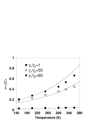
Of course, for an arbitrary temperature the wave function of the oscillator is a superposition of several excited states and the density probability cannot be expressed in the simple form of Eq. (2). However, again for simplicity, we keep this expression and we account for the effect of the temperature by assuming in Eq. (2) given by Eq. (4). Moreover, as a first approximation, we take the mass and the force constant equal for all the oscillators [10], and precisely: Dalton, KJ mole-1 Å-2 [5]. This choice provides K. Thus, the condition is satisfied at room temperature. Figure 1 shows the results of simulations at K of the modulus of the network impedance, , versus time (the modulus has been normalized to its average value). On the other hand, it is well known [1, 9, 11] that loops and terminals are very flexible structures compared with the quite rigid -helices. Therefore, to overcome the crude approximation of a unique force constant for all the oscillators, we consider two different spring elastic constants, and , for oscillators belonging to the -helices and to loops/terminals, respectively, with . Figure 2 shows the relative root mean square fluctuation of the modulus of the impedance, i.e. the root mean square fluctuation, , normalized to the average value of , as a function of the bath temperature. The three sets of data report the results of simulations performed by taking the ratio, , equal (full circles), (open squares) and (full diamonds). In all cases the values of and have been chosen to keep constant the average value of the force constant, , where the sum is performed over the whole number of considered oscillators. The dashed curves in this figure represent the best-fit with exponential functions. We can see that once the higher flexibility of loops and terminals is accounted for, the relative fluctuation of the impedance increases significantly and it becomes strongly sensitive to the temperature. ¿From this study we can conclude that a careful modelization of thermal motion [9, 11] is necessary to provide reliable estimates of the relative fluctuation of the impedance.
Acknowledgments
This work has been performed within the SPOT NOSED project IST-2001-38899 of EC. Partial support from the cofin-03 project “Modelli e misure di rumore in nanostrutture” financed by Italian MIUR is also acknowledged. Authors thank E. Pajot-Augy, R. Salesse and J. Minic (INRA, Jouy en Josas, France) for helpful discussions.
References
- [1] R. J. Lefkowitz. The superfamily of heptahelical receptors. Nature Cell Biology, 2:E133–E136, 2000.
- [2] C. Joachim, J.K. Gimzewski, and A.Aviram. Electronics using hybrid-molecular and mono-molecular devices. Nature, 408:541–548, 2000.
- [3] F. Patolsky, G. Zheng, O. Hayden, M. Lakadamyali, X. Zhuang, and C. M. Lieber. Electrical detection of single viruses. PNAS, 101:14017–14022, 2004.
- [4] C. Pennetta, V. Akimov, E. Alfinito, L. Reggiani, and G. Gomila. Fluctuations of complex networks: Electrical properties of single protein nanodevices. In J. M. Smulko, Y. Blanter, M. I. Dykman, and L. B. Kish, editors, Noise and Information in Nanoelectronics, Sensors and Standards II, number 5472 in Proceedings of SPIE, pages 172–182, Bellingham, 2004. Int. Soc. Opt. Eng.
- [5] A. R. Atilgan, S. R. Durell, R. L. Jernigan, M. C. Demirel, O. Keskin, and I. Bahar. Anisotropy of fluctuation dynamics of proteins with an elestic network model. Biophys. J., 80:505–515, 2001.
- [6] Research Collaboratory for Structural Bioinformatics. Protein data bank. State University of New Jersey, http://www.rcsb.org/pdb, 1.
- [7] R. Albert and A. L. Barabasi. Statistical mechanics of complex networks. Rev. Mod. Phys., 74:47–97, 2002.
- [8] Xueyu Song. An inhomogeneous model of protein dielectric properties: intrinsic polarizabilities of amino acids. J. Chem. Phys., 116:9359–9383, 2002.
- [9] F. G. Parak. Physical aspects of protein dynamics. Rep. Prog. Phys., 66:103–129, 2003.
- [10] M. M. Tirion. Large amplitude elastic motions in proteins from a single-parameter atomic analysis. Phys. Rev. Letl., 77:1905–1908, 1996.
- [11] P. W. Fenimore, H. Frauenfelder, and R. D. Young. Proteins as paradigms complex systems. In S. M. Bezrukov, H. Frauenfelder, and F. Moss, editors, Fluctuations and Noise in Biological, Biophysical and Biomedical Systems, number 5110 in Proceedings of SPIE, pages 1–9, Bellingham, 2003. Int. Soc. Opt. Eng.