Controlled DNA compaction within chromatin: the tail-bridging effect
Abstract
We study the mechanism underlying the attraction between nucleosomes, the fundamental packaging units of DNA inside the chromatin complex. We introduce a simple model of the nucleosome, the eight-tail colloid, consisting of a charged sphere with eight oppositely charged, flexible, grafted chains that represent the terminal histone tails. We demonstrate that our complexes are attracted via the formation of chain bridges and that this attraction can be tuned by changing the fraction of charged monomers on the tails. This suggests a physical mechanism of chromatin compaction where the degree of DNA condensation can be controlled via biochemical means, namely the acetylation and deacetylation of lysines in the histone tails.
pacs:
87.15.He,87.16.Sr,36.20.EyIn eukaryotes (plants and animals) meters of DNA have to be compacted inside micron-sized nuclei. At the same time a considerable fraction of the genetic code has to be accessible. Nature has solved this formidable task by compacting DNA in a hierarchical fashion Schiessel03 . The first step consists of wrapping the DNA two turns around cylinders made from eight histone proteins. This leads to a string of cylindrical DNA spools about 10 nm in diameter and 6 nm in height, each repeating unit being called a nucleosome Luger97 . The chromatin fiber with diameter of about 30 nm is typically posited as the next compaction level which again forms higher order structures such as loops. The density of such structures varies along the fiber and in the course of the cell-cycle and is presumably directly related to the genetic activity with the dense regions corresponding to ”silenced” parts.
It is far from being obvious how nature copes with the challenge of combining high compaction and (selective) accessibility at the same time. Recently – via the combination of experiments and theory – an understanding has begun to emerge of how the nucleosome is meticulously designed to face this challenge. In principle, when DNA is wrapped onto the protein cylinder, it is in a ”closed” state not accessible for DNA binding proteins. But thermal fluctuations open a window of opportunity for such proteins via the unwrapping of either one of the two turns Polach95 ; Kulic04 or via a corkscrew sliding of the octamer along the DNA chain Gottesfeld02 ; Kulic03 . Also remodelling complexes can actively induce nucleosome sliding along DNA Becker02 .
Less clear, however, is the situation at the next levels of compaction. The chromatin fiber has a roughly 40 times shorter contour length than that of the DNA chain it is made from. But at the same time the fiber is much stiffer than the naked chain, so the coil size that would be formed by a chromatin fiber in dilute solution would still be much larger than the diameter of the cell nucleus footnote1 . This clearly calls for the necessity of nucleosome-nucleosome attraction as a further means of compaction. This mechanism should be tunable such that fractions of the fiber are dense and transcriptionally passive, while others are more open and active.
This leads to several important questions: Can nucleosomes attract each other, and what, if so, is the underlying mechanism? Can this interaction be tuned for individual nucleosomes? And can this all be understood in simple physical terms? Recent experiments indeed point towards a simple mechanism that leads to attraction between nucleosomes: the histone tail bridging Mangenot02a ; Mangenot02b ; Bertin04 . The histone tails are flexible extensions of the eight core proteins that carry several positively charged residues Luger97 ; Luger98 . These tails extend considerably outside the globular part of the nucleosome. Mangenot et al. Mangenot02a studied dilute solutions of nucleosome core particles (NCPs; the particles that are left when the non-adsorbed ”linker” DNA is digested away). Via small angle X-ray scattering it was demonstrated that NCPs change their size with increasing salt concentration: At around 50 mM monovalent salt the radius of gyration increases slightly (from 43 Å to 45 Å), but at the same time the maximal extension of the particle increases significantly (from 140 Å to 160 Å). This observation was attributed to the desorption of the cationic histone tails from the NCP, which carries an overall negative charge (cf. Ref. Schiessel03 ). Osmometric measurements Mangenot02b detected around the salt concentration where the tails desorb an attractive contribution to the interaction between the NCPs, manifest in a considerable drop of the second virial coefficient. The coincidence of the ionic strengths for the two effects led Mangenot et al. to suggest that it is the tails that are mainly involved in the attractive interaction footnote2 . This picture was recently supported by another study Bertin04 where it was shown that the attraction disappeared after the tails on the NCPs had been removed.
On the theoretical side the role of histone tails is not clear. Attraction between simplified model nucleosomes has been observed Beard01 ; Boroudjerdi03 , yet both models ignore the tails. In the former study Beard01 the NCP crystal structure has been mimicked by a cylinder with 277 charge patches accounting for charged groups on the surface of the NCP (without tails). In the latter study Boroudjerdi03 the nucleosome was modelled by a negatively charged sphere and a semiflexible cationic chain wrapped around. The interaction between two such complexes (in the ground state approximation) shows an attraction at intermediate salt concentrations leading to a non-monotonic behavior of the second virial coefficient (cf. Fig. 4 in Boroudjerdi03 ). The only theoretical study focusing on tail bridging was recently presented by Podgornik Podgornik_03 . The NCP was modelled by a point-like particle with an oppositely charged flexible chain. This system showed NCP-NCP attraction but there was no non-monotonic behavior of the second virial coefficient. This leads to the question: Is tail bridging responsible for the attraction between NCPs observed at intermediate salt concentrations? Or is this attraction rather based on correlations between charged patches Rouzina96 ? The latter possibility is supported by a recent computer simulation of Allahyarov et al. Allahyarov03 who studied the interaction between spherical model proteins that carry charge patches; the second virial coefficient featured a non-monotonic behavior as a function of ionic strength. Also the non-monotonic interaction found by Boroudjerdi and Netz Boroudjerdi03 belongs to that class of attraction induced by charge correlations Rouzina96 .
The purpose of the present study is fourfold: (i) to introduce a minimal model for an NCP including its tails, (ii) to test whether such a model shows attraction with a non-monotonically varying second virial coefficient, (iii) to put tail bridging on a stronger footing and demonstrate that this effect is qualitatively different from attraction through charge patches, and (iv) to demonstrate how tail bridging can be used to facilitate control of the compaction state of chromatin.
We start with presenting our NCP model, the eight-tail colloid depicted in Fig. 1. It consists of a sphere with eight attached polymer chains. The sphere is a coarse-grained representation of the NCP without the tails, i.e., the globular protein core with the DNA wrapped around. The sphere carries a central charge that represents the net charge of the DNA-octamer complex; since the DNA overcharges the cationic protein core, one has Schiessel03 . Furthermore, the sphere radius is chosen to be with Å being our unit length. The eight histone tails are modelled by flexible chains grafted onto the sphere (at the vertices of a cube). Each chain consists of 28 monomers of size where each third monomer carries a positive unit charge, the rest being neutral. All these parameters have been chosen to match closely the values of the NCP footnote3 . The simulations were performed in a NVT ensemble, using a Langevin thermostat frenkel02b with a time step of 0.01 , and a friction coefficient (Lennard-Jones time unit). The hard cores were modelled with a WCA potential, the chain connectivity with a FENE potential, and the central sphere was allowed to freely rotate (cf. Ref. frank_preprint for details of the implementation). In addition, all charged monomers and the central sphere experience an electrostatic interaction via the standard Debye-Hückel (DH) theory with an inverse screening length , where denotes the monovalent salt concentration and sets the Bjerrum length in water at room temperature (: : electron charge, : dielectric constant of solvent, : thermal energy) mcquarrie . Since we use a DH potential, we need to use an effective value for the central charge to account for charge renormalization Alexander84 .
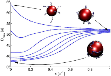
Figure 1 presents results of a Molecular Dynamics (MD) simulation of a single eight-tail colloid. Depicted is the thermally averaged maximal extension of the colloid as a function of for different values of the central sphere charge . For and small values of , i.e., at low ionic strength, the eight tails are extended, radially pointing away from the center of the complex, cf. the example at . For large values of , say, for , and small the tails are condensed onto the sphere, cf. the configuration at and . Increasing the screening leads in both cases finally to structures where the chains form random polymer coils as the ones in the example at . With increasing values of the swelling of initially condensed tails sets in at larger -values. A comparison of our curves for with the experimental ones Mangenot02a shows a qualitatively similar chain unfolding scenario. Furthermore, by choosing we are able to match closely the experimental and the simulation values of at which tail unfolding takes place. In the following we will therefore always use this value as our .
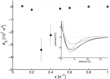
We determined next the interaction between two such complexes by measuring the thermally averaged force at different distances and by interpolating the force-distance curve via a suitable least-square fit. Integration then yields the pair potentials depicted in the inset of Fig. 2 for four different values of . We find an attractive potential with a minimum of a few in all four cases. The depth of the potential shows a non-monotonic dependence on with a maximal value around . This in turn is reflected in a non-monotonic dependence of the second virial coefficient (cf. Fig. 2) with a minimum around the -value where tail unfolding occurs, cf. the curve for in Fig. 1. Again, all these observations are qualitatively similar to the experimental ones Mangenot02b .
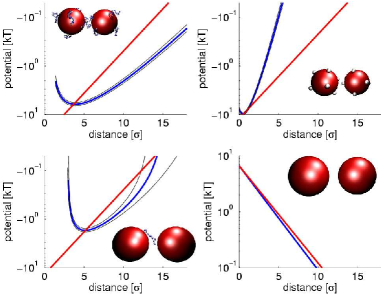
Having a simulation model at hand allows us now to determine whether this attraction can be attributed to the tail-bridging effect. In Fig. 3 we depict a comparison of the full eight-tail model with simplified variants. In all cases we choose , a value close to the one where has its minimal value in Fig. 2; corresponds to 100 mM monovalent salt, i.e., to physiological conditions. In one case (top right) we collapse each chain on a small patch modelled as a grafted monomer that carries the whole chain charge. Inspecting the attractive part of the pair potential we see that this patch model has a very rapidly decaying interaction with a slope larger than the reference line with slope . In sharp contrast, the eight-tail complex has a decay constant that is smaller than (cf. top left of Fig. 3), an effect that can only be attributed to tail bridging. This effect can also be seen for our third variant (bottom left) where 15 of the 16 tails have been removed and has been adjusted so that the net charges of the complexes are unchanged. The remaining one-tail complex is not allowed to rotate so that the grafting point of the chain always faces the other ball. Also in that case the range of attraction is longer than expected from pure screened electrostatics. Finally, on the bottom right we present the trivial case of two charged balls (with the same net charge as the full model) where only a repulsive interaction remains.
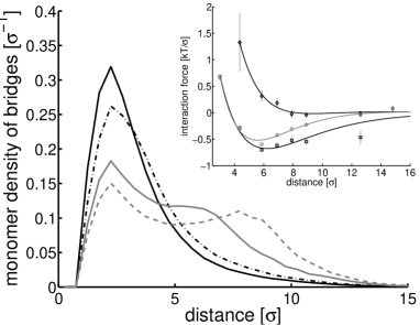
Having established the qualitative difference between tail-induced attraction and attraction via charge patches we take in Fig. 4 a closer look at the tail-bridging effect between two eight-tail colloids, again for . Depicted is the monomer distribution of bridge-forming chains. We define such a chain as a chain that has at least one of its monomers closer than a distance to the surface of the alien core. For very small distances between the colloids there are almost always bridges. Their monomer distribution shows a strong peak around a distance . However, also at much larger distances like and there is still a considerable fraction of configurations that show bridges. Their monomer distribution shows a bimodal distribution with the two peaks clearly reflecting the condensation of monomers on the home core and the alien core. The inset shows the interaction force between two colloids (circles) and the contributions of tail-bridging configurations (squares) and configurations without bridges (diamonds) to this force. It can be clearly seen that the tail-bridging configurations account to an overall attractive force, whereas in the other case the interaction is on average purely repulsive.
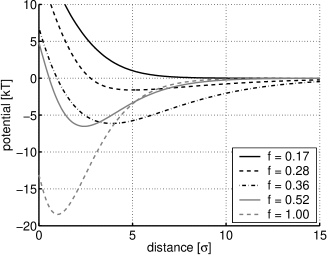
Finally, we speculate how the tail bridging can be used by the cellular machinery to control DNA compaction and genetic activity. We have determined the pair potential between eight-tail complexes for different charge fractions of the tails. As can be seen in Fig. 5, its equilibrium distance goes to larger values and finally disappears when one goes from a charge fraction (the value used above) to . It is in fact known that the cellular machinery is capable of controlling the charge state of the histone tails via the acetylation (the ”discharging”) and deacetylation (the ”charging”) of its lysine groups Horn02 . Active, acetylated regions in chromatin are more open, inactive, deacetylated regions tend to condense locally and on larger scales as well Tse98 . For instance, chromatin fibers tend to form hairpin configurations once a sufficiently strong internucleosomal attraction has been reached Mergell04 ; Grigoryev99 . This suggests a biochemical means by which the degree of chromatin compaction and genetic activity can be controlled via a physical mechanism, the tail-bridging effect.
Acknowledgement: The authors thank M. Deserno, B. Dünweg, K. Kremer, F. Livolant, S. Mangenot and R. Podgornik for helpful discussions.
References
- (1) H. Schiessel, J. Phys.: Condens. Matter 15, R699 (2003).
- (2) K. Luger, A. W. Mader, R. K. Richmond, D. F. Sargent, and T. J. Richmond, Nature 389, 251 (1997).
- (3) K. J. Polach and J. Widom, J. Mol. Biol. 254, 130 (1995).
- (4) I. M. Kulic and H. Schiessel, Phys. Rev. Lett. 92, 228101 (2004).
- (5) J. M. Gottesfeld, J. M. Belitsky, C. Melander, P. B. Dervan, and K. Luger, J. Mol. Biol. 321, 249 (2002).
- (6) I. M. Kulic and H. Schiessel, Phys. Rev. Lett. 91, 148103 (2003); F. Mohammad-Rafiee, I. M. Kulic, and H. Schiessel, J. Mol. Biol. 344, 47 (2004).
- (7) P. B. Becker EMBO J. 21, 4749 (2002).
- (8) The size of a stiff polymer chain in a good solvent scales like (: persistence length, : diameter, contour length) Odijk78 . A human chromosomal DNA chain has cm. This together with nm and nm (assuming physiological ionic conditions) leads to . On the other hand the chromatin fiber has mm, nm Mergell04 and nm leading to . Note that there are 46 chains that have to fit into the nucleus with a diameter of 3 to 10 .
- (9) T. Odijk and A. C. Houwaart, J. Polym. Sci. 16, 627 (1978).
- (10) B. Mergell, R. Everaers, and H. Schiessel, Phys. Rev. E 70, 011915 (2004).
- (11) S. Mangenot, A. Leforestier, P. Vachette, D. Durand, and F. Livolant, Biophys. J. 82, 345 (2002).
- (12) S. Mangenot, E. Raspaud, C. Tribet, L. Belloni, and F. Livolant, Eur. Phys. J. E 7, 221 (2002).
- (13) A. Bertin, A. Leforestier, D. Durand, and F. Livolant, Biochemistry 43, 4773 (2004).
- (14) K. Luger and T. J. Richmond, Curr. Opin. Gen. Dev. 8, 140 (1998).
- (15) Note the opposite effect of colloidal stabilization that occurs when polyelectrolyte chains are grafted densely onto colloids, cf. Pincus91 .
- (16) P. Pincus, Macromolecules 24, 2912 (1991).
- (17) D. A. Beard and T. Schlick, Structure 9, 105 (2001).
- (18) H. Boroudjerdi and R. R. Netz, Europhys. Lett. 64, 413 (2003).
- (19) R. Podgornik, J. Chem. Phys. 118, 11286 (2003).
- (20) I. Rouzina and V. A. Bloomfield, J. Phys. Chem. 100, 9977 (1996).
- (21) E. Allahyarov, H. Löwen, J. P. Hansen, and A. A. Louis, Phys. Rev. E 67, 051404 (2003).
- (22) We choose here the average length of the N-terminal tails whose lengths range from 15 residues (histone H2A) to 44 (H3). The tail bridging effect reported here is very robust: variations in e.g. the anchor positions have only a minor impact on the interaction between our complexes frank_preprint . In the context of the cell some tails (the two H3 tails) might be involved in the interaction with the linker DNA thereby controlling the DNA entry-exit angle as detailed in chapter 3.5 of Ref. Schiessel03 .
- (23) D. Frenkel, B. Smit, Understanding Molecular Simulation (Academic Press, 2nd edition, San Diego, 2002).
- (24) F. Mühlbacher, H. Schiessel, and C. Holm, in preparation
- (25) D. A. McQuarrie, Statistical Mechanics (Harper-Collins, New York, 1976).
- (26) S. Alexander, P. M. Chaikin, P. Grant, G. J. Morales, P. Pincus, and D. Hone, J. Chem. Phys. 80, 5776 (1984).
- (27) P. J. Horn and C. L. Peterson, Science 297, 1824 (2002).
- (28) C. Tse, T. Sera, A. P. Wolffe, and J. C. Hansen, Mol. Cell. Biol. 18, 4629 (1998).
- (29) S. A. Grigoryev, J. Bednar, and C. L. Woodcock, J. Biol. Chem. 274, 5626 (1999).