Theory of Nucleosome Corkscrew Sliding in the Presence of Synthetic DNA Ligands
Abstract
Histone octamers show a heat-induced mobility along DNA. Recent theoretical studies have established two mechanisms that are qualitatively and quantitatively compatible with in vitro experiments on nucleosome sliding: Octamer repositiong through one-basepair twist defects and through ten-basepair bulge defects. A recent experiment demonstrated that the repositioning is strongly suppressed in the presence of minor-groove binding DNA ligands. In the present study we give a quantitative theory for nucleosome repositioning in the presence of such ligands. We show that the experimentally observed octamer mobilities are consistent with the picture of bound ligands blocking the passage of twist defects through the nucleosome. This strongly supports the model of twist defects inducing a corkscrew motion of the nucleosome as the underlying mechanism of nucleosome sliding. We provide a theoretical estimate of the nucleosomal mobility without adjustable parameters, as a function of ligand concentration, binding affinity, binding site orientiation, temperature and DNA anisotropy. Having this mobility at hand we speculate about the interaction between a nucleosome and a transcribing RNA polymerase and suggest a novel mechanism that might account for polymerase induced nucleosome repositioning.
keywords:
nucleosome , DNA ligands , RNA polymerase1 Introduction
The dynamics of folding and unfolding of DNA within the chromatin complex is of vital importance for the regulation of genes. The basic unit of chromatin is the nucleosome where DNA is wound in 1 and 3/4 lefthanded superhelical turns around an octamer of histone proteins [1]. Roughly of all eucaryotic DNA is tightly associated to such protein spools. All that intranucleosomal DNA is usually not accessible to DNA binding proteins [2], leading to the puzzling question of how these proteins can find their hidden target sites. And even more surprising is the fact that most m-RNA coding genes are also covered with tens to hundreds of nucleosomes. So how does a transcribing RNA polymerase deal with all the octamers that it encounters on its way? Can it ”get around” the nucleosomes or have the nucleosomes to be removed before transcription is possible?
An important insight in that respect is the fact that nucleosomes are highly dynamical objects. It has been demonstrated through competitive protein binding that thermal fluctuations induce spontaneous unwrapping of nucleosomal DNA at the ends of its wrapped portion [3, 4]. This leads to a transient opportunity for proteins to bind to nucleosomal DNA. Another important mechanism is nucleosome ”sliding”. It has been observed under well-defined in vitro conditions that nucleosomes spontaneously reposition themselves along DNA [5, 6, 7, 8] transforming nucleosomal DNA into free DNA and vice versa. Heat induced repositioning is, however, a slow process happening on time scales of minutes to hours. The in vivo octamer repositioning has thus to be catalyzed by ATP consuming machines, so-called chromatin remodelling complexes [9, 10].
Repositioning experiments (reviewed in detail in Ref. [11]) are typically performed on short pieces of DNA of length 200 to 400 basepairs (bp) that contain one or two positioning sequences. Repositioning is detected with the help of 2D gel electrophoresis making use of the fact that complexes with octamers close to one of the DNA termini show a higher electrophoretic mobility [5, 6, 7] than complexes where the octamer is associated to the center of the DNA fragment. Another approach [8] uses a chemically modified histone protein that induces a cut on the nucleosomal DNA. The general outcome of these studies is as follows: (1) Heat induced repositioning is a slow process taking place on the time scale of minutes to hours [5, 8] at elevated temperatures (say ) but it is strongly suppressed at lower temperatures (say ). (2) The octamer is found at a preferred position (as mentioned above the DNA contains a positioning sequence!) or multiples of 10 bp (the DNA helical pitch) apart [5, 8]. (3) There is a preference for end positions [5]. (4) For longer DNA segments there is no evidence for a long-range repositioning [6]. (5) In the presence of linker histones (H1 or H5) nucleosome mobility is suppressed [7].
What is the origin of the nucleosome mobility? An ordinary sliding of the DNA on the protein spool is energetically too costly: The interaction between the DNA and the octamer is localized at 14 binding sites where the minor groove of the DNA faces the octamer surface [1], each contributing roughly pure adsorption energy [11] (: thermal energy). A bulk sliding motion would involve the simultaneous breakage of these 14 point contacts, an event that certainly would never occur spontaneously. A rolling motion of the octamer along the DNA is also not possible: Due to the helical wrapping path the cylinder would simply roll off the DNA.
Repositioning must involve intermediate states that have a lower energetic barrier. Two commonly accepted possible mechanisms [11, 12] are based on small defects that spontaneously form in the wrapped DNA portion and propagate through the nucleosome: 10 bp bulges [13, 14] and 1 bp twist defect [15]. The basic idea of bulge defects is as follows: As a first step the DNA unpeels spontaneously from one of the termini of the wrapped portion [3, 4]. Subsequently some DNA is pulled in before the chain readsorbs creating an intranucleosomal DNA bulge that carries some extra length . Once a loop has formed it diffuses along the wrapped DNA portion and finally leaves the nucleosome at either end. If the loop happens to come out at the end where it has been created nothing happens. But if the loop leaves at the other end the extra length has been transported through the nucleosome and the octamer is repositioned by along the DNA. A careful quantitative analysis [14] showed that the cheapest small loop that can be formed has a length of 10 bp. Such a loop is not twisted; the next planar loop, a 20 bp bulge, is much more expensive. However, even the creation of a 10 bp loop is very costly: Its formation requires about desorption and bending energy and thus constitutes a very rare event [11]. As a consequence the corresponding diffusion constant of the octamer along the DNA is very small, namely on the order of . Thus typical repositioning times on a 200 bp DNA fragment are on the order of an hour, in reasonable agreement with the experimental data [5, 8]. The strong temperature dependence and most strikingly the preference for 10 bp steps – corresponding to the extra length stored in the cheapest loops – is also in excellent agreement with the experiments. So at first sight it seems that the loops are in every respect a promising candidate for the mechanism underlying repositioning. There is, however, one serious caveat: We found that larger loops beyond one persistence length of DNA (roughly 150 bp) are easier to form than 10 bp bulges since such loops show a small curvature and have less desorbed binding sites [14]. Of course, for short DNA segments such loops cannot occur. However, experiments with DNA segments of length bp have also not shown any signature of a long range nucleosome repositioning [6].
We therefore reconsidered the underlying mechanism and checked whether the experimental observations would also be consistent with repositioning via twist defects [15]. The basic idea of repositioning via twist defects is that thermally activated defects form spontaneously at the termini of the wrapped DNA portion. There are two types of twist defects: (a) defects with a missing bp requiring the DNA to stretch and overtwist between its two neighboring nucleosomal binding sites and (b) defects with an extra bp thus leading to a compressed and undertwisted piece of DNA between two nucleosomal point contacts. As in the case of bulges a twist defect might diffuse around a nucleosome releasing its stored length (here bp) at the other end. The result of such an event is that the octamer makes a step by one bp and a rotation by around the DNA axis; or vice versa one might say the DNA performs a corresponding corkscrew motion on the nucleosome. The cost of forming a one bp twist defect was estimated to be on the order of [15]. The shorter defect length involved in twist defects as compared to bulges, is thus dramatically overcompensated by their lower activation cost. In fact, we estimated that twist defects lead to a nucleosome diffusion constant on the order of that is 4 orders of magnitude larger than the one predicted by loop defects. The typical repositioning times on a 200 bp piece of DNA are thus predicted to be on the order of a second, a time much shorter than in the experiments! Even worse, the predicted dependence of the dynamics on temperature is much too weak and there is no ”built-in” mechanism for 10 bp steps of the octamer. The experimentally observed preference for positions 10 bp apart manifesting itself in characteristic bands in the products of a gel electrophoresis [5, 6] seems to be inconsistent with this mechanism – at least at first sight.
Here comes into a play an important additional feature of the repositioning experiments, namely that they are typically performed with DNA segments containing positioning sequences, especially the sea urchin 5S positioning element [5, 6, 7]. The characteristic feature of this sequence is that it shows a highly anisotropic bendability of the DNA. If the underlying mechanism of repositioning is a 1 bp twist defect, then the DNA has to bend in the course of a 10 bp shift in all directions, and thus has to go over a barrier. In the case of the standard ”5S-RNA” this barrier is on the order of [16, 17]. The typical repositioning times on a 200 bp DNA segment are now 2 to 3 orders of magnitude longer, i.e., they are on the order of an hour as in the loop case! Now it is a simple matter of equilibrium thermodynamics that the probability of finding the DNA wrapped in its preferred bending direction is much higher than in an unfavorable one. This means, however, that also in the case of 1 bp defects one would find nucleosomes mostly at the optimal position or 10, 20, 30 etc bp apart, i.e., at locations where still most of the positioning sequence is associated with the octamer and this in the preferred bending direction. The bands in the gel electrophoresis experiments have then to be interpreted as reflecting the Boltzmann distribution of the nucleosome positions. In other words, both the 10 bp bulge and the 1 bp twist defect lead in the presence of a rotational positioning sequences to pretty much the same prediction for the experimentally observed repositioning, even though the elementary motion is fundamentally different!
There are many ways to design experiments that could help to identify the mechanism that underlies nucleosomal mobility. The most obvious idea is to use a DNA template with less exotic mechanical properties (in fact, the 5S positioning element is the strongest natural positioning sequence known so far). If nucleosomes move via loop defects a more isotropically bendable DNA should not speed up the dynamics whereas it would have a strong impact on the corkscrew mechanism. In fact, the experiment by Flaus and Richmond [8] goes in that direction. They used a DNA fragment of length 438 bp that featured two positioning sequences where two nucleosomes assembled, each at a unique position. These positions were also found when mononucleosomes were assembled on shorter fragments that included only one of the two positioning elements. The authors studied the degree of repositioning of the mononucleosomes on such shorter fragments (namely nucleosome A on a 242 bp- and nucleosome B on a 219 bp-fragment) as a function of heating time and temperature. It was found that the repositioning rates increase strongly with temperature but also depend on the positioning sequence (and/or length of the fragment). The difference of repositioning for the two sequences is remarkable: at 37∘C one has to wait minutes for the A242 and more than 30 hours for the B219 for having half of the material repositioned. For nucleosome B which showed a slower repositioning the set of new positions were all multiples of 10 bp apart (namely at a 20, 30, 40, 50 bp-distance from the starting position), i.e., they all had the same rotational phase. On the other hand, nucleosome A did not show such a clear preference for the rotational positioning. It was argued that these differences reflect specific features of the underlying base pair sequences involved. Nucleosome B is complexed with a DNA sequence that has AA/AT/TA/TT dinucleotides that show a 10 bp periodicity inducing a bend on the DNA whereas nucleosome A is positioned via homonucleotide tracts. These observations are clearly consistent with the twist defect picture where the corkscrew motion of nucleosome B is suppressed by the anisotropically bendable DNA template.
Another experimental approach was taken recently by Gottesfeld et al. [18]. The authors considered a 216 bp DNA fragment that again contained the sea urchin 5S rDNA nucleosome positioning sequence. They also followed the heat induced nucleosome repositioning but this time in the presence of pyrrole-imidazole polyamides, synthetic minor-groove binding DNA ligands that are designed to bind to specific target sequences. Experiments have been performed in the presence of one of 4 different ligands, each having one binding site on the nucleosomal DNA. The general outcome of this study was as follows: (1) A one-hour incubation at in the absence of any ligand leads to a redistribution of the nucleosomes. (2) In the presence of 100 nM ligands no repositioning of nucleosomes is detected after such an incubation if the target sequence of this specific ligand faces the solution when the DNA is bent in its preferred direction. (3) If a ligand has been added whose binding site faces the octamer in its preferred rotational frame, the ligand has no detectable effect on the reposition dynamics.
This raises the question whether this experiment is capable to distinguish between loop- and twist-defect induced nucleosome mobility. Since the ligands bind into the minor groove (cf. the co-crystal complexes between nucleosomes and such ligands [19]) it is quite likely that a bound ligand will block the overall corkscrew motion of the DNA; the DNA can only rotate on the nucleosome up to a point where the bound ligand comes close to one of the 14 binding sites. A further rotation of the DNA is not possible because of steric hindrance and twist defects are reflected once they encounter the ligand site. In other words: The observed suppression of mobility through ligand binding agrees qualitatively well with the twist defect picture. What about a bulge defect encountering a bound ligand? In this case the answer is not obvious. In a first approximation one should expect that a bound ligand does not hinder bulge diffusion – at least sterically. Of course, the ligand might locally alter the DNA elastic properties so that we cannot give here a definite answer. But obviously the influence of ligand binding on nucleosome mobility supports much more the idea of twist diffusion as the underlying mechanism. The aim of this study is to demonstrate that the twist diffusion picture is indeed compatible with the experimental data presented in Ref. [18]
In the next section we provide a theoretical model for nucleosome repositioning in the presence of DNA ligands. We make use of our recent results on repositioning via twist defect in the absence of ligands [15] that essentially provides us with the nucleosomal diffusion constant as a function of temperature and underlying DNA sequence. Assuming thermodynamic equilibrium we will then calculate the diffusion constant in the presence of ligands. We find – in agreement with the experiments – that in the presence of 100nM ligands the repositioning on the 5S positioning sequence is essentially completely blocked if the ligand binding site prefers to face the solution. On the other hand, when the binding site faces the octamer, the ligands have a negligible influence on the nucleosome mobility.
Knowing the nucleosome mobility in the various cases we speculate in Section 3 what happens when a transcribing RNA polymerase encounters a nucleosome. In fact, this situation has been investigated by Gottesfeld et al. [18] in their study with synthetic ligands. We suggest that in some of the cases the RNA polymerase pushes the nucleosome in front of it in a corkscrew fashion. When the terminus of the DNA template is reached the nucleosome becomes ”undressed” and the other end of the DNA might bind to the exposed binding sites. As a result the RNA polymerase seems to have gotten around the nucleosome with the octamer having been transferred to a location upstream. Our model gives thus an alternative explanation to the popular idea of RNA polymerase transcribing through a nucleosome in a loop [20, 21, 22, 23].
2 Autonomous Repositioning
2.1 Nucleosome Mobility in the Absence of Ligands
Let us first consider the repositioning of nucleosome along DNA induced via 1 bp twist defects in the absence of ligands. This has been recently studied theoretically in detail [15]; here we will restrict ourselves to a short presentation of the results. The basic idea is that a twist defect might form spontaneously at either end of the wrapped DNA portion. Such a defect can carry a missing or an extra bp. A defect is typically localized between two neighboring nucleosomal binding sites, i.e., it is localized within one helical pitch, 10 bp. This short portion of DNA is stretched (compressed) and overtwisted (undertwisted). The energy of a bp twist defects was estimated from the combined stretch and twist moduli of DNA including the (here unfavorable) twist-stretch coupling to be on the order of [15]. That means that one finds for a given time a twist defect only on one of around thousand nucleosomes.
Once a twist defect has formed, it can diffuse through the wrapped DNA portion. The nucleosome provides in total 13 positions for the defect between the 14 binding sites. A defect (say a ”hole” with a missing bp) can move from one position to the next in the fashion of an earthworm creep motion: The bp that is in contact with a binding site moves towards the defect, leading to an intermediate state where the defect is stretched out over 20 bp, releasing elastic energy but paying desorption energy. When the next bp binds to the nucleosome the twist defect has moved to the neighboring location. During this process the kink goes over a barrier; its energy was estimated from the adsorption energy per contact and the DNA elasticity to be on the order of [15]. Of course, not all twist defects that have formed will reach the other end of the nucleosome, most fall off at the terminus at which they have been created. In fact, one can show – assuming that all 13 possible defect locations are energetically equivalent – that only of the defects are ”successful”, only this fraction contributing to the nucleosomal mobility.
Putting all these points together we were able to estimate the diffusion constant of the nucleosome along DNA to be . As mentioned in the introduction this is surprisingly fast – especially much faster than the experimental values [5, 6, 7, 8]. We explained this discrepancy by the mechanical properties of the underlying DNA template. A nucleosome performing a (random) corkscrew motion might encounter a very bumpy energy landscape. Especially, positioning sequences like the commonly used 5S sequence show an anisotropic bendability. In that case the elastic energy of the bent DNA is a periodic function of the nucleosome position with the helical pitch being the period. We approximated this energy by an idealized potential of the form with being the number of the bp say at the dyad axis and denoting the difference in elastic energy between the optimal and the worst rotational setting. Of course, these oscillations die out completely when the nucleosome leaves the positioning sequence, i.e., if it has moved around 140 bp. Since the templates are usually quite short (for instance, 216 bp [18]) the nucleosome will always feel the rotational signal from the positioning sequence so that our elastic energy should provide a reasonable description. In that case the nucleosomal diffusion constant is reduced as follows [15]:
| (1) |
with being the modified Bessel function and denoting the diffusion constant for homogenously bendable DNA, . For the sea urchin 5S positioning element one has [16, 17] leading to a reduced mobility with .
2.2 Nucleosome-Ligand Cocomplex: Equilibrium Properities
In this subsection we determine the equilibrium properties of a nucleosome in the presence of a finite concentration of a synthetic ligand targeting one specific site on the nucleosomal DNA. In Fig. 1 we represent the different possible states by nodes and the possible pathways from one state to the next state by connecting lines. Fig. 1(a) shows the case of a DNA template with an isotropic bendability. The upper row of symbols represent nucleosomes at different positions without the ligand being bound. Each circle with a hole (state ”1”) represents a state where a ligand can bind, i.e., states where the ligand binding site (assumed to be located on the wrapped DNA portion) faces away from the octamer. In this case a ligand can bind leading to a state that is represented by an open circle, the nucleosome, with a ”bound” black circle, the ligand (state ”0”). We assume that in this case the nucleosome looses its mobility, i.e., we have no line connecting this state to a neighboring state. Before the nucleosome can ”slide” to a neighboring position the ligand has to unbind, i.e., one has to go back to state ”1”. If the nucleosome is in a position where the ligand binding site faces the octamer (open circle, state ”2”) the site is blocked. At these positions the nucleosome mobility is not affected by the ligands. For simplicity, we will assume here that always 5 consecutive bp positions (correspond to one half turn of the corkscrew motion) have the ligand binding site exposed to the solvent and represent these 5 position by one circle with hole. Likewise the other 5 positions have been lumped together into the open circle.
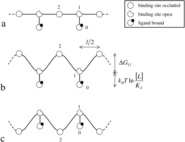
Fig. 1(b) and (c) show the case of a rotational positioning sequence as used in the experiment [18]. In case (b) the situation is such that the ligand can bind when the DNA sits on the nucleosome in its preferred bending direction. In this case the states ”1” sit in the potential wells of the elastic energy landscape. We call the difference in the DNA bending energy between the top and the bottom . The case correspond to the situation where a ligand can most effectively bind to the nucleosome and block the repositioning (in the experiment this corresponds to the ligands 1 and 4 [18]). Fig. 1(c) depicts the other extreme where the binding site faces the octamer in the preferred rotational frame (this corresponds to ligands 2 and 3 in the experiment [18]).
We denote by the probability for the nucleosome to be in state , . Detailed balance relates these probabilities as follows:
| (2) |
and
| (3) |
Here denotes the transition rate from state to state , the Boltzmann weight between state 1 and 2, the equilibrium constant for the ligand and its dissociation constant. Eqs. 2 and 3 together with yield immediately the occupation probabilities for the three states:
| (4) |
| (5) |
and
| (6) |
Let us consider some special cases. In the absence of ligands one has and . For a homogeneous template one finds in addition the Boltzmann weight (defined in Eq. 2) to equal unity. Then and, of course, . Using a rotational positioning sequence the nucleosome prefers to be in its optimal rotational frame. Then for one finds . For the 5S positioning element one has so that state ”1” is populated with a roughly 10000 times higher probability then state ”2”. This might explain the band structure with a 10 bp periodicity as observed in most repositioning experiments [5, 8]. The presence of ligands changes the relative weight of the different states. Consider, for instance, a rotational positioning sequence with the ligand binding site facing inwards for the preferred rotational frame ( and hence , cf. Fig. 1(c)). The relative weight of finding the nucleosome at its mechanically unfavorable position, state ”0” and ”1”, will then increase in the presence of ligands. This leads to the intriguing possibility that the nucleosome changes its preferred position, i.e., . This is in fact the case if . This means that for sufficiently high concentration and affinity ligands can in principle overrule a positioning sequence! The ligands used in the experiment [18] have dissociation constants ranging from to . For a strong positioning sequence is much too large for the above inequality to hold for ligand concentration typically used in the experiment, say . However, for less strong sequences this might play a role. Also in the case of a 146 bp template corresponding to the total wrapping length as consider by Suto et al. [19] it might well be that ligands shift the preferred centered DNA position to an off-centered position. A 1-9 bp shift would cost the opening of one binding site but might allow ligands to bind more effectively, especially if the binding site(s) at the centered DNA positions are (partially) occluded. In one case [19] (polyamid 2) such an effect might have been indeed observed, cf. Fig. 5 in that paper.
2.3 Nucleosome Mobility in the Presence of Ligands
We are now in the position to determine the diffusion constant of a nucleosome along DNA in the various cases. The diffusion constant can be determined from the average of the diffusion constant for the nucleosome to jump from state ”1” to one of the two neighboring states ”1” and that of going from ”2” to neighboring ”2’s”. Let us denote by the rate to go from a given state ”1” to the next position ”1” to the right and by the rate of jumps to the right from ”2” to ”2”. Then the diffusion constant is given by
| (7) |
where is the jump length, here . Using Kramers’ rate theory [24] it can be shown that
| (8) |
with the attempt frequency being given by for and for .
Using Eqs. 5 to 8 we arrive at the final formula for the diffusion constant for the case (i.e. ):
| (9) |
In the opposite case, (i.e. ), we find
| (10) |
Let us now consider special cases:
(i) homogeneous DNA bendability, no ligands (, ): In that case , . Both formulas, Eqs. 9 and 10, reduce to .
(ii) homogeneous DNA bendability but ligands present (, ), cf. Fig. 1(a): leads to
| (11) |
(iii) rotational positioning sequence, no ligands present (, ): Equations 9 and 10 reduce to Eq. 1 with , the case that has been already discussed before [15].
(iv) rotational positioning sequence, ligands present with binding site exposed in the preferred orientational frame (, ), cf. Fig. 1(b): Using we obtain from Eq. 9
| (12) |
(v) rotational positioning sequence, ligands present with binding site occluded in the preferred orientational frame (, ), cf. Fig. 1(c): Here and from Eq. 10 we find
| (13) |
We are now in the position to check how effectively ligands reduce repositioning in the various cases. We estimate in the following the typical equilibration time on a 216 bp long template (as it has been used in Ref. [18]) to be . Let us start with case (i) where . This leads to the typical time . Adding now a ligand with and (case (ii)) this leads to a 50 fold reduction of the diffusion constant, , cf. Eq. 11, and to an equilibration time . If one uses a positioning sequence instead with one finds in the absence of ligands (case (iii)) from Eq. 1 and . Repositioning experiments on such sequences are thus typically performed on time scale of an hour to ensure equilibration [5, 18]. Adding now a ligand with and and having its binding site facing the solution in the preferred rotational frame (case (iv), Fig. 1(b)) we predict from Eq. 12 an additional dramatic reduction of the diffusion constant by a factor of 100: and . In other words, in this situation one does not observe any repositioning of the nucleosomes on the time scale of an hour. This is in accordance with the experimental observations, cf. Fig. 5, lane 1 and 4 in the study by Gottesfeld et al. [18]. On the other hand, for the case of a ligand with same affinity and concentration but with the binding site in the unfavorable orientation (case (v), Fig. 1(c)) one finds hardly any effect; in fact the diffusion constant as compared to the ligand free case, case(iii), is reduced by approximately 1 percent, cf. Eq. 13. In the experiment [18] these two cases were indeed indistinguishable as seen in Fig. 5, lane 0, 2 and 3 in that paper.
3 Transcription Induced Sliding
Gottesfeld et al. [18] also studied how nucleosomes affect transcription. For that purpose the 216 bp DNA fragment contained a T7 promoter in addition to the 5S positioning element. The transcription reaction of the naked 216 bp fragment with T7 RNA polymerase produced the 199 bp full-length RNA transcript. Importantly this reaction was not affected by the presence of any of the ligands. Also the nucleosome templates produced full length transcripts with a very high yield, indicating that the RNA polymerase was able to overcome the nucleosomal barrier. This was also the case in the presence of ligands 2 and 3 whose binding site face the octamer in the preferred rotational frame. Remarkably the addition of ligand 1 or 4 blocked the transcription. In fact, single round transcription assays showed that the polymerase got stuck just within the major nucleosome position. Moreover, an inspection of the nucleosome positions showed that in the absence of any ligand or in the presence of ligand 2 or 3 nucleosome repositioning took place. In other words, transcription did not result in a loss of the nucleosome but in its repositioning instead.
In the previous section we have shown that nucleosomes in the presence of ligands 1 or 4 show a dramatic reduction of their diffusion constant, cf. Eq. 12. The Einstein relation provides a link between nucleosomal mobility and diffusion constant – in case of thermodynamic equilibrium. It is tempting to speculate that it is this difference in nucleosomal mobility that is responsible for the different outcome of the transcription experiment described in Ref. [18].
Let us first consider the case of a long DNA template with a nucleosome positioned far from any of the DNA termini. Suppose that an elongating RNA polymerase encounters such a nucleosome. If the mobility of the nucleosome is large enough the RNA polymerase would be able to push the nucleosome in front of it – by pulling the DNA in corkscrew fashion. In the most simple mean-field-type approach the nucleosome will begin to slide with a constant speed as a result of the imposed external load as follows:
| (14) |
The polymerase slows down as a result of the force that it has to exert on the nucleosome. According to Wang et al. [25] (cf. also related studies [26, 27]) the force-velocity relation of RNA polymerase has typically the following functional form
| (15) |
where is the velocity of the elongating complex in the absence of an external load and is the load at which the speed of the RNA polymerase is reduced to . is a dimensionless fit parameter.
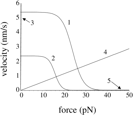
Equating Eqs. 14 and 15 we can determine the average speed of an RNA polymerase that pushes a nucleosome in front of it. The solution is found graphically in Fig. 2 by determining the point of intersection between the corresponding curves. Curves ”1” and ”2” in Fig. 2 give the force-velocity relation of RNA polymerase, Eq. 15, at two different concentrations of pyrophosphate (), namely curve ”1” is for where , and and curve ”2” corresponds to for which , and . In both cases there is 1 mM nucleoside triphosphates (NTPs). Note that these numbers give a good fit to the data of Wang et al. [25] for the case of Escherichia Coli RNA polymerase. As mentioned above in the experiment of Gottesfeld et al. [18] a T7 RNA polymerase has been used and the concentration of NTPs was . This means that curves ”1” and ”2” can only be considered as rough estimates for the force-velocity characteristics of the T7 RNA polymerase. The other curves, ”3” to ”5”, give the force-velocity relation, Eq. 14, for the nucleosomes in various cases. Curve ”3” corresponds to the case when a nucleosome slides along an isotropic DNA segment in the absence of any ligands (case (i) in the previous section). Curve ”4” represents corkscrew sliding along an anisotropic DNA with a barrier height as it is the case for the 5S positioning sequence (case (iii)). And finally, curve ”5” corresponds to the case where in addition to such an anisotropic bendability the mobility is slowed down by the presence of ligands with the ligand binding site facing the solution in the preferred DNA bending direction, case (iv).
By inspecting the points of intersection between the curves we come to the conclusion that RNA polymerase would be hardly slowed down by the presence of a nucleosome on a homogenous track of DNA, cf. point of intersection between line ”3” with curve ”1” (or ”2”) in Fig. 2. We expect that the polymerase would easily push the nucleosome in front of it without being slowed down. On the other hand, the 5S positioning element should affect the transcription rate by a considerable amount (cf. line ”4” and curve ”1” and ”2”); still the RNA polymerase might be able to push the nucleosome ahead of it. Finally, in the case of added ligands the nucleosome blocks the way of the nucleosome: the point of intersection between curve ”5” and ”1” (or ”2”) is close to a vanishing transcription velocity.
In the experiment [18] there is, however, an additional complication: The nucleosome is positioned at the 3’-end of the template. That means as soon as the polymerase encounters the nucleosome (here after it has transcribed the first bp) it would have to push the nucleosome off the DNA template. What is the energetic cost of this process? There are 14 binding sites between the DNA and the octamer, with a 10 bp distance between neighboring ones. It can be estimated [11] that the detachment of any of these 14 nucleosomal binding sites costs . However, the overall energetic cost of undressing the nucleosome is smaller: When pulling 10 bp off the octamer one binding site is opened but 10 bp are released on the other side that gain roughly elastic energy by going from the wrapped, bent state to the straight state. In total, a shift of the DNA by 10 bp cost therefore only which corresponds to a force of just 2 pN. This additional force can be easily supplied by the RNA polymerase.
Therefore our calculation leads to the prediction of the following effect of the RNA polymerase on the nucleosome: (1) In the ligand-free case the RNA polymerase is able to produce the full-length transcript pushing the nucleosome off the template. (2) If a ligand is bound to the nuclesomal DNA the nucleosome is immobile and the polymerase stalls as soon as it encounters the nucleosome. Whereas the second prediction is indeed in agreement with the experimental observations, the first one is not. In that case transcription does not lead to the loss of the nucleosome but instead to its repositioning on the template [18]. The experimental findings even indicate that the nucleosome – as a result of the transcription – is effectively moving upstream! In fact, such effects have been studied in detail before and let to proposition of a spooling mechanism [20, 21, 22, 23]; we will here, however, delegate a discussion of these experiments and their interpretation to our discussion section.

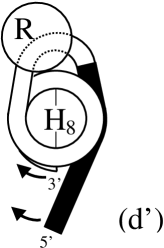
In order to explain the experimental observations of Ref. [18] we propose a new mechanism that is depicted in Fig. 3. (a) At the beginning of the transcription (the first 54 bp in Ref. [18]) the RNA polymerase walks along the free DNA section (shown in black) in a corkscrew fashion. (b) The polymerase comes into contact with the nucleosome. At this stage the polymerase gets stuck if the nucleosome is immobile. (c) If the nucleosome is mobile the polymerase pulls on the DNA, undressing the nucleosome at the other end (the 3’ end). During this process the polymerase and the octamer (H8) are not moving with respect to each other and it is only the DNA that is performing a corkscrew motion. (d) After enough nucleosomal contact points (at the 3’ end) are exposed to the solvent the 5’ end might adsorb on these contact points, forming an extranucleosomal loop. (e) The DNA continues to circle around the polymerase-nucleosome complex via the corkscrew mechanism. (f) When the 3’ end reaches the polymerase this end is released from the nucleosome. As a result one has again an end-positioned nucleosome but now it is the promoter end that is wrapped on the nucleosome. A section of the original positioning sequence (shown in white) forms now the free tail.
This mechanism always transfers the nucleosome from one end of the DNA template to the other. In principle, it is also possible that a smaller loop forms with the 5’ end forming an overhanging tail, cf. Fig. 4. Such a small loop might be possible since the RNA polymerase induces a bend on the DNA. The RNA polymerase will then again pull the DNA around via the corkscrew mechanism. Due to the presence of the loop the 5’ tail might only be able to adsorb beyond the dyad after the 3’ end is released. At this point the nucleosome has effectively made a step upstream. The step length is the sum of the length stored in the loop plus the number of bp of the 3’ end that were still adsorbed at the point of its release. It is possible that the 3’ is released at a point where it was still associated with a few binding sites (each binding site just contributes on the order of ). The typical upstream step length is then a few tens of bp. An interesting feature of this variant of the model is that the step length should not depend on the length of the originally free DNA portion (shown in black in Fig. 4). In other words: If the nucleosome was initially positioned at one end of the template (due to some positioning sequence), after transcription it is shifted upstream to a new position by a distance that is independent of the length of the DNA template.
4 Discussion
We provide here a critical discussion of our results, mainly focusing on alternative mechanisms that allow RNA polymerase to get around nucleosomes. Before doing so we mention an important assumption on which our whole analysis is based: Bound ligands do not act across the two turns. If the ligand would somehow bind to both turns of the nucleosome, it could effectively block any nucleosome dynamics, whether it is induced by bulge or twist defects. In fact, such a situation occurs in the presence of linker histones H1 or H5 that bind the in- and outgoing DNA together in a stem-like region. It has been observed that there is no nucleosome repositioning in this case [7]. However, the cocrystal structure of a nucleosome core particle with bound ligands [19] indicates that ligands bind locally to one turn without affecting the other one. This supports our assumption – namely that a ligand blocks twist defects only.
This brings us to the discussion of polymerase-induced nucleosome repositioning. The experiment by Gottesfeld et al. [18] showed that nucleosomes survive transcription but it was not possible to deduce from the data whether transcription through a nucleosome leads to its repositioning along DNA. There is, however, a long series of experiments that have focused on this point [20, 21, 22, 23]. Also in these experiments a bacteriophage RNA polymerase has been used, namely that of SP6. The standard 227 bp template includes an SP6 promoter and a nucleosome positioning sequence [20]. Typically the nucleosome is positioned at the promoter distant end. Transcription results in an upstream displacement to the other end, i.e. by 80 bp [20]. Whether this step length reflects a built-in step length of the repositioning process or whether the nucleosome is displaced from one end to the other has been checked by adding an extra length to the DNA template at either end. Adding extra 50 bp at the promoter side (the 5’ end) the upstream step is typically 90 bp, i.e., it does not increase much. This might indeed indicate that the displacement process has a natural 80 to 90 bp step length. On the other hand, addition of 35 bp to the 3’ end has surprisingly also an effect on the upstream step length that shows now three smaller values, namely 40, 60 and 75 bp [20]. Finally, going to a much larger template by adding 126 bp at the promoter end led to another surprise: In this case the nucleosome is transferred from one end to the other as a result of the transcription [21].
How can these observations be rationalized? Studitsky et al. [20] introduced the ”spooling” mechanism, cf. their Fig. 7: As the polymerase encounters the nucleosome it continues to transcribe by prying off the DNA from the octamer. After the polymerase has proceeded far enough into the nucleosomal DNA the DNA behind might attach to the now exposed nucleosomal binding sites. This results in an intranucleosomal loop. The polymerase travels around the nucleosome inside this loop. When reaching the other end the loop disappears and as a result the nucleosome steps upstream by the extra DNA length that has been stored in that loop. The step lengths observed in the experiments have then to be interpreted as the loop sizes. A preferred value would then be around 80 bp. Studitsky et al. explained the much shorter step lengths observed in the case of a template with a DNA extension on the promoter distant site as a result of ”octamer slippage” before the spooling mechanism comes into play with the usual 80 bp upstream step. Finally, the end to end transfer on the long 353 bp template indicates a large loop that stores 180 to 200 bp [21].
In fact, these observations and their explanation are entirely consistent. One should nevertheless ask whether our extranucleosomal loop model provides also a picture consistent with these experimental facts. In fact, the model depicted in Fig. 3 predicts an end-to-end transfer of the nucleosome as it has been observed for the longest template discussed above. The modified model with a small extranucleosomal loop as depicted in Fig. 4 leads to a smaller upstream step of the octamer whose value depends on microscopical details but should be on the order of a few tens of bp. So this picture could in fact also explain the typical 80 bp shifts observed in several cases. This leads us to the surprising conclusion that either mechanism, the extra- and the intranucleosomal one, is consistent with the observations. It is only the smaller steps where Studitsky et al. suggested octamer slippage to occur that might ironically speak in favor of their model. When the nucleosome steps back by 40 bp it might have first slid 35 bp to the 3’ end and then go back by 80 bp with either mechanism. However, the fact that after transcription some nucleosomes were found 60 and 75 bp upstream might support the intranucleosomal loop picture: First the nucleosome slides a short distance (but not up to the DNA terminus) and then steps back by 80 bp in an intranucleosomal loop. Nevertheless it seems impossible to exclude from these experimental observations one or the other mechanism and it might well be the case that both play a role.
Another feature that has been observed during the transcription ”through” nucleosomes is a characteristic pausing pattern of the polymerase [21, 28]. Studitsky et al. [21] reported for their SP6 system pausing with a 10 bp periodicity to occur up to the dyad where it disappears. Also Protacio et al. [28] find pausing with this periodicity, however, also extending far beyond the dyad. The ladder system uses T7 RNA polymerase and the 5S positioning element as in Ref. [18]. Studitsky et al. interpret their observations with their spooling model: Once the loop has formed the polymerase might not be able to continue with elongating since it would have to corkscrew through the loop and this process might be too costly if not even sterically forbidden. Instead pausing occurs up to the point when the loop reopens through a spontaneous fluctuation. The loop formation (and the concomitant pausing) might happen with a 10 bp periodicity since the bend induced by the polymerase might help the loop formation every 10 bp. Once the dyad has been reached the last loop forms that is finally broken ahead of the polymerase, allowing the polymerase to transcribe from now on without interference from the octamer. Further support for this idea was given by removal of DNA behind elongating complexes that have been arrested just at the nucleosomal border. Resuming transcription the polymerase was able to elongate into the nucleosome much further without pausing before it encountered a first pausing side. This was interpreted again as a fact supporting the spooling model [21]: The formation of the loop was only possible when enough DNA was available at the 5’ end.
We believe that these observations are also consistent with the extranucleosomal loop picture. The 10 bp pausing pattern might reflect the 10 bp periodicity of the bending energy of the positioning sequence. Enhanced pausing might occur once the loop has formed due to an enhanced friction of the corkscrewing DNA. And the disappearance of pausing sites beyond the dyad (which is not for all situations the case, cf. Ref. [28]) might reflect the termination of an interaction between the polymerase and DNA wrapped close to the dyad. In case of the 5’ end forming a tail, as shown in Fig. 4, this end might not be able to adsorb beyond the dyad as long as the intranucleosomal loop is present so that the friction or entanglement between the components decreases once the polymerase passes the dyad.
This brings us to the next point of our discussion. One might wonder whether such intra- or extranucleosomal loops could be directly ”seen” in electron micrographs. In fact, cryomicroscopy has been performed for such complexes [23]. Unfortunately, also here the situation is rather complex. When the polymerasewas arrested after transcribing 23 bp into the nucleosome the electron cryomicrographs showed complexes with one DNA tail. The length of that tail was considerably longer than the tail in the absence of RNA polymerase. This was interpreted as being due to a polymerase-induced DNA unwrapping. Interestingly our corkscrew sliding scenario also leads to a tail lengthening without the necessity of DNA unpeeling, cf. Fig. 3(c). The polymerase was also arrested further into the nucleosome (42 bp), a location at which intra- or extranucleosomal loops should be expected. Loops were, however, not observed (at least not large ones), instead there was a considerable fraction of two-tailed intermediate states. These closed transcription intermediates were interpreted as states that resulted from the collapse of an internucleosomal loop, cf. Fig. 7 in Ref. [23]. In our opinion such an explanation (being an attempt to reconcile the spooling model with the two-tail intermediates) is not obvious, even though this picture cannot be excluded. On the other hand, when the polymerase is stalled after a small extranucleosomal loop has formed, two-tail intermediates should in fact be expected. In Fig. 4 the 5’ end is forming the only tail. But it is also possible that the 3’ desorbs up to the dyad where the loop blocks further unpeeling. This leads to two-tail complexes where both ends form tails of varying lengths.
In conclusion we agree that the experiments of Studitsky et al. [20, 21, 22, 23] are indeed compatible to their spooling model. However, we have shown that also our extranucleosomal loop mechanism gives a consistent explanation of these experiments. If the recent experiments by Gottesfeld et al. [18] had been performed for the same system under identical conditions, then their observation of transcription blockage via ligands would speak in favor of our model. As it stands it is not clear which mechanism is responsible for transcription through nucleosomes in the various cases.
We should mention that the two different scenarios involving intra- and extranucleosomal loops lead to dramatically different pictures for transcription on multinucleosomal templates. Whereas the elongating RNA polymerase could easily get around all the nucleosomes via intranucleosomal loops, our extranuclesomal variant relies on the finite length of the DNA. This mechanism would cease to work for the multinucleosomal situation. In fact, transcription on reconstituted multinucleosomal templates showed that T7 RNA polymerase is under certain conditions capable of disrupting completely the nucleosomal cores [29, 30]. Electron micrographs show the transcribed section to be freed of nucleosomes and parts of the histones being transferred to the nascent RNA chain [30]. Interestingly upon addition of some nuclear extract the nucleosomal template seem to survive during transcription [29]. This shows that the in vivo situation might be rather complex involving additional factors mediating between polymerase and nucleosomes.
5 Acknowledgements
We are thankful to Karolin Luger for sharing experimental results [19] prior to publication. We thank R. Bruinsma and R. Golestanian for discussions. F.M. acknowledges the hospitality of the MPIP in Mainz where this work was initiated.
References
- [1] Luger, K., Mäder, A. W., Richmond, R. K., Sargent, D. F. & Richmond, T. J. (1997). Crystal Structure of the nucleosome core particle at 2.8 Å resolution. Nature, 389, 251-260.
- [2] Workman, J. L. & Kingston. R. E. (1998). Alteration of nucleosome structure as a mechanism of transcriptional regulation. Annu. Rev. Biochem. 67, 545-579.
- [3] Polach, K. J. & Widom, J. (1995). Mechanism of protein access to specific DNA sequences in chromatin: A dynamic equilibrium model for gene regulation. J. Mol. Biol. 254, 130-149.
- [4] Anderson, J. D. & Widom, J. (2000). Sequence and position dependence of the equilibrium accessibility of nucleosomal DNA target sites. J. Mol. Biol. 296, 279-287.
- [5] Pennings, S., Meersseman, G. & Bradbury, E. M. (1991). Mobility of positioned nucleosomes on 5S rDNA. J. Mol. Biol. 220, 101-110.
- [6] Meersseman, G., Pennings, S. & Bradbury, E. M. (1992). Mobile nucleosomes-a general behavior. EMBO J. 11, 2951-2959.
- [7] Pennings, S., Meersseman, G. & Bradbury, E. M. (1994). Linker histones H1 and H5 prevent the mobility of positioned nucleosomes. Proc. Natl. Acad. Sci. 91, 10275-10279.
- [8] Flaus, A. & Richmond, T. J. (1998). J. Mol. Biol. 275, 427.
- [9] Kornberg, R. D. & Lorch, Y. (1999). Twenty-five years of the nuclesome, fundamental particle of the eukaryote chromosome. Cell, 98, 285-294.
- [10] Becker, P. B. (2002). Nucleosome sliding: facts and fiction. EMBO J. 21, 4749-4753.
- [11] Schiessel, H. (2003). The physics of chromatin. J. Phys.: Condens. Matter, 15, R699-R774.
- [12] Flaus, A. & Owen-Hughes, T. (2003). Mechanisms for nucleosome mobilization. Biopolymers, 68, 563-578.
- [13] Schiessel, H., Widom, J., Bruinsma, R. F. & Gelbart, W. M. (2001). Polymer reptation and nucleosome repositioning. Phys. Rev. Lett. 86, 4414-4417; (2002). Erratum. Phys. Rev. Lett. 88, 129902.
- [14] Kulić, I. & Schiessel, H. (2003). Nucleosome repositioning via loop formation. Biophys. J. 84, 3197-3211.
- [15] Kulić, I. & Schiessel, H. (2003). Chromatin dynamics: nucleosomes go mobile through twist defects. Phys. Rev. Lett. 91, 148103.
- [16] Anselmi, C., Bocchinfuso, G., De Santis, P., Savino, M. & Scipioni, A. (2000). A theoretical model for the prediction of sequence-dependent nucleosome thermodynamic stability. Biophys. J. 79, 601-613.
- [17] Mattei, S., Sampaolese, B., De Santis, P. & Savino, M. (2002). Biophys. Chem. 97, 173.
- [18] Gottesfeld, J. M., Belitsky, J. M., Melander, C., Dervan, P. B. & Luger, K. (2002). Blocking transcription through a nucleosome with synthetic DNA ligands. J. Mol. Biol. 321, 249-263.
- [19] Suto, R. K., Edayathumangalam, R. S., White, C. L., Melander, C., Gottesfeld, J. M., Dervan, P. B. & Luger, K. (2003). Crystal structures of the nucleosome core particles in complex with minor groove DNA-binding ligands. J. Mol. Biol. 326, 271-280.
- [20] Studitsky, V. M., Clark, D. J. & Felsenfeld, G. (1994). A histone octamer can step around a transcribing polymerase without leaving the template Cell, 76, 371-382.
- [21] Studitsky, V. M., Clark, D. J. & Felsenfeld, G. (1995). Overcoming a nucleosomal barrier to transcription. Cell, 83, 19-27.
- [22] Studitsky, V. M., Kassavetis, G. A., Geiduschek, E. P. & Felsenfeld, G. (1997). Mechanism of transcription through the nucleosome by eukaryotic RNA polymerase. Science, 278, 1960-1963.
- [23] Bednar, J., Studitsky, V. M., Gregoryev, S. A., Felsenfeld, G. & Woodcock, C. L. (1999). The nature of the nucleosomal barrier to transcription: Direct observation of paused intermediates by electron cryomicroscopy. Molecular Cell, 4, 377-386.
- [24] Hänggi, P., Talkner, P. & Borkovec, M. (1990). Reaction-rate theory: fifty years after Kramers. Rev. Mod. Phys. 62, 251-341.
- [25] Wang, M. D., Schnitzer, M. J., Yin, H., Landick, R., Gelles, J. & Block, S. M. (1998). Force and velocity measured for single molecules of RNA polymerase. Science, 282, 902-907.
- [26] Jülicher, F. & Bruinsma, R. (1998). Motion of RNA polymerase along DNA: A stochastic model. Biophys. J. 74, 1169-1185.
- [27] Wang, H.-Y., Elston, T., Mogilner, A. & Oster G. (1998). Force generation of RNA polymerase. Biophys. J. 74, 1186-1202.
- [28] Protacio, R. U. & Widom J. (1996). Nucleosome transcription studied in a real time synchronius system: Test of the lexosome model and direct measurement of effects due to histone octamer. J. Mol. Biol. 256, 458-472.
- [29] ten Heggeler-Bodier, B., Schild-Poulter, C., Chapel, S. & Wahli, W. (1995). Fate of linear and supercoiled multinucleosomic templates during transcription. EMBO J. 14, 2561-2569.
- [30] ten Heggeler-Bodier, B., Muller, S., Monestier, M. & Wahli, W. (2000). An immuno-electron microscopical analysis of transcribing multinucleosomal templates: What happens to the histones?. J. Mol. Biol. 299, 853-858.