Nucleosome interactions in chromatin: fiber stiffening and hairpin formation
Abstract
We use Monte Carlo simulations to study attractive and excluded volume interactions between nucleosome core particles in 30 nm-chromatin fibers. The nucleosomes are treated as disk-like objects having an excluded volume and short range attraction modelled by a variant of the Gay-Berne potential. The nucleosomes are connected via bendable and twistable linker DNA in the crossed linker fashion. We investigate the influence of the nucleosomal excluded volume on the stiffness of the fiber. For parameter values that correspond to chicken erythrocyte chromatin we find that the persistence length is governed to a large extent by that excluded volume whereas the soft linker backbone elasticity plays only a minor role. We further find that internucleosomal attraction can induce the formation of hairpin configurations. Tension-induced opening of such configurations into straight fibers manifests itself in a quasi-plateau in the force-extension curve that resembles results from recent micromanipulation experiments. Such hairpins may play a role in the formation of higher order structures in chromosomes like chromonema fibers.
pacs:
87.14.Gg,87.15.Aa,87.15.La,61.41.+eI Introduction
DNA of all eucaryotic organisms is wrapped around millions of cylindrical protein spools, so-called histone octamers. Each complex has a radius of 5 nm and a height of 6 nm and – together with the stretch of linker DNA connecting to the next such spool – is called nucleosome, the basic unit of the chromatin complex Holde_89 . In a next step of DNA compaction the string of nucleosomes organizes itself into the chromatin fiber. For low salt concentrations a ’beads-on-a-string’ structure is observed, sometimes referred to as the 10-nm fiber Thoma_jcellb_79 . For higher salt concentrations ( 40 mM) the fiber appears to thicken into a condensed structure with a diameter of roughly nm Widom_jmb_86 . The degree of compaction also depends strongly on the presence of linker histones. They cause the in- and outcoming DNA to form a short stem-like structure Bednar_pnas_98 . In the absence of linker histones the entry-exit angle of the in- and outcoming DNA is larger, leading to more open structures.
While the structure of the nucleosome core particle (the protein spool with the 2 turns of wrapped DNA) is known up to atomistic resolution Luger_nat_97 there is still considerable controversy about the details of the structure of the 30-nm chromatin fiber Holde_89 ; Widom_arbc_89 ; Holde_jbc_95 ; Holde_pnas_96 ; Langowski_03 . There are essentially two competing classes of models: (i) the solenoid models Thoma_jcellb_79 ; Finch_pnas_76 ; Widom_cell_85 and (ii) the zig-zag- or crossed-linker models Woodcock_pnas_93 ; Bednar_pnas_98 ; Schiessel_bpj_01 . In the solenoid models one assumes that the successive nucleosomes form a helix with the normal vectors of the protein spools (the axis of the superhelical wrapping path of the DNA) being perpendicular to the solenoidal axis. The entry-exit sides of the nucleosomal DNA face inward towards the solenoidal axis and the linker DNA must bend in order to connect neighboring nucleosomes. In contrast the linker DNA in crossed-linker models remains straight and connects nucleosomes that sit on opposite sides of the fiber.
Chromatin fibers have been studied for various salt concentrations using electron cryo-microscopy Bednar_jcb_95 ; Bednar_pnas_98 , atomic force microscopy Leuba_pnas_94 ; zlatanova_bpj_98 , neutron scattering and scanning transmission electron microscopy Gerchman_pnas_87 . Structural parameters such as the mass density (number of nucleosomes per 11 nm) and the linker entry-exit angle are measured to characterize the state of compaction. For low salt concentrations all these studies support the picture of an open zig-zag-like fiber structure. Similarly the force-extension curves of single chromatin fibers measured under these conditions Cui_pnas_00 (for a recent review see Ref. Zlatanova_jmb_03 ) are in good agreement with the results from computer simulations Katritch_jmb_00 and analytical approaches Schiessel_bpj_01 ; Haim_pre_01 ; Haim_physicaA_02 based on a crossed-linker geometries of the fiber (for a recent review see Ref. Helmut_03 ).
The situation is less clear at physiological salt concentrations. The internal structure of the dense 30-nm fiber could not yet be resolved despite enormous experimental efforts, especially X-ray diffraction studies (cf. Ref. zlatanova_jmb_95 for a critical discussion). The interpretation of related studies on di- and trinucleosomes is also still controversial Holde_pnas_96 ; Bednar_jcb_95 ; Yao_pnas_90 ; Butler_jmb_98 . Even less is known about how chromatin folds into chromosomes on larger scales. Theories for the elastic properties of the DNA linker backbone predict rather flexible fibers with persistence lengths on the order of 10-20 nm (compared to 50 nm for uncomplexed DNA). In contrast, Langowski and coworkers Munkel_pre_98 ; Wedemann_bpj_02 have presented convincing evidence from experiments and simulations for rather large persistence length of the order of 300 nm. At the same time, the quasi-plateau at 5 pN that Cui and Bustamante observed in their stretching experiments at physiological salt concentrations points to rather delicate features in the fiber structure (for comparison, the B-S transition in DNA is observed at a critical force of 65 pN Smith_cosb_00 ).
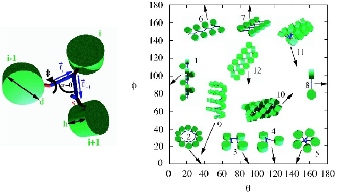
In the present paper we use computer simulations to study the consequence of internucleosome attractive and excluded volume interactions in dense crossed-linker fibers. Our chromatin fiber model is depicted on the lhs of Fig. 1. We model the protein spools as ellipsoidal disks and assume that linker histones induce the in- and outcoming DNA to form stem-like structures Bednar_pnas_98 . Our opening and rotation angles and are compatible with experimentally observed values for the 30 nm fibers Bednar_pnas_98 ; Widom_pnas_92 (see the fiber “10” on the rhs of Fig. 1). As a consequence, the local fiber geometry remains basically unchanged when we vary the strength of the attractive interaction. Local condensation-decondensation transitions in chromatin fibers Schiessel_bpj_01 ; Helmut_03 will be investigated in a forthcoming study everaers_prep .
With respect to the degree of coarse-graining of the nucleosome structure our model is comparable to the one employed by Katritch et al. Katritch_jmb_00 . The main difference is their use of quenched, randomly distributed -angles along the chain. Since we are interested in rather generic features of the dense 30 nm fiber with a narrow distribution of linker lengths (and hence twist angles) Widom_pnas_92 , we focus on the ideal case of a constant -angle for all linkers. Furthermore we follow Wedemann and Langowski Wedemann_bpj_02 in modelling the nucleosomes as ellipsoids (as opposed to spheres as in Ref.Katritch_jmb_00 ) interacting via a Gay-Berne potential. However, we have not included Debye-Hückel interactions between different parts of the linker DNA Wedemann_bpj_02 , since the Debye-Hückel screening length is smaller than the diameter of the DNA double helix at physiological salt concentrations. As a consequence, intra and inter-linker electrostatic interactions can be accounted for by renormalized bending rigidities Barrat_acp_96 and opening angles Schiessel_el_02 respectively.
Problematic for all attempts to model chromatin are the soft parts of the nucleosome. The charges on the linker histones H1/H5 and the histone tails are under biochemical control, allowing the cell to regulate the stem formation and the attractive interactions between nucleosomes Holde_pnas_96 ; Mangenot_bpj_02 . While simulations investigating this ”tail-bridging” effect Mangenot_epje_02 ; Podgornik_jcp_03 between nucleosomes are on the way muhlbacher_03 , we prefer a generic nucleosome interaction potential to a model which neglects the tails but accounts in detail for the surface charges on the nucleosome core Beard_structure_01 . Similarly, little is known about the stem structure (see the cryo micrographs in Ref. Bednar_pnas_98 ) and for simplicity we have assumed that the short axis of the nucleosomal disk (the axis of the superhelical wrapping path) is oriented perpendicular to the plane defined by the in- and outgoing DNA linker.
The paper is organized as follows. Section II introduces our chromatin model and the methods used. We present our results for the mechanical fiber properties in section III. Section IV provides a discussions of the observed hairpin structures and possible biological implications of our findings. Finally we give a conclusion in Section V.
II Model and Methods
II.1 Definition of the chromatin model
Our chromatin fiber model is depicted on the lhs of Fig. 1. We model the protein spools as ellipsoidal disks with a diameter of 10 nm and a height of 6 nm corresponding to the experimental values Livolant_bpj_97 ; Luger_nat_97 . We assume that the in- and outcoming DNA are glued together in rigid stem-like structures that mimic the nucleosomal structures observed in the electron cryo micrographs in the presence of linker histones Bednar_pnas_98 . The stem portion is assumed to end 7 nm from the center of the corresponding protein spool. From this point on the entering and exiting linker DNA portions of length connect to the next nucleosomes.
The presence of a stem has to be taken into account when one calculates the length of the free linker DNA from the nucleosomal DNA repeat length, a number that varies not only from organism to organism but even from tissue to tissue of the same organism Holde_89 . It is known that 147 base pairs (bp) are closely associated with the protein spool wrapping the histone core in 1.65 turns Luger_nat_97 . We assume here that the presence of the linker histone forces the DNA to wrap two full turns corresponding to 177 bp. Together with the 2 nm stem this makes 189 bp that are associated with the core and linker histones. For instance, in the case of chicken erythrocyte chromatin the nucleosomal repeat length amounts to about 210 bp Holde_89 so that there are roughly 21 bp, i.e. nm, of free linker DNA. In the following simulation runs we will always use this value for .
There are two angles that determine the fiber geometry: the deflection angle and the rotational angle . The former angle characterizes the entry/exit angle of the linker DNA at the stem, the latter angle describes the rotational setting of the nucleosomal disks with respect to the DNA. Some example configurations are depicted on the rhs of Fig. 1 with the arrows pointing to their location in the -space. Note that some configurations are forbidden since they would lead to overlapping nucleosomes; a systematic investigation of the boundary between allowed and forbidden structures will be provided in a forthcoming study everaers_prep .
In the present study we use a canonical value of which has been estimated experimentally Bednar_pnas_98 at 80 mM salt concentration. Having fixed and we have chosen values of that lead to a reasonable nucleosome line density. Based on Eq. (78) in Ref. Helmut_03 we expect 6.5 nucleosomes per 11 nm for and 6.1 nucleosomes per 11 nm for compared to experimental estimates of 6 - 7 nucleosomes per 11 nm Gerchman_pnas_87 ; Bednar_pnas_98 .
The only non-rigid elements in our model are the linker DNA portions. We discretize the linkers into four segments in order to allow for the bending and torsional deformations. To each segment we attach a local set of basis vectors where denotes the tangent vector, the normal, and the binormal vector. The elastic energy of the linkers is then described by
| (1) |
where and are the DNA bending and twist persistence lengths respectively, is the number of nucleosomes in our fiber and denotes the segment length (i.e. ). refers to the bending angle between two neighboring segments and denotes the twist angle. The spontaneous bending angle is equal to for those segment pairs which are connected to a stem and is zero otherwise. everywhere (except at the kink where ) enforces the right-handed helicity of the DNA which in turn gives rise for the fiber twist angle .
The position and orientation of the nucleosomes is calculated from the linker positions with the help of a set of three orthonormal basis vectors . is the normal vector perpendicular to the disk plane that we assume to be given by
| (2) |
Here the set denotes the vectors that connect the stems of neighboring nucleosomes (see Fig. 1). points from the tip of the stem towards the center of the nucleosomal disk, i.e.
| (3) |
Finally, is defined as
| (4) |
For the interactions of the nucleosome core particles we use the same variant of the Gay-Berne (GB) potential for ellipsoids of arbitrary shape Berardi_cpl_98 ; ralf_GB as in a recent study of a stacked-ellipsoid model of DNA mergell_pre_03 . Here we set the structure matrix (Eq. (15) in Ref. mergell_pre_03 ) equal to
| (5) |
where is the diameter of the nucleosome core particle with a canonical value for the core particle height of nm Luger_nat_97 . The effective distance of closest approach between two ellipsoids is calculated using Eqs. (17)-(19) in Ref. mergell_pre_03 . In cases where we study purely repulsive hard core interactions, we reject all Monte Carlo moves leading to values . In cases where we study attractive interactions, the distance dependent part of the interaction potential Eq. (14) in Ref. mergell_pre_03 is given by a modified Lennard-Jones potential
| (6) |
The orientation dependent parts are defined in Eqs. (20)-(25) in Ref. mergell_pre_03 . This parameterization of the GB potential leads to a preferred lateral spacing of about and a vertical spacing of about of the disks. The values are in good agreement with experimental results for columnar phases of nucleosome core particles Livolant_bpj_97 . In this paper we characterize the strength of the nucleosome-nucleosome attraction by the depth of the attractive well for two parallel disks instead of the prefactor in Eq. (6).
II.2 Methods
We use a Monte-Carlo scheme to simulate the chromatin fiber which relies on the following three moves: (i) a local move where one randomly chooses a nucleosome that is rotated around an axis determined by two points on the in- and out-coming linker DNA by a small random angle, (ii) a non-local pivot move where a random segment point is chosen at which the shorter part of the chain is rotated around a random axis by a random angle and (iii) a non-local crankshaft move where two random points along the DNA segments define the axis of rotation around which the inner part of the chain is rotated. The moves are accepted or rejected according to the Metropolis scheme Metropolis_jcp_53 .
We simulated either fibers with (simulations with applied stretching force) or nucleosomes corresponding to 10 and 20 kbp of DNA respectively. The number of nucleosomes in pulling experiments by Cui and Bustamante Cui_pnas_00 is on the same order ( nucleosomes). Each simulation run consists of 200000 MC sweeps, where one sweep corresponds to trials with being the number of DNA segments. The amplitudes are chosen such that the acceptance rate equals approximately 50%. Every 20 sweeps we save a configuration. As initial conformation we used the relaxed () fiber structure. In order to determine the longest relaxation time of the system, we measured the ’time’ correlation functions of the energy, the mass density, the end-to-end distance and the twist. We typically find that . Note, however, that strong attractive interactions can cause a glasslike trapping of the fiber in local energy minima. As a consequence, only a limited range of attractive well depths can be studied meaningfully in simulations. In the present case, the simulation runs for were clearly non-ergodic for low stretching forces. In this case we averaged the results over 10 independent simulation runs.
The calculation of the elastic constants of the simulated fiber is performed as follows. First we determine the fiber axis by calculating the centers of mass of groups of neighboring nucleosomes. is chosen to match approximately one or two helical turns. Then we calculate the autocorrelation function of the tangent vectors of the fiber axis
| (7) |
and extract the persistence length of the fiber from an exponential fit
| (8) |
The stretching modulus can be estimated via
| (9) |
with being the mean deviation from the average contour length of the fiber (defined as the length of the fiber axis ). It should be noted that depending on the estimated values of the contour length, the persistence length and the stretching modulus vary. In case of being too large the stretching modulus is underestimated since bending fluctuations within and are averaged out so that the contour length of the fiber appears to be smaller. On the other hand, values of which are too small lead to a helicoidal fiber axis, and the contour length of the fiber is overestimated. This entails a systematic error which must be minimized. We found that values of corresponding to one or two helical turns lead to reasonable estimates.
III Results
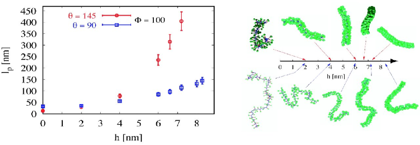
In order to quantify the influence of the nucleosome excluded volume on the fiber stiffness we performed a series of simulations with purely repulsive interactions between the nucleosome core particles, constant linker geometry and variable core particle size. The lhs of Fig. 2 shows our results for the persistence length for two fiber geometries; the corresponding snapshots in the rhs of the figure illustrate the observed fiber stiffening with increasing core particle size.
To be more specific, we studied two different sets of angles, namely and as well as and . In both cases, starting at (i.e., no nucleosomes present) we observe the theoretically expected values for the persistence length of the linker backbone (Eq. (97) in Ref. Helmut_03 ), namely nm and nm, respectively. We verified this formula also for many other values of and (data not shown). However, with increasing the value of increases and reaches values of nm and nm, respectively, at the canonical value of nm. This corresponds to a 17-fold and 3-fold increase respectively of the fiber stiffness relative to the theoretical prediction. The different degrees of stiffness (and stiffening) directly reflect the different nucleosome densities in the two cases: in the -fiber the nucleosomes are always quite close to each other whereas the -fiber is still much more open.
We also performed simulations of stretched fibers with and . The resulting force-elongation curves (red symbols in Fig. 3) and histograms of the end-to-end distance distribution (red symbols in Fig. 4) show the typical behavior of extensible worm-like chains. The entropic small force regime is strongly suppressed, since the contour length, nm, of fibers containing 50 nucleosomes is much smaller than their persistence length of nm. The effective stretching modulus nm-1 at finite extensions is smaller than the value nm-1 deduced from the analysis of the length fluctuations (see Eq. 9) and larger than the the theoretical prediction nm-1 which is based on the linker mechanics alone.
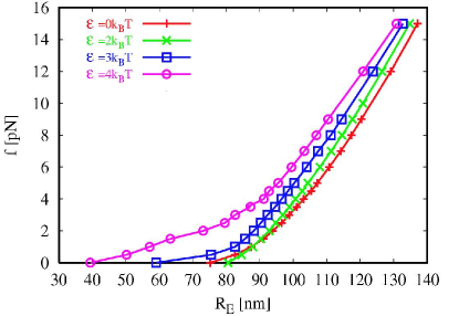
For our study of the effect of attractive interactions between core particles we focused on linker backbone geometries which by themselves already lead to relatively dense fibers ( and ). As a consequence, the local fiber geometry remains basically unchanged when we vary the strength of the attractive interaction from (the case of purely repulsive interactions discussed above) to . For example, the observed attraction induced reductions in the fiber contour lengths are only of the order of 10%.
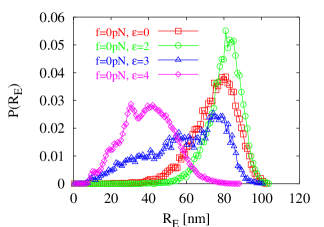
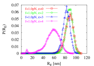
Weak attraction up to (green symbols in Figs. 3 and 4) has only a small effect on the observed fiber properties. While the contour length decreases to about nm, there is a corresponding small increase in both the fiber stiffness and its stretching modulus, yielding an overall small increase in the end-to-end distance. The situation changes dramatically for larger values of the attractive well depth. In the absence of external forces fibers with and, in particular, with (blue/magenta symbols in Figs. 3 and 4) have considerably smaller and more broadly distributed end-to-end distances . For small forces the fiber shows a quasi-plateau in the force-extension curve.
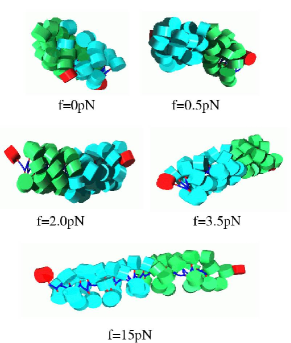
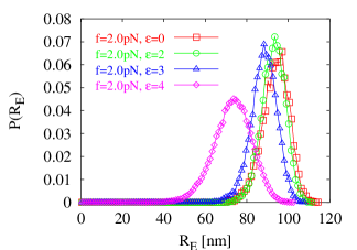
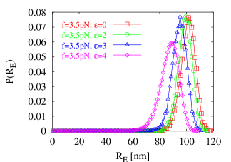
The origin of this peculiar behavior can be identified by inspecting snapshots of the fiber (Fig. 3): At pN the fiber forms a hairpin structure where the end nucleosomes (in red) are located at the same end of the structure. The hairpin persists at pN but opens up into a straight configuration around pN. For larger forces all observed force elongation curves show qualitatively the same WLC behavior.
IV Discussion
Our results show that the properties of dense chromatin fibers are dominated by the interactions between the nucleosome core particles, while the mechanical properties of the linker backbone play a less important role. The dominant effect of excluded volume interactions is to stiffen the fiber. The persistence lengths of the order of 200 - 250 nm observed in the present study are in good agreement with the values obtained by Langowski et al. Munkel_pre_98 ; Wedemann_bpj_02 . The surprising result of our simulations is that attractive interactions of a few per nucleosome pair are sufficient to bend fibers with a contour length of about of the persistence length into dense hairpin configurations. Judging from Fig. 5 in Ref. Katritch_jmb_00 hairpins also occurred in the simulations by Katritch et al. – even though the authors did not discuss this issue in their paper. Since the observed force-elongation curves show qualitatively similar features in all three cases, it is tempting to speculate that similar effects also occurred in the stretching experiment by Cui and Bustamante Cui_pnas_00 . In the following, we will discuss the condensation of semi-flexible filaments, the local structure of the hairpins observed in our simulations, the signature of hairpin opening in stretching experiments, and possible implications of our observations for the folding of chromatin into chromosomes.
IV.1 Condensation of semi-flexible filaments
The condensation of semi-flexible filaments due to short-range attractive interactions has recently been treated in detail by Schnurr et al. Schnurr_pre_02 . They introduced a condensation length (: attraction energy per length) by balancing expressions for typical bending and surface energies, . Filaments with a contour length remain extended, while longer chains aggregate into structures with a typical size of the order of . So-called “racquet” states consist of a straight stem where the chain is folded back on itself several times and where U-turns are shaped like the head of a tennis racquet. A racquet state with one turning point resembles our hairpin structure. Longer fibers lower their energy by having multiple turning points. For infinitely thin filaments Schnurr et al. were able to show that the kinetically preferred racquet states Schnurr_epl_00 have slightly higher energies than toroidal structures.
In the present case, we measured a typical attraction energy per length of for hairpin conformations of the -fiber in good agreement with an estimate of for the attractive energy per nucleosome pair of height . Using this estimate and a persistence length nm, chromatin fibers with weak attractive interactions should start to condense, if they are longer than about 400 nm. Note that the above argument breaks down for , when the predicted racquet head or torus radii become smaller than the radius of the 30 nm-fiber.
Can the structure of the hairpins we observe really be understood in the framework of the WLC model? Or does one have to consider the relatively sharp turning points with the curvature radii of the order of the fiber radius as localized kink defects? Following the analysis of Schnurr et al. Schnurr_pre_02 , the fibers in our simulations are too short to fold back on themselves. The WLC estimate of the bending energy for a U-turn with radius 15nm is , while the onset of hairpin formation at in 80 nm long fibers suggest that the actual energy penalty for the kinks is as small as . We note that the combination of finite fiber radii and relatively small kink energies should stabilize racquet structures relative to toroids.
IV.2 Hairpin structure
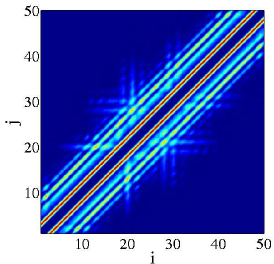
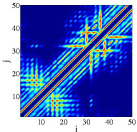
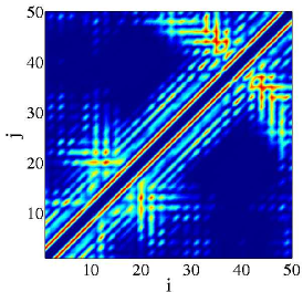
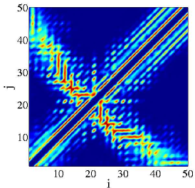
The internal structure of hairpin configurations shows up very clearly in the contact matrix of the fiber that we depict in Fig. 5. For a given configuration the contact matrix is defined as follows: If a pair of nucleosomes and is in contact (center-to-center distance smaller than ) one has , otherwise . By taking the average of the matrix elements for many configurations of a given simulation run we obtain two-dimensional histograms as it is shown in Fig. 5. For large forces no kinks are present and the nucleosomes of the fiber form a quite regular crossed-linker structure. We find two pronounced diagonals at the positions that correspond to ”short-ranged” excluded volume interaction. Less pronounced side stripes parallel to the main diagonal are also observed and describe interactions between nucleosomes of neighboring turns, especially at and at . Hairpins manifest themselves by crosslike patterns in the contact matrices with branches along the secondary diagonal. The location of the kink corresponds to the point where the two diagonals cross each other. Closer inspection of the histograms shows that the hairpin arms outside the kink region are partially interdigitated but not strongly perturbed. However the effect is strong enough to effectively prevent any possibility of sliding of the two arms with respect to each other on the time scale of our simulations. Therefore a hairpin structure, once formed, can be very stable with a frozen in position of the turning point. With respect to the fiber structure at the turning point, Fig. 5(d) in particular shows disruptions of the internal order of the nucleosomes: The diagonals are broken at that position, presumably due to broken contacts of nucleosomes located at the outside of the turning point. This observation supports the picture of a localized kink defect.
IV.3 Large scale vs. local decondensation in stretching experiments
Toroidal or racquet-like condensates become unstable under the influence of a stretching force, if the applied force exceeds a value on the order of the attractive energy per unit length, . Neglecting kinetic barriers (for a discussion of DNA unwrapping from nucleosomes or toroidal aggregates see kulic_04 ), the corresponding force plateau in the force-extension curves for chromatin fibers should be observed for forces of pN. In order to experimentally distinguish this scenario from local structural changes which are expected to occur at similar force levels Schiessel_bpj_01 , one should plot force-relative extension curves for fibers reconstituted under similar conditions on DNA strands of different length. In the case of local structural changes, the obtained curves coincide, while for a global decondensation the size of the unperturbed state is of order independently of the fiber length.
IV.4 Biological implications
As a final point we discuss possible biological implications of fiber stiffening and hairpin formation. Both effects are associated with characteristic length scales; fiber stiffening with a persistence length on the order of 300 nm, hairpin formation with a condensation length on the order of a few tens of nm.
We note again that the biochemical control of the effective interaction between nucleosomes is essential for the condensation and decondensation of chromosomes during the cell cycle Horn_sci_02 . There is indirect experimental evidence for the formation of chromosomal DNA loops having a size of about 50 kilobases CalladineDrew99 (corresponding to about 250 nucleosomes), e.g. from the comparison of separation patterns of excised large DNA fragments by pulsed field electrophoresis with the patterns obtained by DNA cleavage by topoisomerase II of the nuclear matrix of the cell Razin_jbc_95 ; Razin_pnas_95 . Using a nucleosome density of 6/(11 nm), 250 nucleosomes correspond to a fiber length of 460 nm, i.e. about twice the persistence length of 240 nm found in our simulations.
There are two scenarios linking the persistence length to the loop size; both are based on the observation that the free energy cost for the formation of a loop is smallest for semi-flexible chains with a length of about twice the persistence length. For shorter chains the bending energy is high, for larger chains the entropy of chain conformations counteracts loops formation. In the first (equilibrium) scenario, the chromatin fiber loops around strongly attracting organizing centers. Spherical centers could induce the formation of rosette structures Schiessel_epl_00 . Filamentous organizing centers could lead to structures resembling cartoons of chromosomes often found in biological textbooks, cf. e.g. Ref. cell . The second scenario which links the persistence length to the loop size is closer to what we observed in our simulations: if the attraction between fibers is strong enough to prevent structural reorganization after the two halves of the fiber have tightly closed the gap between the point of first contact and the hairpin defect, then loops with the typical size of the fiber persistence length are preferred for kinetic reasons. Hairpins have indeed been observed in cryo-EM pictures of chromatin fibers in the presence of MENT Grigoryev_jbc_99 , a protein that is involved in the formation of dense, transcriptionally inert sections of chromatin, the so-called heterochromatin.
If, on the other hand, structural reorganization is still possible in collapsed fibers, then the size of the aggregates should be controlled by the smaller condensation length and not by the persistence length of the 30-nm fiber. Fibers with diameters ranging between 60 to 130 nm diameter have indeed been observed in mitotic and in G1 chromosomes and are called chromonema fibers Belmont_jcb_94 , cf. also Ref. Horn_sci_02 . However, the folding of 30-nm fibers into superfibers with a diameter on the order of the condensation length requires a mechanism that prevents the formation of globular aggregates.
V Conclusion
We have used computer simulations of model chromatin fibers to investigate the influence of excluded volume and attractive interactions between the nucleosomes on the large-scale structure and elasticity of the 30 nm chromatin fiber. Our results shed light (i) on the discrepancy between the theoretically expected and the observed persistence length of 200-300 nm as well as (ii) on the somewhat counterintuitive observation that such fibers nevertheless seem to be able to curl up into chromonema fibers with diameters of 60 - 130 nm.
Our results clearly show that the stiffness of dense fibers is dominated by excluded volume interactions between nucleosomes. The observed persistence lengths exceed estimates based on the linker backbone elasticity by one order of magnitude. With respect to internucleosomal attraction we have concentrated on generic, non-local aspects of fiber condensation. The 2 pN force plateau observed in our simulations reflects a structural feature beyond the 30 nm fiber: the opening of a hairpin. Our results suggest that aligned (anti-) parallel fibers are only weakly perturbed, but that the fiber geometry is locally disrupted at the turning point. The crucial point is that the energetic cost of this localized defect is considerably smaller than for a smoothly bending semi-flexible filament with the same bending persistence length as chromatin. This effect could be important for an understanding of chromatin folding in chromosomes.
VI Acknowledgments
We thank K. Kremer, I. M. Kulic, J. Langowski and J. Widom for valuable discussions, M. R. Ejtehadi for providing parts of the simulation code and a referee for useful comments on how to structure the material presented in a first draft of the present manuscript. BM and RE gratefully acknowledge financial support from an Emmy-Noether grant of the DFG.
References
- (1) K. E. van Holde, Chromatin, Springer, New York, 1989.
- (2) F. Thoma, T. Koller, and A. Klug, J. Cell. Biol. 83, 404 (1979).
- (3) J. Widom, J. Mol. Biol. 190, 411 (1986).
- (4) J. Bednar et al., Proc. Natl. Acad. Sci. USA 95, 14173 (1998).
- (5) K. Luger, A. W. Mader, R. K. Richmond, D. F. Sargent, and T. J. Richmond, Nature 389, 251 (1997).
- (6) J. Widom, Annu. Rev. Biophys. Chem. 18, 365 (1989).
- (7) K. V. Holde and J. Zlatanova, J. Biol. Chem. 270, 8373 (1995).
- (8) K. V. Holde and J. Zlatanova, Proc. Natl. Acad. Sci. USA 93, 10548 (1996).
- (9) J. Langowski and H. Schiessel, New Comprehensive Biochemistry , in press.
- (10) J. T. Finch and A. Klug, Proc. Natl. Acad. Sci. USA 73, 1897 (1976).
- (11) J. Widom and A. Klug, Annu. Rev. Biophys. Chem. 43, 207 (1985).
- (12) C. L. Woodcock, S. A. Grigoryev, R. A. Horowitz, and N. Whitaker, Proc. Natl. Acad. Sci. USA 90, 9021 (1993).
- (13) H. Schiessel, W. M. Gelbart, and R. Bruinsma, Biophys. J. 80, 1940 (2001).
- (14) J. Bednar, R. A. Horowitz, J. Dubochet, and C. L. Woodcock, J. Cell. Biol. 131, 1365 (1998).
- (15) S. H. Leuba et al., Proc. Natl. Acad. Sci. USA 91, 11621 (1994).
- (16) J. Zlatanova, S. H. Leuba, and K. van Holde, Biophys. J. 74, 2554 (1998).
- (17) S. E. Gerchman and V. Ramakrishhnan, Proc. Natl. Acad. Sci. USA 84, 7802 (1987).
- (18) Y. Cui and C. Bustamante, Proc. Natl. Acad. Sci. USA 97, 127 (2000).
- (19) J. Zlatanova and S. H. Leuba, J. Mol. Biol. 331, 1 (2003).
- (20) V. Katritch, C. Bustamante, and W. K. Olson, J. Mol. Biol. 295, 29 (2000).
- (21) E. Ben-Haim, A. Lesne, and J.-M. Victor, Phys. Rev. E 64, 051921 (2001).
- (22) E. Ben-Haim, A. Lesne, and J.-M. Victor, Physica A 314, 592 (2002).
- (23) H. Schiessel, J. Phys.: Condens. Matter 15, R699 (2003).
- (24) K. van Holde and J. Zlatanova, J. Mol. Biol. 93, 8373 (1995).
- (25) J. Yao, P. T. Lowary, and J. Widom, Proc. Natl. Acad. Sci. USA 87, 7603 (1990).
- (26) P. J. G. Butler and J. O. Thomas, J. Mol. Biol. 281, 401 (1998).
- (27) C. Münkel and J. Langowski, Phys. Rev. E 57, 5888 (1998).
- (28) G. Wedemann and J. Langowski, Biophys. J. 82, 2847 (2002).
- (29) C. Bustamante, S. B. Smith, J. Liphardt, and D. Smith, Current Opinion in Structural Biology 10, 279 (2000).
- (30) J. Widom, Proc. Natl. Acad. Sci. USA 89, 1095 (1992).
- (31) R. Everaers and H. Schiessel, in preparation.
- (32) J.-L. Barrat and J.-F. Joanny, Adv. Chem. Phys. 94, 1 (1996).
- (33) H. Schiessel, Europhys. Lett. 58, 140 (2002).
- (34) S. Mangenot, A. Leforestier, P. Vachette, D. Durand, and F. Livolant, Biophys. J. 82, 345 (2002).
- (35) S. Mangenot, E. Raspaud, C. Tribet, L. Belloni, and F. Livolant, Eur. Phys. J. E 7, 221 (2002).
- (36) R. Podgornik, J. Chem. Phys. 118, 11286 (2003).
- (37) F. Mühlbacher, C. Holm, and H. Schiessel, in preparation.
- (38) D. A. Beard and T. Schlick, Structure 9, 105 (2001).
- (39) A. Leforestier and F. Livolant, Biophys. J. 73, 1771 (1997).
- (40) R. Berardi, C. Fava, and C. Zannoni, Chem. Phys. Lett. 297, 8 (1998).
- (41) R. Everaers and M. R. Ejtehadi, Phys. Rev. E 67, 041710 (2003).
- (42) B. Mergell, M. R. Ejtehadi, and R. Everaers, Phys. Rev. E 68, 021911 (2003).
- (43) N. Metropolis, A. W. Rosenbluth, M. N. Rosenbluth, A. N. Teller, and E. Teller, J. Chem. Phys. 21, 1087 (1953).
- (44) B. Schnurr, F. Gittes, and F. C. MacKintosh, Phys. Rev. E 65, 061904 (2002).
- (45) B. Schnurr, F. C. MacKintosh, and D. R. M. Williams, Europhys. Lett. 51, 279 (2000).
- (46) I. M. Kulic and H. Schiessel, Phys. Rev. Lett. , in press.
- (47) P. J. Horn and C. L. Peterson, Science 297, 1824 (2002).
- (48) C. R. Calladine and H. R. Drew, Understanding DNA: The molecule and how it works, Academic Press, 1999.
- (49) M. A. Lagarkova, O. V. Iarovaia, and S. V. Razin, J. Biol. Chem. 270, 20239 (1995).
- (50) I. I. Gromova, B. Thomsen, and S. V. Razin, Proc. Natl. Acad. Sci. USA 92, 102 (1995).
- (51) H. Schiessel, J. Rudnick, R. Bruinsma, and W. M. Gelbart, Europhys. Lett. 51, 237 (2000).
- (52) B. Alberts et al., Molecular Biology of the Cell, Garland Publishing, Inc., New York and London, 1994.
- (53) S. A. Grigoryev, J. Bednar, and C. L. Woodcock, J. Biol. Chem. 274, 5626 (1999).
- (54) A. S. Belmont and K. Bruce, J. Cell. Biol. 127, 287 (1994).