Force steps during viral DNA packaging ?
Prashant K. Purohit∗, Jané Kondev†, and Rob Phillips∗
∗Division of Engineering and Applied Science, California
Institute of Technology, Pasadena, CA 91125.
†Physics Department, Brandeis University, Waltham, MA 02454.
Abstract
Biophysicists and structural biologists increasingly acknowledge the role played by the mechanical properties of macromolecules as a critical element in many biological processes. This change has been brought about, in part, by the advent of single molecule biophysics techniques that have made it possible to exert piconewton forces on key macromolecules and observe their deformations at nanometer length scales, as well as to observe the mechanical action of macromolecules such as molecular motors. This has opened up immense possibilities for a new generation of mechanical investigations that will respond to such measurements in an attempt to develop a coherent theory for the mechanical behavior of macromolecules under conditions where thermal and chemical effects are on an equal footing with deterministic forces. This paper presents an application of the principles of mechanics to the problem of DNA packaging, one of the key events in the life cycle of bacterial viruses with special reference to the nature of the internal forces that are built up during the DNA packaging process.
1 Introduction
Mechanics has a long and rich tradition of reaching out to other fields. For example, the mechanical analysis of thin films has revealed insights into applications ranging from microelectronics to MEMS (see Freund and Suresh (2003)). Mechanics arguments have demonstrated that dislocations and surface instabilities play a key role in determining the mechanical and electrical properties of such films. While the study of thin films is a relatively new area to have been influenced by mechanics there are others, like geology and metallurgy, that have drawn on mechanics for decades. The most recent developments in intersonic crack propagation (Rosakis (2002), Rice (2001)) have direct analogues in the study of motion along geological fault lines. Geophysicists have relied heavily on data emerging from shock compression of solids to model the thermomechanical behavior of planets (see Meyers (1994)). In addition, the impact of dislocation mechanics, phase transformations and fracture mechanics on the science of metallurgy can scarcely be overestimated.
Another field that promises to be visited by mechanicians with increasing regularity is biology, though the use of mechanical principles for both fluids and solids in understanding biological processes is not at all new. For example, slender body theory from fluid mechanics has been fruitfully applied to the study of motion in bacteria and other microorganisms (see Bray (2001)). In addition, variational principles for determining optimal shapes have been used for the study of red-blood cells (see Boal (2002)). Evans and Skalak (1980) show how lipid bilayer vesicles can be modeled as shells and membranes with no shear resistance. The flagella of bacteria have been idealized as rods in an effort to study how their rotation can give rise to propulsion (Goldstein et al. (1998)). The cytoskeletal framework of the cell is studied as a three dimensional network of elements to obtain estimates of the response of cells to external forces (see Boal (2002)). In this paper we restrict our attention to problems in biological nanomechanics with the observation that there are a range of fascinating connections between mechanics and biology at larger scales as well. One of the most intriguiging features of problems in biological nanomechanics is that they involve a rich interplay of statistical and deterministic forces as will be seen below.
It is only recently, however, that structural biologists have begun to understand the significance of mechanical properties for the macromolecular processes that sustain life. The role of bending stiffness of DNA in gene regulation (Widom (2001)) and the stiffness of substrate in cell migration (Lo et al. (2000)) are two examples. Such insights have been garnered with the aid of sophisticated experiments that have made it possible to apply piconewton forces on nanometer size objects and measure the resulting deformations. The data from these experiments point to a rich interplay of thermal and chemical forces with electrical and mechanical forces. In this paper we show how experimental insights can be combined with mechanical principles to construct a simple and quantitative mechanical theory of DNA packaging in bacterial viruses that suggests a new round of experiments.
2 Mechanics and the Viral Life Cycle
Viruses have been studied extensively (see Alberts et al. (1997)) in the last three decades not just with the goal of understanding their pathogenic nature but also for gaining insights into structural biology of proteins and nucleic acids and for studying gene regulation. Even though viruses are perhaps the simplest entities from a biological perspective, they are nanotechnological marvels that embody a wealth of physics at the nanometer scale. The principles operating at these scales are being rapidly unravelled through ingenious experiments, such as that by Smith et al. (2001) on DNA packaging in the 29 virus, which serves as the primary motivation for the theoretical analysis presented below.
Before going into the details of the experiment it is useful to get a glimpse of the life-cycle of a virus (see fig. 1). We are especially interested in the class of viruses known as bacteriophage and which infect bacteria such as the well-known E.coli. In simplest terms, a bacteriophage is nothing but a protein coat, the capsid, filled with nucleic acid such as DNA or RNA. The capsids of bacteriophage attach to the surface of the bacterial cell which is under attack. The genetic material of the virus is ejected into the bacterium leaving the capsid behind. Though the process of DNA ejection is not completely understood, it is argued that a mature bacteriophage capsid is highly pressurised (see Smith et al. (2001)) and the genome is forcefully released into the host cell whose contents are effectively at a much lower pressure.
Once the viral DNA is inside the bacterium it hijacks the protein production machinery of the bacterium to synthesize its own proteins such as those that will ultimately make up the capsid. Copies of the viral DNA are also made. Here again, experiments have shed light on the mechanics and kinetics of the processes of transcription of the genome and its translation into proteins. The process of transcription is mediated by a large protein called RNA polymerase. This protein attaches to the DNA and then moves along its length transcribing the DNA into a molecule known as mRNA. As it moves along, the protein exerts forces on the DNA molecule. These forces have been measured experimentally (Wang et al. (1998)) and it has been found that the protein can be stalled by exerting a force of roughly 25pN. In fact, the rate of transcription is heavily dependent on the force exerted. The type of data emerging from such experiments unmistakably points to a plethora of mechanics problems still to be resolved at the level of macromolecular assemblies.
After a sufficient quantity of the viral proteins have been synthesized they begin to self-assemble into hollow capsids. In the case of the 29 virus, the capsids have a portal at one end with an attached protein motor. The motor proteins identify the appropriate end of the as-yet unpackaged viral DNA and push it into the capsid. As more of the DNA gets pushed into the capsid the motor has to perform work against an increasing resistive force due to confinement of the packaged DNA. This causes the motor to slow down as more DNA fills up, but ultimately all the DNA is packed and the remaining proteins attach themselves to the capsid thus making it ready to infect another bacterium. Once the capsids are fully assembled an enzyme is released that breaks up the bacterial cell membrane and releases the mature viruses so that they can repeat their evil action elsewhere (see Ptashne (1992)).
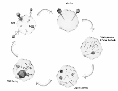
As noted above, a critical step in the life cycle (see fig. 1) of the virus is the packaging of DNA into the capsid by the portal motor. This step was the focus of the experiment by Smith et al. (2001) in which they used optical tweezers to measure the force exerted by the portal motor of the 29 bacteriophage on the DNA as a function of the amount of DNA already packed in the capsid. One end of the DNA was attached to a silica bead which was held in an optical trap while the motor tugged on the other end. The capsid itself was attached to a second silica bead that was held by a glass pipette thus immobilizing the capsid during the packaging process. Through a series of careful experiments, the average force exerted by the portal motor was measured and plotted against the fraction of packed DNA. The rate at which the DNA is packed was also measured as a function of the fraction packed. It was found that during the early stages of packing the motor packs at a rate of roughly 100 base pairs per second which correponds to nm per second. However, the rate drops considerably after about % of the DNA has been packed and it slows to a crawl as the packaging nears completion. We note that the experiment was conducted in vitro in a solution which contained mM NaCl, mM MgCl2 and tris-HCl buffer at pH 7.8. These are not the conditions prevailing in a bacterium and one of the interesting outcomes of the type of model being described here is the recognition that the ionic strength is a key control parameter in governing the physics of DNA packaging. There are a variety of interesting mechanics issues to be confronted relating to those stages of the viral life-cycle involving both DNA packaging and ejection. One such issue involves determining what is the maximum sustainable pressure in a viral capsid (Purohit et al. (2003)). Another concerns determining the amount of DNA ejected by a bacteriophage as a function of the osmotic pressure in the exterior solution (Evilevitch et al. (2003)).
This paper puts forth a simple mechanical model of the packaging process in an endeavour to understand the nature of the forces being exerted on the DNA. Previously, we described results of an analysis based on a continuum approximation to the mechanical model of the packaging force (Purohit et al. (2003)). Here we analyze a discrete version of that same model which reveals interesting new effects which could be accessible experimentally.
3 The Free Energy Function for Packed DNA
In order to estimate the energetics of viral DNA packing, we take a structural hint from the cryo-electron microscopy images of capsids (see, for example, Cerritelli et al. (1997)) in various stages of the packing process. It has been observed for several different viruses that the packed DNA is arranged in a series of circular hoops (or a spool). Each hoop finds itself at the center of a hexagon formed by nearly parallel segments of the same overall strand. The exception are the hoops hugging the surface of the capsid, or those at the inner surfaces of the spool, which are surrounded on average by three nearest neighbors. We note that the geometry is more subtle than the hoop packing adopted here since the packing involves a helical pitch and hence the adjacent strands are probably not perfectly parallel. Following earlier work (see Riemer and Bloomfield (1978), Odijk (1998) and Kindt et al. (2001)), the basic idea is to write a free energy function which characterizes the energetics of structures like those described above as a sum of elastic terms and interaction terms. It is well known that DNA has a considerable bending stiffness at length scales of a few tens of nanometers. This is evident from the fact that the persistence length of DNA is about nm. The persistence length is defined as the distance over which the tangents to a fiber are correlated. The more flexible a fiber is the smaller its persistence length. Polymer physicists use the persistence length as a measure of the stiffness of a chain (see Doi and Edwards (1988)). Since the
persistence length of DNA is of the same order as the size of viral capsids we expect the bending energy to play an important part in the packaging process. Such a description of the elasticity of DNA is now quite firmly established in literature and is found to be useful in understanding key biological processes related to genetic regulation and DNA condensation in chromosomes of eucaryotic cells.
Beyond its explicit mechanical properties another relevant characteristic of DNA is its strong acidity. In particular, in an aqueous solution the backbone of the molecule is highly charged, with two units of negative charge per base pair. As a result there are electrostatic penalties involved in bringing two strands in close proximity. In the salty solutions found in cells the picture is more complicated because charged ions screen these interactions. Another complication arises from the fact that close packing involves the ejection of water molecules from the vicinity of the strands, which make sizable contributions to the free energy. All of these complex interactions of the DNA with the ambient solution and with itself result in an effective interaction energy. The importance of these effects is immediately evident when we look at the physics of DNA condensation in a solutions containing trivalent and tetravalent cations. In particular, DNA is known to form hexagonally packed toroidal structures when immersed in a solution containing these ions (Raspaud et al. (1998)). The aim of the calculations to follow is to model viral packing, taking into account both elastic and interaction energies.
3.1 Elastic Contribution to the Free Energy
We begin by idealizing DNA as an elastic rod capable of sustaining bending deformations. The centerline of the rod is parametrized by the reference coordinate . The bending energy stored in length of this rod is given by
| (1) |
where is the radius of curvature at reference position along the rod and is a bending modulus which we assume to be independent of the reference position though we note that the
sequence dependence of the elasticity of DNA is one of its most
intriguing features. For a rod bent into a hoop, the radius of curvature is just the radius of the hoop , and the length is , and so the integral reduces to
| (2) |
The modulus is usually taken to be where is the Young’s modulus of the material and is the moment of inertia of the cross-section. However, it is convenient to express the elastic properties of molecules differently since thermal oscillations play a dominant role in their deformation. In particular, the deformation at a given point on a molecule may be uncorrelated with the deformation at another location because of the randomness caused by these thermal motions. As noted above, the length at which the importance of bending energy is comparable to that due to thermal vibrations is called the ‘bend persistence length’ and is denoted by . More precisely, is determined by balancing the bending energy and the thermal energy and results in , where is the persistence length, is the temperature and is Boltzmann’s constant; nm for DNA and pNnm at K. Using this description of the elastic properties of DNA and adding up the contributions due to all of the hoops in the spool, the total bending energy is given by
| (3) |
where is the number of hoops in a column of radius . Note that we neglect the fact that the DNA is packed in a helical arrangement with an associated contribution to the curvature due to the helical pitch. As noted in our earlier paper (Purohit et al. (2003)), this effect is negligible. For analytical simplicity the discrete expression given above can be converted into an integral and written as
| (4) |
where is the spacing between adjacent hoops, is the radius of the capsid and is the radius of the innermost set of hoops. The factor of appears since the spacing between two adjacent columns of hoops is in a hexagonal array. The integral approximation has the virtue that it leads to closed-form analytical expressions for the energies and forces in the viral packing problem, at least for simple geometries like the cylinder and the sphere. An interesting set of conclusions to be discussed below are the differences between the discrete and continuum descriptions of this problem, and their possible experimental consequences.
3.2 Free Energy of Interaction
In addition to the role played by elastic bending we must also consider the effect of the interaction between adjacent strands of DNA. For this we appeal to the experiments of Parsegian et al. (1986) and Rau et al. (1984). In these experiments, osmotic pressure was applied on hexagonally packed DNA and the interstrand separation was measured as a function of the pressure for a variety of different solvent conditions. For conditions comparable to those of Smith et al. (2001), it was found that the osmotic pressure could be related to the interstrand spacing as
| (5) |
where is a constant whose magnitude depends on the type and strength of the ionic solution, and nm is a decay length which is roughly constant over a range of ionic conditions. For a solution containing mM NaCl at 298K, measurements reveal pN/nm2. To this piece of empirical evidence we add the assumption that parallel strands interact through a pair potential per unit length and that interactions are limited only to nearest neighbors. Given this description of the total energy and the measured pressure vs interstrand spacing, we can deduce the functional form of .
In particular, consider parallel strands of length each packed in a hexagonal array with a spacing . The total interaction energy of this arrangement is
| (6) |
where the factor of appears because each strand interacts with nearest neighbours (ignoring surface effects) and we multiply by to account for double counting. The total volume of the assemblage is obtained by adding together the (prismatic) volume occupied by each strand, thus
| (7) |
We now use the thermodynamic identity to relate the expression for pressure obtained from experiments to our simple model. We note that and , so that
| (8) |
where is the force per unit length on the strands. We now substitute the experimental result, namely and solve the differential equation above with the boundary condition . This results in the potential
| (9) |
If this result is now exploited in the context of eqn. (6) the interaction energy can be written as
| (10) |
where is the total length of the strands.
Now we are in a position to write the total energy of the packaged DNA as a sum of the bending energy and the interaction energy as follows
| (11) |
In the continuum approximation this takes the form
| (12) |
We still need to express the length in terms of the radii of the hoops. To do so, we simply add up the accumulated length of all the hoops as,
| (13) |
In the continuum approximation this can be rewritten as
| (14) |
With both the elastic and interaction contribution to the total energy of packed DNA in hand, we turn now to a concrete investigation of the implications of this model.
4 Geometry and Energetics of Packed DNA
The calculation of the free energy associated with the DNA packed in a partially filled capsid is predicated on key insights gained from experiments. We assume throughout that the DNA adopts an inverse-spool geometry like that described above, with the proviso that the class of minimum free energy structures we consider is constrained to the spool-type and that there are perhaps more complex structures with lower free energy. We also assume that the strands have local six-fold coordination. As a result, the only variable that remains to be determined is the spacing between the strands as a function of the amount of DNA packed. In the regime where the interaction between the DNA strands is repulsive the interaction term dictates that the strands be as far apart as possible. On the other hand, since the outer dimensions of the capsid are fixed, larger spacings imply smaller radii of curvature which results in a steep rise in the bending energy cost. The competition between these two contributions to the free energy determines the geometry of the packed DNA (Reimer and Bloomfield (1978), Odijk (1998), Kindt et al. (2001)). We illustrate this through two examples and also make a comparison with the experimental data of Smith et al. (2001).
For simplicity we begin by considering a cylindrical capsid which is a first approximation to the geometry of the 29 virus considered in the experiments of Smith et al. (2001). In reality, the 29 virus is shaped like a hollow oblong spheroid (Tao et al. (1998) and Wikoff and Johnson (1999)) which has an outer radius of nm and a height of about nm. Its protein coat is about nm thick (on average) so that the radius of the capsid volume available to the DNA is nm while its height is roughly nm. The capsid has other interesting geometrical features, but for the purposes of the present calculation we idealize it as a cylinder. The dimensions of this idealized cylinder are given in fig. 2 where the key point is selecting the height of the cylinder so as to gaurantee that the volume available for packing is identical to that of the real virus. The figure shows a second geometry that will be used later as a more refined model of the 29 capsid.
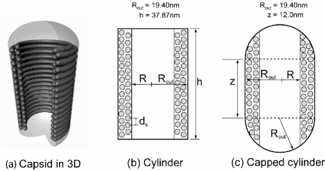
The 29 capsid encloses about m of double-stranded DNA corresponding to a genome of kilobase pairs. As noted above for our cylindrical model we take the height of the cylinder as nm to ensure that the internal volume is the same as that of the actual capsid. For such a capsid, the number of hoops at radius is given by where is the separation between the strands and is the height of the cylinder as shown in fig. 2. One of the key themes of the present paper is the comparison of the discrete and continuous treatments of the structure and energetics of viral DNA packing. To that end, it is of interest to examine the length of packaged DNA in the discrete setting. Assuming that there are columns of stacked hoops (see fig. 2 where we show two completed columns of hoops, i.e. ), the length of the packed DNA is
| (15) | |||||
This equation results from adding up the accumulated length of the hoops at each radius as demanded by eqn. (13). The hexagonal coordination of the strands appears once again through the distance between adjacent columns of hoops which is . As a concrete example, we use the numbers for the 29 phage and compute how many columns of stacked hoops it contains when fully packed. X-ray measurements on packed 29 indicate that the spacing between the strands in a fully packed 29 capsid is about nm (Earnshaw and Casjens (1980)). The length of the genome is nm, and eqn. (15) implies corresponding to columns of completely stacked hoops and one innermost column of hoops that is only about full. There is a hollow cylindrical region inside whose radius is nm. This calculation reveals that there are not a large number of columns of hoops even in a completely filled capsid and as a result, caution should be exercised in making the continuum approximation in converting the discrete sums in equations (4) and (14) to integrals.
We now turn to the more detailed aspects of the geometry and see how by minimizing the free energy we can obtain experimentally falsifiable insights into the packing process.
4.1 Interstrand Spacing in Packed DNA Capsids: Discrete Model
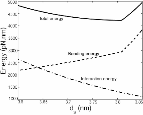
We mentioned earlier that the spacing between the strands is determined by the competition between bending and interaction energies. We demonstrate this minimization procedure with a very simple calculation. The model as set forth above has one free parameter, namely, , the parameter characterizing the strength of the repulsion between adjacent DNA strands. We fix pN/nm2 since, as shown below, this leads to the best fit to the experiment of Smith et al. (2001). We note that the ion concentration in their experiment is small compared to the values reported by Rau et al. (1984), which implies that the repulsive forces will be stronger than those measured by Rau et al. (1984) resulting, in turn, in a larger value of . We continue with the cylindrical capsid geometry with the same internal volume as the 29 phage. The goal is to determine the spacing between the strands for a given length of genome packed. To that end, we minimize the free energy with respect to at fixed length . In particular, we set to a fixed value in eqn. (15) and then solve for the number of columns of hoops this implies while holding the length fixed. We then have all the information needed to calculate the energy using eqn. (11). We repeat these steps for a range of different values of to get a plot of energy vs . Finally, we use this plot to determine the corresponding to minimum energy at a given length of DNA. One such plot is shown in fig. 3. The interaction energy decreases as the spacing increases. This is expected since the interaction energy is a decaying exponential and increasing implies that the interactions come from deeper in the tail of the pair potential. The bending energy rises as the spacing increases since the radii of the innermost strands become successively smaller. The total energy is dominated by the interaction part at smaller spacings and by the bending energy at higher spacings. For the case shown in fig. 3 we can clearly identify a minimum corresponding to nm in the total energy.
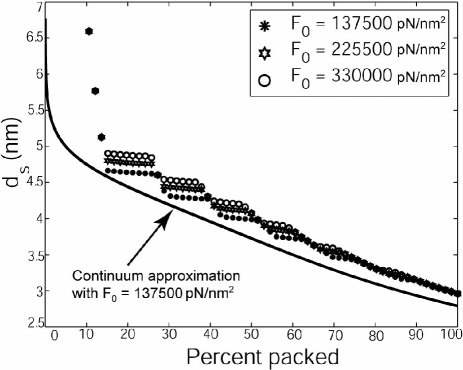
More generally to find the trends for all values of the length packed, this same procedure is repeated and then we plot the optimal (at minimum energy) against the fraction of DNA packed for the 29 phage as shown in fig. 4 for three different values of . We note that in the later stages of packing all the curves collapse onto a single curve, though we also note that the discrete and the continuous curves have different values in the large packing limit. As shown in our earlier work (Purohit et al. (2003)), in the large packing limit is dictated by packing the DNA in such a way as to maximize the distance between adjacent strands and should be seen as a geometric limit.
On the basis of the simple model presented above, fig. 4 provides several distinct predictions. First, on a gross level, it gives the history of the variation of interstrand spacing during the packing process. This is a verifiable prediction since it is possible to perform the viral packaging reaction for different ionic concentrations of the ambient solution and for different values of the genome length. Such experiments have been carried out for the T7 phage for three different genome lengths by Cerritelli et al. (1997). Similar experiments have also been performed for the -phage (see Earnshaw and Harrison (1977)). The second more subtle outcome of the model is the possibility that there are actually discrete effects in the packing process due to the packing of the DNA at a finite set of discrete radii. It would be of interest to determine whether these effects in the packing spacing (and a related force signature to be described below) are present in experiments.
4.2 Interstrand Spacing in Packed Capsids: Continuum Model
One of the interesting features of the calculations presented here is the ability to contrast discrete and continuous descriptions of the energetics of DNA packing. To that end, we now repeat the energy minimization argument that was used to determine the interstrand separation in the discrete setting in the continuum approximation. In particular, we minimize the free energy given in eqn. (12) with respect to , but subject to the constraint that is constant. In concrete terms this amounts to
| (16) |
where we have used the constraint which takes the form
| (17) |
We substitute for using eqn. (14) and then set to get the following general equation
| (18) |
This equation represents a competition between the interaction terms (on the left) and the bending terms (on the right). Note that the effect of the capsid geometry appears through the second term on the right hand side of eqn. (18). For a cylinder, , a constant, and eqn. (18) takes the form
| (19) |
Eqn. (14) is similarly specialized for a cylinder to yield
| (20) |
Eqns. (19) and (20) feature two unknowns, and , and we solve for them using the Newton-Raphson method. The result of this computation is a history of the interstrand spacing as a function of the length of DNA packed. This has been plotted as a thick line in fig. 4 for pN/nm2. It is evident that while the continuum version captures the trend quite well, it underestimates the value of for the cylindrical geometry in comparison with the more exact discrete version. Also, details captured by the discrete version, such as the series of steps and jumps, are smoothed out in the continuum approximation.
5 Forces during DNA packaging
One of the key outcomes of the model presented here is the internal force that builds up during DNA packing. The force vs percent packed curves should be seen as a second set of experimental implications for this mechanical model which complements predictions of the spacing vs percent packed.
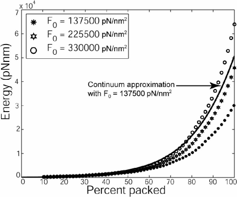
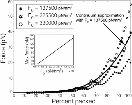
Indeed, once we have the spacing as a function of the amount of DNA packed we can determine the energy using eqn. (11) or (12) depending on whether we have adopted the discrete or continuum version of the free energy. The variation of the free energy has been plotted for the three values in fig. 5. Recall that the choice of reflects the ionic conditions present during the packaging process. It is evident that the total energy increases at any given value of the length packed as the strength of the repulsive interactions (the value of ) is increased. Figure 5 also shows the energy obtained using the continuum approximation which overestimates the energy but correctly captures the trend. The force required to pack DNA is simply the derivative of the energy with respect to the length packed. We have calculated this derivative numerically and the results are plotted in fig. 6.
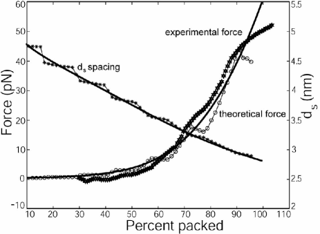
The behavior of the force curves is very much as expected, with higher forces for larger values of . We note that the strong dependence of the maximum packing force on is amenable to experimental observation and is worth further attention. In particular, we note that the maximum packing force can change by as much as a factor of two depending upon the ionic conditions (and the related value of ). Perhaps the most striking feature of the force curves is the appearance of steps. The same steps are seen in the plots of as a function of fraction packed. They occur when a new column (stack) of hoops starts to form on the inside of the spool. Curiously enough, the experimental force curve reproduced in fig. 7 also seems to show discontinuities of the slope at 4-5 different points, which might correspond to the five columns of completely stacked hoops needed to fill the 29 capsid with its DNA. However, to establish that this actually occurs during the packaging of a virus would require a more detailed analysis of the kinetics of packing and more careful experiments. The presence of such steps in the model results from implicitly assuming that the dynamics of packing follows the sort of organized column-by-column hoop packing described above. Therefore, the observation of such steps in experiment would be not only a confirmation of the model, but a provocative hint about the dynamics of packing.
6 Application to the 29 virus
As a final calculation we go beyond the analytic simplicity offered by the cylindrical capsid and consider the second approximate geometry shown in fig. 2 namely, the capped cylinder. Though the cylindrical geometry offers considerable insight into the mechanics of viral packaging it clearly represents an oversimplification of the 29 capsid. The 29 capsid is shaped like an oblong spheroid and we approximate it as a cylinder with hemispherical caps in order to make comparisons with the experimental results of Smith et al. (2001). The radius of the cylinder (and the hemispheres) is taken to be nm (see fig. 2). The height of the cylindrical portion (also called the ‘waist’) is nm. It is useful to study this geometry because one can obtain expressions for spherical capsids (which are very good approximations to icosahedral viruses) merely by setting the height of the waist to zero. The expression for is now a bit more complicated and is given by
| (21) |
Using this formula in eqn. (13) results in the following expression for the length of DNA packed:
| (22) | |||||
Unlike the cylindrical geometry, this expression is not analytically tractable. However, the same procedure can be used for determining the interstrand spacing, and the energy and force as a function of percent packed. The results of numerical calculations for this geometry, both in the discrete and the continuous setting, are plotted in fig. 7. We find that pN/nm2 results in a good fit to the experimental data. Moreover, the continuum and discrete versions of the theory are in better agreement for this geometry than they were for cylindrical capsids.
It is of interest to examine the effect of ionic concentration on the forces exerted by the portal motor on the DNA. The ionic concentration affects the value of and we have already seen that larger values of imply larger values of the force. In the inset to fig. 6, the maximum force as a function of the parameter is shown. This information can be potentially useful in the following way. It is known (see Smith et al. (2001)) that the portal motor of the 29 virus stalls at a force of pN. In other words, if one were to conduct the packaging reaction in a solution with ionic conditions such that pN/nm2 then the genome would not be completely packed since the motor would stall prior to complete packing. We are hopeful that experiments carried out with different ionic concentrations would permit an investigation of such predictions. We have made similar calculations to those presented here for viruses other than 29 and find interesting variations in the maximum packing force from one virus to the next which should be similarly accessible experimentally.
7 Conclusion
Recent advances in the development of tools for single molecule manipulation have permitted the direct mechanical investigation of a variety of biological processes. One intriguing recent example involves the direct measurement of mechanical forces during DNA packaging in viruses. We have developed a simple, analytically tractable model which responds to these experiments. The model emphasizes the role of elastic and electrostatic effects in the packaging process and is in good quantitative agreement with experimental data. More importantly, the model makes a series of predictions that we hope will inspire and guide future experiments. In particular, we predict the appearance of steps in the force versus percent of DNA packed curve. There may be some evidence of steps in the existing experimental data, but further experimentation is needed. The model also predicts a growth of the maximum force required for packaging as the ion concentration is decreased; the underlying cause being the stronger electrostatic repulsion between the DNA strands inside the capsid. This points to a new series of experiments that would look for incomplete packaging of the viral DNA at lower salt concentrations than used previously. An alternative approach would be to work at the same ion concentration but with longer DNA constructs.
The biggest shortcoming of the model as presently stated is that it does not address dynamical issues associated with the packaging process. The fundamental experimental observation is that as the packaging process proceeds the packaging rate falls from the initial 100 base pairs per second to zero. We note that the appearance of force steps described in this paper implicitly assumes a precise dynamical pathway resulting in a helical packing of the DNA in a series of helices of ever decreasing radius and are hopeful that the future work will shed further light on the dynamics of viral packing.
8 Acknowledgements
We are grateful to Ron Hockersmith, Paul Grayson, Dwight Anderson and Carlos Bustamante for useful suggestions. We have benefitted from a long series of stimulating discussions with Bill Gelbart, Alex Evilevitch and Chuck Knobler. RP and PP acknowledge support of the NSF through grant number CMS-0301657, the NSF supported CIMMS center and the support of the Keck Foundation. JK is supported by the NSF under grant number DMR-9984471, and is a Cottrell Scholar of Research Corporation.
References
- [1] Alberts B., Bray D., Johnson A., Lewis J., Raff M., Roberts K. and Walter P., (1997) Essential Cell Biology, Garland Publishing, Inc., New York.
- [2] Boal D., (2002) Mechanics of the cell, Cambridge University press, Cambridge.
- [3] Bray D. (2001) Cell movements, Garland Publishing, Inc., New York.
- [4] Bustamante C., Macosko J. C. and Wuite G. J. (2000), Grabbing the cat by the tail: Manipulating molecules one by one, Nat. Rev. Mol. Cell Biol. 1, 130-136.
- [5] Cerritelli, M. E., Cheng, N., Rosenberg A. H., McPherson C. E., Booy, F. P., and Alasdair C. Steven, A. C., (1997), Encapsidated conformation of bacteriophage T7 DNA, Cell, 91, 271-280.
- [6] Doi M., and Edwards S. F., (1988), The theory of polymer dynamics, Oxford University Press, Oxford.
- [7] Evans E. and Skalak R., (1980), Mechanics and thermodynamics of biomembranes, CRC reviews in Bioeng., CRC Press, Boca Raton, Florida, 1-254.
- [8] Earnshaw W. C. and Casjens S. R. (1980), DNA packaging by double-stranded DNA bacteriophages, Cell, 21, 319-331.
- [9] Earnshaw W. C. and Harrison S. C. (1977), DNA arrangement in isometric phage heads, Nature, 268, 598-602.
- [10] Evilevitch A., Lavelle L., Knobler C. M., Raspaud E. and Gelbart W. G., (2003), Osmotic pressure inhibition of DNA ejection from phage, Proc. Natl. Acad. Sci., in press.
- [11] Freund L. B. and Suresh S. (2003), Thin film materials: Stress, defect formation and surface evolution, Cambridge University Press, Cambridge.
- [12] Goldstein R. E., Powers T. R. and Wiggins C. H., (1998) Viscous non-linear dynamics of twist and writhe, Phys. Rev. Lett., 80, 5232-5235.
- [13] Kanamaru S., Leiman P. G., Kostyuchenko V. A., Chipman P. R., Mesyanzhinov V. M., Arisaka F. and Rossmann M. G., (2002), Structure of the cell-puncturing device of bacteriophage T4, Nature, 415, 553.
- [14] Kindt, J.T., Tzlil, S., Ben-Shaul, A., and Gelbart, W. (2001), DNA packaging and ejection forces in bacteriophage, Proc. Nat. Acad. Sci., 98, 13671-13674.
- [15] Lo C. M., Wang H. B., Dembo M., Wang Y. L. (2000), Cell movement is guided by the rigidity of the substrate, Biophys. J. 79, 144-152.
- [16] Meyers M. A., (1994), Dynamic Behavior of Materials, John Wiley & Sons.
- [17] Odijk, T., (1998), Hexagonally packed DNA within bacteriophage T7 stabilised by curvature stress, Biophys. J. 75, 1223-1227 .
- [18] Parsegian V. A., Rand R. P., Fuller N. L., and Rau D. C., (1986), Osmotic stress for the direct measurement of intermolecular forces, Methods in Enzymology, 127, 400-416.
- [19] Ptashne, M., (1992), A genetic switch, Blackwell Science (UK).
- [20] Purohit P. K., Kondev J., and Phillips R., (2003), Mechanics of DNA packaging in viruses, Proc. Nat. Acad. Sci., 100(6), 3173-3178.
- [21] Raspaud E., de la Cruz M. O., Sikorav J. L., Livolant F., (1998), Precipitation of DNA by polyamines: A polyelectrolyte behavior, Biophys. J., 74(1), 381-393.
- [22] Rau, D. C., Lee, B., and Parsegian, V. A., (1984), Measurement of the repulsive force between the poly-electrolyte molecules in ionic solution - hydration forces between parallel DNA helices, Proc. Natl. Acad. Sci., 81, 2621-2625.
- [23] Rau, D. C. and Parsegian, V. A.,(1992), Direct measurement of the intermolecular forces between counterion-condensed DNA helices - evidence of long-range attractive hydration forces, Biophys. J. 61 , 246-259.
- [24] Riemer, S. C. and Bloomfield, V. A., (1978), Packaging of DNA in bacteriophage heads: Some considerations on energetics, Biopolymers 17, 785-794.
- [25] Rice J. R., Lapusta N. and Ranjith K., (2001), Rate and state dependent friction and the stability of sliding between elastically deformable solids, J. Mech. Phys. Sol., 49(9), 1865-1898.
- [26] Rosakis A. J. (2002) Intersonic shear cracks and fault ruptures, Adv. Phys., 51(4), 1189-1257.
- [27] Smith D. E., Tans S. J., Smith S. B., Grimes S., Anderson D. L. and Bustamante C., (2001), The bacteriophage 29 portal motor can package DNA against a large internal force, Nature, 413, 748.
- [28] Simpson A. A., Tao Y., Leiman P. G., Badasso M. O., He Y., Jardine P. J., Olson N. H., Morais M. C., Grimes S., Anderson D. L., Baker T. S. and Rossmann M. G., (2000), Structure of the bacteriophage 29 DNA packaging motor, Nature, 408, 745.
- [29] Seifert U., (1997), Configurations of fluid membranes and vesicles, Adv. Phys., 46, 13.
- [30] Tao, Y., Olson N. H., Xu, W., Anderson, D. L., Rossman, M. G., and Baker, T. S., (1998), Assembly of tailed bacterial virus and its genome release studied in three dimensions, Cell, 95, 431-437.
- [31] Vogel S., (1988), Life’s Devices: The physical world of plants and animals, Princeton University press, Princeton, New Jersey.
- [32] Wang M. D., Schnitzer S. J, Yin H., Landick R., Gelles J. and Block S. M. (1998), Force and velocity measured for single molecules of RNA polymerase, Science, 282, 902-907.
- [33] Widom J., (2001), Role of DNA sequence in nucleosome stability and dynamics, Q. Rev. of Biophys., 34, 269-324.
- [34] Wikoff W. R. and Johnson J. E., (1999), Imaging a molecular machine, Curr. Bio., 9(8), R296-R300.