Effect of histamine on the electric activities of cerebellar Purkinje cell
Abstract
The effect of histamine (HA) on the electric activities of Purkinje cell (PC) is studied on the cerebellum slice. We find that: (1) HA’s main effect on PC is excitative (72.9%); there are also a small amount of PC showing inhibitive (10.2%) or no (16.9%) response to HA. (2) Different from the conventional opinion, HA’s excitative effect on PC is mutually conducted by H1 and H2 receptors; the antagonist for H1 receptor could weaken HA’s excitative effect on PC, while the antagonist for H2 receptor could weaken or even block the excitative effect of HA on PC. (3) PC’s reaction to HA is related to its intrinsic discharge frequency; there exists a frequency at which PC is highly sensitive to HA, and well above this frequency PC becomes stable against HA. These results indicate that the histaminergic afferent fibre can adjust PC’s electric activities by releasing HA, and thereby influence the global function of the cerebellar cortex; and that just like the region of cerebrum, cerebellum may also have some sort of characteristic frequency.
PACS number: 87.10.+e
Key words: cerebellar cortex; Purkinje cell; histamine; receptor; cerebellum slice
I Introduction
Cerebellum deserves more extensive studies than was conventionally realized. Historically, cerebellum was thought of as mainly a motor control organ, while recent researches reveal that it has many other functions, and it has more intimate connections to other parts of the brain [1]. Histamine (HA), a neurotransmitter or neuromodulator in the brain, plays an important role in the functions and interactions of various parts of the brain, and also in the studies of these functions and interactions. For instance, the neuroanatamic researches revealed the existence of the hypothalamus-cerebellum histaminergic path [2, 3], and shows that hypothalamus has great influence on the cerebellar activities and hence plays an important role in coordinating the functions of the body and viscera. Besides, HA has the possible role of controlling the cerebellar circulation, and HA receptors are also found in the neurons of the cerebellar cortex [2, 3].
Cerebellum is made up of the outer dark matter (cortex), inner white matter, and three pairs of deep nuclei lying in the heart of the white matter. These nuclei are the fastigial nucleus FN, interposed nucleus IN, and dentate nucleus DN. The afferent fibres to the cerebellum are mainly from the vestibule, spinal cord, and the cerebral cortex. They form synaptic connections with the neurons in the cerebellar deep nuclei and cortex. The synapses of Purkinje cell (PC) make the efferent fibres of the cerebellar cortex, they are mainly projected into the deep nuclei, then the neurons there stick out fibres which make the cerebellar output; a small amount of PC synapses are directly projected into the vestibular nucleus.
The cerebellar cortex can the divided into three layers: the (out-most) molecular layer, PC layer, and the granular layer. It contains three kinds of afferent fibres (musciform fibre MF, crawl fibre CF, and monoaminergic fibre), and five kinds of neurons (PC, granular cell GR, basket cell BA, star-like cell ST, and Golgi cell GO). Hence we see that the cerebellar afferent fibres and the intermediate neurons, with PC acting as the core, constitute the basic neural circular that is responsible for the sensory function of the cerebellar cortex, and the cerebellar cortex together with the deep nuclei undertake various functions of the cerebellum.
In our laboratory there has been studies of HA’s effects on certain neurons of the cerebellum, such as the granular cells [3, 4], while HA’s effect on the neurons in the cerebellar cortex is not yet intensively studied. Considering that PC is the only efferent neuron of the cerebellar cortex, we are going to investigate HA’s effect on the electric activities of PC, based on our accumulated experiences in studying the influences of aminergic materials such as norepinephrine (NA) and serotonin (5-HT) on the spontaneous and induced discharge activities of the cerebellar PC; so as to learn more about the role of aminergic afferent system in the process of information treatment in the cerebellar cortex.
II Experimental material and method
We use for our experiments 19 mature SD rats (200-250g). Anaesthetize a rat by injecting betchloramines hydrochloride (4mg/100g) into the abdominal cavity, take out the cerebellum right after cutting the head, wash the cerebellum with frozen artificial cerebrospinal fluid (ACSF, C), stick it onto the operating table of a microtome (at which the cerebellum is soaked in frozen ACSF), and cut a 400 thick of arrow-like slice from the vermis. The process of making the slice should be done within 20 minutes. Then move the so obtained slice into a recording trough, and begin the experiment after 15 minutes of hatching. The recording trough is continuously irrigated (3ml/min) by ACSF (33), and is aerated with the mixed air of 95%O2 +5%CO2. The concentrations of various elements in ACSF are (mmol/l): NaCl 124, KCl 5, KH2PO4 1.2, MgSO4 1.3, CaCl2 2.4, NaHCO3 26, glucose 10. Put a tiny glass electrode (filled in with colored conducting fluid) at the PC layer of the X leaflet of the cerebellar cortex to make out-cell records of PC’s discharge activity. This is because the X leaflet received the least mechanical wound in making the slice, and hence the cerebellum slice should be so put in the recording trough that the X leaflet be well hatched by the ACSF and the mixed air.
We base on the following criteria to single out PC discharge signals: (1) position of the electrode: PCs in the cerebellar cortex are of linear type, namely, the cell bodies concentrate at one end and the dendrites stick to the other end, thereby form a PC layer in the cerebellar cortex. We put the recording electrode at the out side of the PC layer and near to the molecular layer, therefore there is very little chance to catch a discharge signal of the granular cell; (2) discharge wave shape: the PC action potential has large magnitude (0.7000.143) and thick contour [5], moreover, the effective recording distance of PC discharge is 50m, much longer than that of the granular cell (20-30m) [4]; (3) cell numbers: in the cerebellar cortex there are much less GO, ST, and BA than PC, and hence the discharge signals from these cells may be ignored.
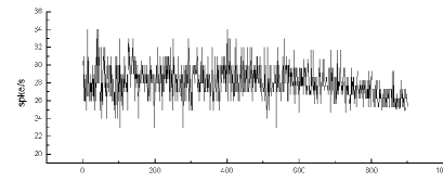
Because the nerve cells in the brain have the character of continuously producing impulses, and often at the frequency of tens of times per second, the physiological status of a neuron is usually symbolized by its discharge frequency. Under the environmental stimulation, the discharge frequencies of central neurons will variate, and the intrinsic potentials will also deviate. Hence we magnify the PC discharge signal, choose a relatively small time constant for the recording system, so as to differentiate the input signal and transform the slowly increasing wave into a sharp high peak wave, then use a pulse discriminator to convert the peak signal into TTL pulse and input it into the computer, and then draw the post-stimulus histogram. Thus we can describe PC’s reaction to chemicals by the post-stimulus histogram of its discharge frequency.
All the drugs here are contemporarily compounded using ACSF, and are used to irrigate the cerebellum slice separately. When doing the experiments of studying the receptor mechanism of HA’s effect on PC, the slice is continuously irrigated by the receptor antagonist for over 10 minutes before by HA.
III Results
A The effect of HA on PC’s electric activities
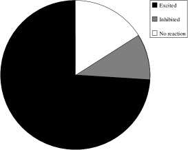
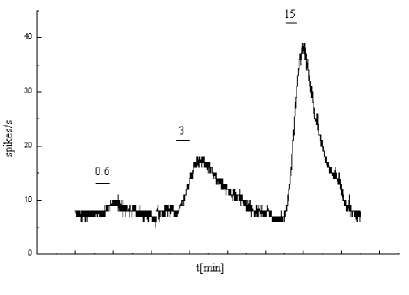
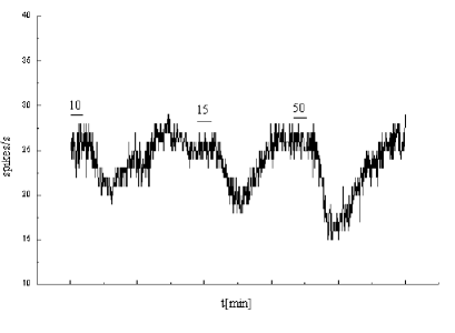
Because intermittent discharge is often due to the ill state of PC or indistinguishability of cell discharge signal from other signals, we choose in 29 cerebellum slices 59 cells with relatively stable spontaneous discharge for our study. Their spontaneous discharge frequencies range from 4.48 to 78.32 Hz. We find that of the 59 cells, 83.1% (49/59) are affected by HA, the other 16.9% (10/59) show no response. And of the 49 responsive ones, 87.8% (43/49) are excited, the other 12.2% (6/49) are inhibited. (See Fig. 1) Therefore HA’s effect on PC is mainly excitative. From Figs. 1B, 1C, we can see that there is an evident increase of the variation of the cell’s discharge frequency as the irrigating concentration of HA increases.
|
A
 |
B
 |
C
Another important observation is that whether or not a PC shows response to HA may have some connection with its spontaneous discharge frequency. (cf. Fig. 2) The spontaneous discharge frequencies of the 59 tested cells are 4.48-78.32 Hz, while that of the 49 cells which show response to HA is 21.3412.69 Hz (MSD), this is significantly lower than the average frequency (37.007.73 Hz) of the 10 cells unaffected by HA. However, whether a cell is excited or inhibited by HA seems to have nothing to do with its intrinsic frequency. The average frequencies of the 43 excited cells and of the 6 inhibited cells are 21.5313.20 and 20.008.80 Hz, respectively, which are not much different (P0.5, t test). This differs from how HA’s effect on the cerebellar granular cells is related to their intrinsic frequencies [3]. We also find that among the 49 responsive cells, those who have higher intrinsic discharge frequencies are more sensitive to HA (there are 8 responsive cells with intrinsic frequencies higher than 30 Hz, and 7 of them show response to HA at the lowest HA concentration of less than 30mol/l).
 |
A
 |
B
B The receptor conducting mechanism of HA’s excitative effect on PC
It is known that there are three sub-types of HA receptors in brain: H1, H2, and H3, therefore it is necessary to study the receptor conducting mechanism of HA’s effect on PC. Since HA’s main effect on PC is excitative, we make study for the excitative effect. For this purpose we observe the influences of Triprolidine (hyper-specific antagonist for H1 receptor) and Ranitidine (hyper-specific antagonist for H2 receptor) on PC’s excitative reaction to HA, and find that low-concentration Triprolidine (0.5-1.0mol/l) could weaken HA’s excitative effect on PC, while low-concentration Ranitidine (0.9-1.0mol/l) could weaken or even block HA’s excitative effect on PC. (cf. Fig. 3).
H3 receptor is a presynaptic self-receptor, it adjusts the presynaptic release of HA [2]. In this research we have not investigated its role in HA’s excitative effect on PC.
 |
A
 |
B
 |
C
IV Discussions
A Data-taking system
In this experiment we used the data-taking system: oscillometer-pulse discriminator-computer, and drawn the post-stimulus histogram, then evaluate PC’s reaction to chemicals by the post-stimulus histogram of the discharge frequency. The sampling bin width is 3, point=900, interval=1000, one data represents the discharge times of a cell in one second. Since a cell discharges at very high frequency and its electric signals transmit at very high speed, it gives very rich information in one second. We know that the information carried by a neuron is represented by its frequency distribution over the action potential, therefore if we can obtain the frequency spectrum, and make the combined analyses of the frequency and power spectra, we will get greater amount information. Besides, what we take in this experiment is out-cell record of a cell’s discharge activities, and PC is in connection with various other cells in the cerebellum slice, the measurement in such case may be disturbed by the environment, and the excitative or inhibitive reaction of PC may actually be a combined result of various influences including those from the environment. These are to be improved in future studies.
B HA’s effect on PC
Histamine is an important neurotransmitter or neuromodulator, and acts as an inter-cell messenger. HA can hardly penetrate the blood-brain barrier, hence must be produced by the histaminergic neurons. It can be released through depolarization and by means of calcium dependence. The hypothalamus-cerebellum histaminergic fibres in the brain are casted by the hypothalamus tuberous mastoid nucleus (HA pericaryon) into the cerebellar cortex and the cerebellar deep nuclei (HA nerve end) [2, 3]. Cerebellum-hypothalamus histaminergic system can bring about many functions via the inter-cell messenger HA. The hypothalamus-cerebellum histaminergic projecting fibres end at the cerebellar cortex as spreading multi-layer fibres, and their nerve ends mainly spreadingly adjust the functions of the peripheral neurons in the varix form; these are much like the serotoninergic and norepinephrinergic afferent fibres of the cerebellum [2, 4].
Purkinje cell is the only kind of efferent neuron of the cerebellar cortex, it is a deformation of the multi-pole cell, and has very strong dentrite. Our research shows that PC’s discharge activity is significantly affected by HA. From our observations in Sec. IIIA, and associating how the reaction of cerebellar granular cells to HA is related to their intrinsic discharge frequencies [3], we speculate that the cerebellar afferent fibres might be mainly responsible for adjusting the basic discharge levels of the cerebellar neurons: as the discharge frequency of a target cell lies in a certain region it loses sensitivity to HA; and as the discharge frequency lies out of this region the cell reacts to HA, and thereby its frequency tends to that region. Moreover, the high-frequency region is a low-sensitivity region to HA, while for the responsive cells, those with higher frequency are highly sensitive to HA. We therefore speculate that there exists a critical frequency, a cell with intrinsic frequency near to this critical point will significantly change its states by only a small amount of HA, while as its frequency goes well above this point, it becomes stable. In connection with the influence of NA and 5-HA on the spontaneous and induced electric activities of cerebellar PC [6], we go further to speculate that histaminergic and other aminergic afferent systems might, through their coordinations and/or antagonism, adjust the synaptic transmission efficiency of MF-PF (parallel fibre)-PC and CF-PC, or adjust PC’s sensitivity to signals from MF and CF, and thereby take part in the the global process of the sense movement of the cerebellar neuron net. That many kinds of neural active materials mutually act on the neuron is a common pattern of signal transmission of the nerve system; such mutual action may happen at the presynapse, postsynapse, or even postreceptor level. HA’s effect on the cerebellar PC is to adjust the excitativity level of the neuron and the neuron’s sensitivity to input information from outside of the cerebellum, and adjust the sleep or awake state of the cortex. The aminergic afferent fibres have synaptic or non-synaptic chemical transmission effect on the cerebellar cortex, their non-synaptic transmission may not encode the fast phase-like information, but hyperfinely adjusts the membrane potential and basic discharge level or the target neuron.
C The receptor conducting mechanism of HA’s excitative effect on PC
Of the three sub-types of HA receptors in the brain (H1, H2, and H3), H3 receptor is a presynaptic self-receptor. The acting mechanism of H1 receptor is through some sub-type of G protein to activate PLC, hydrolize 4,5-PIP2 to be 1,4,5-IP3 and DG, these two secondary messengers go further to trigger various biological effects, such as stimulating endoplasmic reticulum to release Ca+and thence trigger the ion channels [2]. The H2 receptor brings effect mainly via G protein-AC-cAMP: HA combines with the H2 receptor of the cell membrane, the H2 receptor is then deformed and triggers G protein to give off its and sub-radicals, and on the sub-radical GDP is replaced by GTP, then AC is activated and catalyzes ATP to transform to cAMP, cAMP acts as a secondary messenger and triggers a series of reactions, such as activating PKA and then triggering ion channels, or directly triggering ion channels. Because many highly efficient enzymes take part in these reactions, small signals are magnified step by step.
It is usually thought that H1 and H2 receptors conduct HA’s excitative and inhibitive effects on neurons respectively [2]. However, this is in contradiction with our report in Sec. IIIB that low-concentration H1 receptor antagonist could weaken HA’s excitative effect on PC, while low-concentration H2 receptor antagonist could weaken or even block HA’s excitative effect on PC. This makes us to conjecture that both H1 and H2 receptors involve in HA’s excitative effect on PC, and H2 receptor is the main conductor. There has been report that HA’s excitative effect on the granular cells of the cerebellar cortex and on the cells of the vestibular inner-side nucleus is mutually conducted by H1 and H2 receptors [2]. Histochemistry researches also reveal that in the cerebellar cortex of rat the density of H2 receptor is higher than that of H1 receptor. All these partly support our conjecture. This may suggest that the H2-related signal transmission chain in cell differs from one type of neuron to another, and such difference determines whether the H2 receptor conducts excitative or inhibitive effect; but the particular mechanism of signal transmission is still to be studied.
D Mathematical simulation
To systematically study the biophysical or biochemical mechanisms of the reactions of cells, it is often helpful to make mathematical simulations of the reactions. Here we give a simulation for the relation between PC’s (excitative) reaction to HA (measured by its maximum variation of discharge frequency under the action of HA at a certain irrigating concentration) and its intrinsic discharge frequency . To be concrete, we choose a fixed frequency variation , and denote by the irrigating concentration of HA at which the maximum change of a cell’s frequency would be , then study the relation between and . For this purpose we first look at how the maximum frequency of a cell under the action of HA is related to HA’s irrigating concentration .
As we noted in Sec. IIIA, PC’s reaction to HA has something to do with its intrinsic discharge frequency: high-frequency cells are insensitive to HA, while for the cells affected by HA, the higher-frequency ones are highly sensitive to HA, and at low intrinsic frequencies increases steadily with . We therefore assume that is an -like function of :
| (1) |
As , is just the intrinsic frequency :
| (2) |
According to our above explanations, we write
| (3) |
From Eqs. (2,3) we can derive the - relation:
| (4) |
Here the physiological meaning of is the maximum discharge frequency of the cell, and is an arbitrarily chosen minimum frequency variation that can be observed in the experiment. It is straightforward to check that has a minimum value at an intermediate , just as the experimental results show. Eq. (4) is much like the relation in the Hopfield model. But further study of the connection to the Hopfield model would require more detailed knowledge about the relation between and , which is not possible in the present experiment, for PC cannot live through a long enough time to allow tests for various HA concentrations; hence the simulation here is merely a rough one.
E Characteristic frequency in the cerebellum?
Eqs. (1,4) show a pattern very similar the characteristic frequency of the cerebrum (the region of around 40 Hz): there exist a particular frequency at which the target cell is highly sensitive to HA, and well above this frequency the cell loses sensitivity to HA. For PC this insensitive region is 25.00-46.24 Hz, with the average 37.007.72, which is close to the region. These suggest that cerebellum might also exhibit some sort of characteristic frequency to external stimulations. Further studies of such characteristic frequency would necessarily take into account of the interactions of various cell, such as in the HR model [7]; these we hope to accomplish in the future.
V Acknowledgements
The author owes special thanks to Prof. Jiang-Jun Wang and Prof. Bing Zhu for valuable instructions, to Le Tian and Jie Ma for collaborations in this experiment, and to Feng Dong, Qin Xi, and Guo-Ning Hu for kind help and suggestions.
REFERENCES
- [1] See, eg., J.H. Gao, L.M. Parsons, J.M. Bower, J. Xiong, J. Li, P.T. Fox, Science 272, 545 (1996); Thompson, Science 238, 1729 (1987); and references therein.
- [2] J.C. Schwartz et al, Physio. Rev. 71, 1 (1991).
- [3] W.C. Li, J.J. Wang et al, Science Bulletin 41, 2269 (1996) (in Chinese).
- [4] W.C. Li, J.J. Wang et al, to be published.
- [5] C.M. Huang, H. Mu and C.F. Hsiao, Brain Res. 619, 313 (1993).
- [6] J.J. Wang, et al, J. Physio. 48, 581 (1996) (in Chinese).
- [7] R.R. Llinas, Science 242, 1654 (1988).