INTERFACIAL TENSION IN WATER AT SOLID SURFACES***Published in proceedings of the Third International Symposium on Cavitation, vol. 1, p87-90 (1998).
Abstract
A model for the formation of cavitation nuclei in liquids has recently been presented with basis in interfacial liquid tension at non-planar solid surfaces of concave form. In the present paper investigations of water-solid interfaces by atomic force microscopy are reported to illuminate experimentally effects of interfacial liquid tension. The results support that such tension occurs and that voids develop at solid-liquid interfaces.
NOMENCLATURE
| , | coordinates on specimen surface in scan direction |
|---|---|
| and perpendicular to this direction | |
| coordinate along surface normal | |
| equilibrium pressure | |
| mean radius of surface corrugation | |
| tensile strength of liquid | |
| amplitude of surface corrugation |
I INTRODUCTION
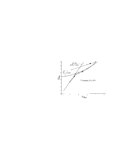
In cavitation research the formation and stabilization of cavitation nuclei has always been an intriguing problem which has made calculations of cavitation inception highly problematic. Subcritical gas cavities in water are inherently unstable and go into solution [1] or they drift to surfaces due to buoyancy. Therefore, stabilization must take place at liquid- solid interfaces. A model was proposed by Harvey et al. [2]. Though able to explain some of the experimental results of inception research, and during half a century the only reasonably realistic model, it is insufficient. A new model was proposed by Mørch [3], and recently it has been improved to allow quantitative calculations [4]. According to this model interfacial tension in the liquid adjacent to solid surfaces opens the possibility of detachment of the liquid, i.e. void formation, at surface elements of concave form. At sufficiently high curvature the voids may develop spontaneously, but at moderate and low curvatures the content of gas being in solution in the liquid and reaching the interface by diffusion is important for breaking liquid-solid bonds which are strained by the interfacial tension in the liquid. It is predicted that a void grows until the contact line between detached liquid and liquid still in contact with the solid reaches the locus of balance between the tensile stress due to interfacial liquid tension and the pressure in the bulk of liquid. It seems possible to explain qualitatively the most significant results of experimental research from this model. Quantitatively the measurements of tensile strength of tap water vs. increasing equilibrium pressure by Strasberg [5] can be simulated from assuming that solid particles in tap water have shallow corrugations of sinusoidal cross section, axially symmetric around their bottom and of mean radius and with relative amplitude , and small, relatively deeper ones with , , FIG. 1.
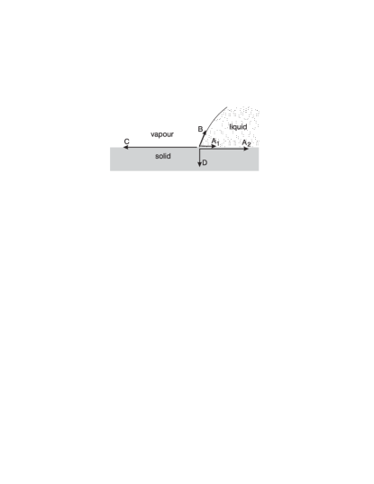
The interfacial tension present at the interface of two substances in contact is normally given as a single quantity. However, as the solid-liquid interfaces we consider are not planar it is suitable to split this interface tension into two components, one for the liquid, , and one for the solid, , to obtain information of the influence of curvature on the liquid-solid bonding. In FIG. 2 the balance of forces is shown at a solid-liquid-vapour contact point. In addition to the interface tension forces , , and at the three interfaces this balance demands also an adhesion force (van der Waals’ force) between the liquid and the solid perpendicular to the solid surface. These forces give the contact angle of the liquid- vapour interface. In water where hydrogen bonds dominate the intermolecular forces an appreciable interfacial liquid tension () is to be expected adjacent to solid surfaces as a result of a stabilized interfacial liquid structure. Experimentally effects of an orderly structured water layer have been measured near a mica surface [6] and by computer simulations it has been shown that at platinum surfaces the interfacial layer of water has an essentially ice-like solid structure [7]. These results support the hypothesis that water generally exhibits a more or less stabilized structure at solid surfaces. The interfacial liquid tension expected to result from this structure is the crucial parameter in the model of void formation [4] and thus for the formation of cavitation nuclei in liquids. It is the object of the present paper to verify its existence experimentally.
II EXPERIMENTAL TECHNIQUE AND RESULTS
Experimental techniques available for investigating the local interfacial tension in the liquid adjacent to a solid surface are very few - at present only atomic force microscopy (AFM) seems available [8]. This technique is basically used to give information of the surface topography of a solid object, and resolution to atomic scale is available for crystallographically planar surfaces. However, it can be used also for local force spectroscopy. In AFM a pointed tip, usually of pyramidal form and of height and base dimensions and with a tip radius of curvature , which is mounted close to the free end of a thin cantilever of length about , is approached to the surface which is to be investigated. When the distance between the tip apex and the surface becomes sufficiently small interatomic forces between the tip and the object attract the tip, and the cantilever is bent. This is detected by the deflection of a laser beam being reflected from the cantilever surface opposite to the tip. The deflection is a measure of the force on the tip. If the tip is approached further to the surface contact is achieved and the resulting force shifts into repulsion. This so-called contact mode is the one generally used for topographic investigations. Here a suitable repulsive deflection is chosen and the tip is scanned in the - and - directions across the specimen while its height is regulated by a feedback circuit to maintain the deflection chosen, independent of surface corrugations. Thus the voltage in the feedback circuit is a measure of the topographic changes.
In the force spectroscopy mode the tip is stationary in the - and -directions, and the tip deflection is measured while the cantilever base is moved along the -axis at constant speed towards the specimen until a suitable repulsive deflection is achieved, then its motion is reversed. These investigations can be made in vacuum, in gas, and in (optically transparent) liquid. In vacuum only interatomic forces between tip and specimen (van der Waals’ forces) affect the deflection. In gas (usually atmospheric air) also forces between molecules adsorbed to the surfaces are important. In particular water molecules forming adsorbed water layers on the tip and specimen surfaces are important because surface tension forces cause strong attraction when these layers get in contact. The surface tension forces and the van der Waals’ forces result in a transient ”snap-in” of the tip at approch just before contact is obtained. At operation in liquids it is generally assumed that snap-in is absent because the liquid is taken to have bulk structure right to the liquid-solid interface. The present results indicate that this is not correct.
For the experiments a TopoMetrix TMX 2000 Explorer AFM was used with V-shaped cantilevers of nominal spring constant and tip radius of curvature about .
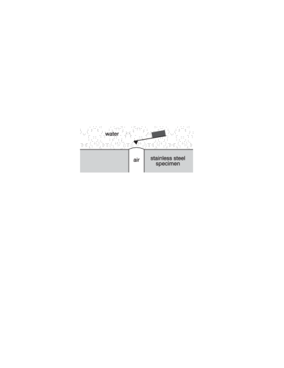
The interfaces to be considered here are distilled water-air interfaces which were approached from the liquid space, i.e. with the tip and cantilever fully submerged, and diamond polished stainless steel surfaces submerged in distilled water, and an air-gold interface. The setup with cantilever and tip submerged in water is shown in FIG. 3. At suitable air pressure in the central bore of the specimen a stable water-air interface of form as a spherical segment is created at the top of the bore, and it can be approached with the tip and cantilever fully submerged in water. At lateral translation of the specimen the water-stainless steel interface can be investigated.

When the tip approaches the water-air interface from the liquid space and get in contact with the interfacial water it is strongly attracted to the interface and crashes through it in a violent snap-in. The process is interpreted to result from the interaction of the orderly structured liquid at the water-air interface with that at the water-tip interface. The initial interaction results in an increased order in the zone of liquid around the tip apex, and an attractive, but unbalanced force between the tip and the water air-interface is set up by the interfacial liquid tension in the structured zone, FIG. 4a. A balance is then obtained by local elevation of the water-air interface and bending of the cantilever. As a consequence the tip breaks through the interface, FIG. 4b. With the soft cantilever used in the present experiments balance was not achieved until the interface reached the cantilever itself. It was not possible to record the event as the dynamical range of the microscope () was greatly exceeded.
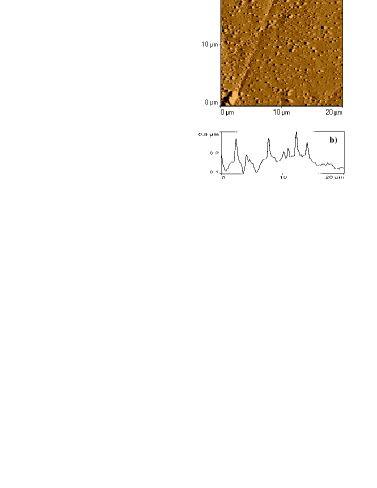
When subsequently the specimen was moved laterally to allow investigation of the water-stainless steel interface the topography of an area on the steel surface could be recorded as shown in FIG. 5a. A cross section along a single line, , is shown in FIG. 5b. The surface appears slightly wavy with localized micro-hills in the range.
By force spectroscopy it is found that the snap-in at approach as well as the snap-out at the subsequent retraction depend strongly on the location. Very often the sn ap-in is quite small, just a few nm, and at retraction the tip sticks to the solid surface until the cantilever base has retracted about corresponding to an attractive force of . Then the tip escapes from the specimen surface, but it does not return to the non-deflected condition until the cantilever base has moved another during which the tip relaxes in two steps, FIG. 6, found in repeated cases. This may be related to the quantized adhesion reported in [9], though in the present case the changes occur at a much larger scale.
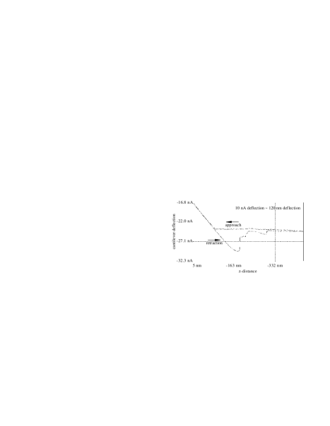
In other cases a very large snap-in occurs reproducibly, as shown in FIG. 7, where the snap-in is about , and at retraction the tip remains in contact until the cantilever base has moved about . Then the cantilever returns to non- deflected condition in a single jump.
The interpretation we give to these results is as follows: at locations on the specimen surface where the liquid is in direct contact with the solid surface there is an orderly structured liquid layer of thickness about adjacent to the solid, and when the tip with its own orderly structured interface layer of liquid, also of thickness about , approaches the solid surface these interface layers merge and set up an attractive force on the tip, which in
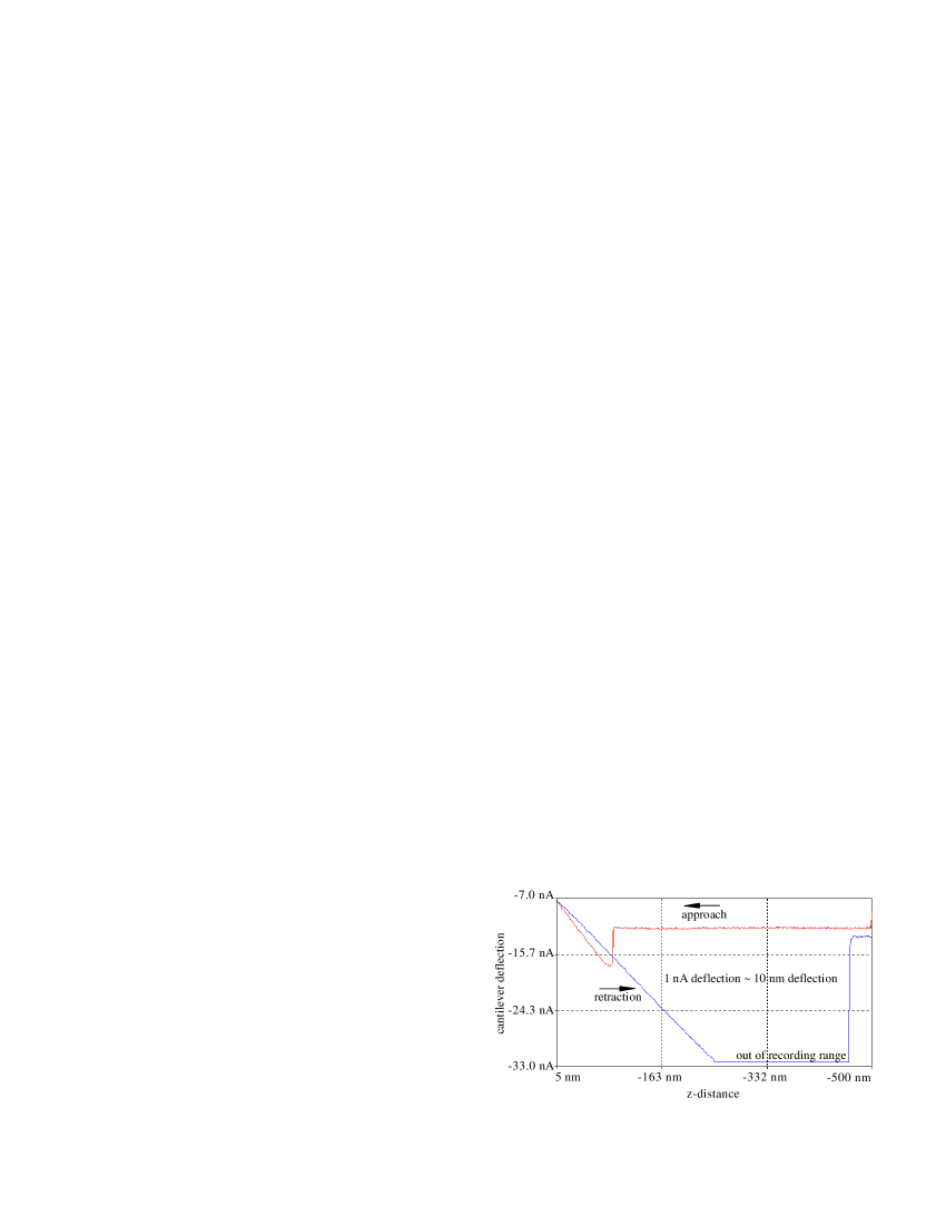
combination with the van der Waals’ forces between tip and sample, being of range typically about a few nanometer [10], result in a tip snap-in of less than , as actually apparent from FIG. 6. When the snap-in brings the tip in contact with the solid surface the van der Waals’ forces are strongly enhanced. Therefore, a larger force is required to withdraw the tip from contact. This is also evident from FIG. 6 where retraction of the cantilever base over a distance of about is demanded to set up the force needed for the tip to escape the surface itself. However, it appears that a bending force on the cantilever remains. We suppose this is a consequence of a nanovoid being formed between the tip apex and the sample when contact between the two solid surfaces is broken. The surface tension at the double curved liquid-vapour interface which connects the tip and sample and bounds the void prevents that liquid flows into the gap and at the same time it establishes an attractive force between tip and sample. Therefore, the tip is not totally free of interaction with the specimen until the distance becomes so large that the void collapses. This appears to happen after a further withdrawal, though in steps. The intermediate jumps may be related to discontinuous changes of the loci of contact of the liquid surface to the tip and sample surfaces. Apparently this takes place when the force imposed by the bent cantilever exceeds about .
In cases of a significant snap-in, as in FIG. 7, the event cannot be attributed to neither contact between the structured interfacial liquid at the tip and specimen surfaces nor to the van der Waals’ forces, as these do not extend beyond at most . In liquid the range of the van der Waals’ forces is actually reduced compared to their range in air [11]. However, if a stable interfacial void has grown on the specimen surface due to the local characteristic features of this surface, as modelled in [4], the tip meets a water-gas interface during the approach. As described above such an interface attracts the tip strongly and makes it penetrate deeply, i.e. in the present case it penetrates until tip-solid contact prevents further penetration, and a significant repulsive force between tip and specimen surface may then occur. This interpretation is supported by the large retraction distance of , corresponding to an attractive force of , observed before snap-out occurs, and the tip now escapes the solid surface as well as the supposed surface-attached void in a single large jump. This force considerably exceeds the van der Waals’ forces on the tip, which must be smaller than the found from FIG. 6. Thus it reveals strong interfacial forces in the liquid adjacent to a water steel interface.
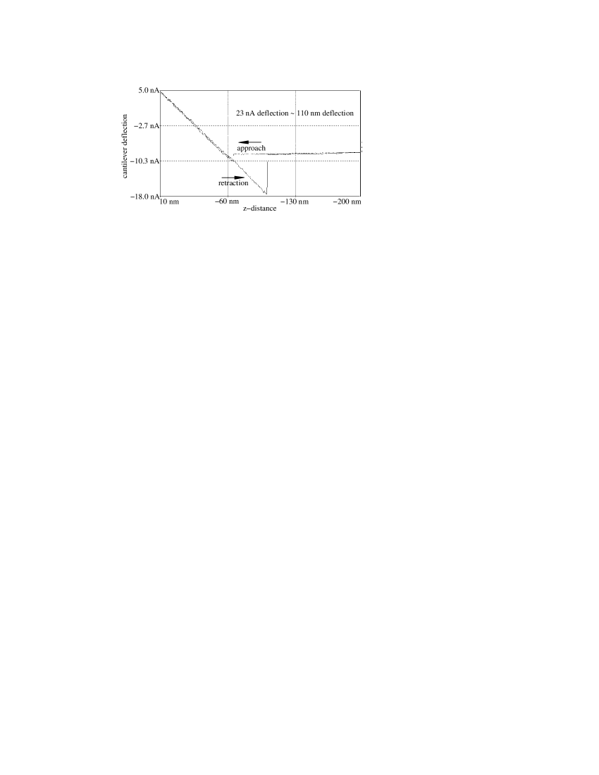
It is of interest to compare the above results from stainless steel surfaces submerged in water with observations in air of a solid surface which does not adsorb water. In such a case the water adsorbed to the tip is of no significance as water is not attracted to the non-adsorbing solid surface. Gold does not adsorb water to any significant extent, and in FIG. 8 force spectroscopy on an air-gold interface is shown. The snap-in is about , and it can be ascribed to van der Waals’ forces which are unscreened due to the absence of water on the gold surface. At retraction the snap-out occurs in a single jump after withdrawal of the cantilever base, corresponding to van der Waal’s forces of only .
If we compare the force spectroscopy of the submerged stain- less steel surface in contact with water, FIG. 6, with that of the air-gold interface, we notice that at the submerged water- stainless steel interface the van der Waals’ forces are notably weaker at snap in, probably due to the screening effect of water. At snap out however, the upper limit of the van der Waals’ forces can be estimated from the air-gold experiment, and they are clearly insufficient to explain the force needed for the first snap-out. Therefore, by the first snap-out in FIG. 6 already a major part of the attractive force can be ascribed to liquid interfacial tension.
III CONCLUSION
AFM force spectroscopy investigations at solid as well as gaseous interfaces with water, probed from the liquid space, reveal characteristic attractive forces which can only be attributed to liquid tension in the interfacial water. This brings experimental support to the model of void formation at liquid- solid interfaces [4] in which the interfacial liquid tension is a basic assumption. Further, the presence of interfacial voids is actually experimentally supported. Such voids are sources of cavity formation when single-phase liquids are exposed to tensile stress.
REFERENCES
- [1] Epstein P.S., Plesset M.S., 1950, J. Chem. Phys., 18:1505- 1509.
- [2] Harvey E.N., Barnes D.K., McElroy W.D.,Peace D.C., Cooper K.W., 1944, J. Cell. Comp. Physiol., 24:1-22.
- [3] Mørch K.A., 1992, ”A molecular approach to cavitation inception”. 2é me Journees CAVITATION, Paris, France, March 1992.
- [4] Mørch K.A., 1997, ”Void formation at water-solid interfaces due to interfacial liquid tension”. Submitted to J. Fluid Mech.
- [5] Strasberg M., 1959, J. Acoust. Soc. Am., 31: 163-176.
- [6] Israelachvili J.N., Pashley R.M., 1983, Nature, 306: 249- 250.
- [7] Xia X., Perera L., Essmann U., Berkowitz M.L., 1995, Surface Science, 335:401-415.
- [8] Gü ntherodt H.J., Wiesendanger R., 1992 & 1993, Scanning Tunneling Microscopy II & III, Springer Verlag.
- [9] Hoh J.H., Cleveland J.P., Prater C.P., Revel J.-P., Hansma P.K., 1992, J. Am. Chem. Soc. 114: 4917-4918.
- [10] Sørensen, A.H., 1997, ”Scanning Probe Microscopy Investigations of Landed Clusters and Electrostatic Forces”, Ph.D. Thesis, Institute of Physics, Tech. Univ. Denmark.
- [11] Israelachvili J.N., 1992, Intermolecular & Surface Forces, Academic Press.