[
Instability and ‘Sausage-String’ Appearance
in Blood Vessels during High Blood Pressure
Abstract
A new Rayleigh-type instability is proposed to explain the ‘sausage-string’ pattern of alternating constrictions and dilatations formed in blood vessels under influence of a vasoconstricting agent. Our theory involves the nonlinear elasticity characteristics of the vessel wall, and provides predictions for the conditions under which the cylindrical form of a blood vessel becomes unstable.
]
High blood pressure can experimentally be induced by intravenous infusion of a vasoconstricting agent like angiotensin II (regulates the contraction of the smooth muscle cells surrounding the blood vessel) [1, 2, 3]. As the infusion is continued, a substantial narrowing of the smaller blood vessels is observed, and suddenly the narrowed vessels develop a peculiar pattern consisting of alternating regions of constrictions and dilatations, giving the vessels the appearance of sausages on a string (Fig. 1). The ‘sausage-string’ pattern may cause severe damages to the blood vessels because plasma and macromolecules are transported into the vessel wall in the dilated regions. The sausage-string pattern has been observed in small vessels from many organs, including the brain, the gut, and the kidney [4].
Despite several decades of research, the mechanism causing the ‘sausage-string’ pattern has remained unknown [4]. It has been suggested that it represents a ‘blow out’ of the vessel wall due to the high blood pressure [5], but this seems unlikely for several reasons. The sausage-string pattern occurs in the smaller vessels (small arteries and large arterioles), and here the pressure elevation is relatively small compared to that in the larger arteries. Secondly, the phenomenon is highly reproducible [2]. If the infusion of the vasoconstricting agent is stopped, the normal, uniform ‘cylindrical’ structure is restored. Restoring the infusion causes again an extreme, uniform vasoconstriction followed by the reappearance of the sausage-string pattern. A third spectacular feature of the phenomenon is its periodicity with constrictions and dilatations occurring in a regular and repetitive pattern.
In this Letter we present a simple anisotropic, elastic model of the vessel wall. We show that under certain hypertensive conditions an instability occurs which leads to a periodic pattern of constrictions and dilatations along the vessel. Our theory provides predictions for the conditions under which the cylindrical form of a blood vessel becomes unstable.
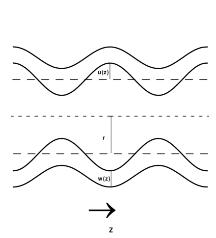
To be specific, a cylindrical shaped blood vessel is unstable if a small axial symmetric perturbation of the inner radius, , grows (Fig. 2). To determine the stability, we must therefore know the dynamic equation for the perturbation . To this end, we invoke the continuity equation, , associating a local change of the cross-sectional area at a downstream site with a fluid flux . The flux is related to the transmural pressure , by , where is the vascular conductance [6]. From the continuity equation and the flux-pressure relation, the dynamic equation to lowest order in the perturbation follows,
| (1) |
As a simple illustration, consider first a very thin vessel wall, for which the pressure is given by the Laplace form [7] , where and are the tensions circumferential to and parallel with the cylinder axis , and and are the curvatures in the corresponding directions [8]. Assume furthermore that the tensions are constant and identical, . Inserting the above expression for the pressure into Eq. (1) and retaining only first order terms in , we get
| (2) |
For a given periodic perturbation, , we have , where
| (3) |
Thus the vessel wall is unstable to modes with . The dominant mode, where is maximal, is at .
The above instability is the well-known Rayleigh instability [9, 10]. The theory explains why a cylindrical column of water with surface tension is unstable at all radii. However, cylindrical structures may be stable due to a reluctance against bending [11]. Still an instability may occur if the tension can be brought to exceed a critical value of order , being the bending modulus [11]. This is demonstrated by the so-called ‘pearling’ instability, recently observed by Bar-Ziv and Moses [12] in tubular lipid membranes.
For blood vessels the width of the vessel wall cannot be neglected. Furthermore, the stress is highly nonlinear and strongly dependent on the strain [7]. Taking the width of the blood vessel into account, the Laplacian form for the pressure is replaced by an integral,
| (4) |
where is the angular stress, and is the stress along the vessel. The stresses, defined as the forces per actual cross-sectional area, are related to the experimentally measured idealized stresses and , defined as the forces per relaxed cross-sectional area, and [13]. Here and are the normalized lengths [14] in the angular and vessel direction. Since the length of a vessel remains almost constant during a contraction, is here assumed to be constant, . Correspondingly, the stress is replaced by a constant . We note that the width of the vessel wall changes when the inner radius changes (Fig. 2). Assuming that the cross-sectional area of the vessel wall is constant, the radius dependence of is given, when the inner radius and wall thickness are known for the angularly relaxed state () [15].
For small perturbations, the relevant expression for the pressure reduces to
| (5) |
For the angular direction, the stress depends on the normalized length . To first order in the perturbation , we find
| (6) |
where
| (7) | |||||
| (8) | |||||
| (9) |
and
| (10) |
The partial derivatives of with respect to and are related, , and can be expressed in terms of the normalized length ,
| (11) |
where is the normalized inner radius, and is the normalized outer radius. We note that is not singular at , where also . Inserting Eq. (6) into Eq. (1) we get for a given periodic perturbation, , that , where
| (12) |
The value of is always positive. Thus, it is the sign of that determines the stability of the vessel wall. If is positive the cylindrical shape is stable for all modes. If is negative, the cylindrical shape is unstable to modes with .
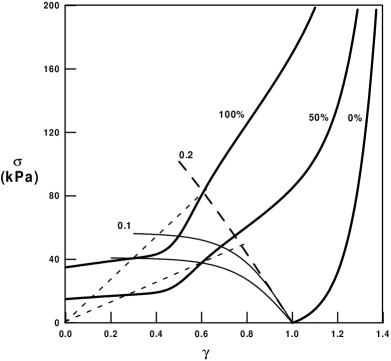
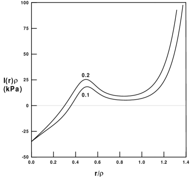
As seen from the expression for , Eq. (11), the important quantity is . The point of instability is where calculated at the inner radius equals the value of at the outer radius. This can be illustrated geometrically by drawing a line in the plot of versus (Fig. 3) from through . If the point lies above this line, is positive and the cylindrical form is stable. If however the point lies below this line, is negative, leading to an instability of the cylindrical form.
Under normal physiological conditions, the angular stress in blood vessels increases linearly to exponentially with the normalized length [7, 16] (Fig. 3), and the value of is therefore positive (Fig. 4). This ensures that the blood vessel keeps its cylindrical shape – no bending arguments are needed to explain the stability of the cylindrical shape of a blood vessel. However, when acute hypertension is induced by infusion of a strong vasoconstricting agent like angiotensin II, there will be a substantial reduction of the inner radius in small arteries and large arterioles due to contraction of the smooth muscle cells. The operating point for the vessel will now be on a less steep part of the curve (Fig. 3), and when the radius is reduced below a certain inner radius at which , the value of becomes negative (Fig. 4). This will result in an instability of the cylindrical form, giving rise to the ‘sausage-string’ pattern. The dominant (fastest growing) mode is given by , which will correspond to ‘sausages’ of length . Insertion of typical values [17] for the various parameters of the model yields , hence the length of the ‘sausages’ will be 5-10 times the radius of the relaxed vessel. The theory therefore predicts that the ‘sausages’ will have an elongated shape with a length that decreases as the vessel radius gets smaller. This is in good agreement with experimental observations [2] (Fig. 1).
A way to view the basic phenomenon underlying the instability is to note that, when becomes negative, the pressure at slightly larger radii is smaller than at slightly smaller radii. Accordingly, the resulting flow will be directed from low-radii regions to high-radii regions, causing the small radii to become even smaller, and the large radii to become larger. This continues until the pressure stabilizes at a value which is the same for both the large radius and the small radius . The stabilization is only possible, because the pressure for radii above the instability, , again increases with . The theory allows an estimate of the radius in the dilated regions, . Assuming that is small (close to zero), we can estimate the final value of by the condition . Interestingly, the almost linear stress function in the region above (Fig. 3) gives rise to a decay of in the same region (Fig. 4). As a consequence can become quite large. However, close to , the stress increases exponentially due to the elastic properties of the vessel wall [7], and the value of will increase rapidly. This will effectively prevent from attaining a value substantially larger than the relaxed radius, , of the vessel. However, may be larger than the working radius of the vessel under normal physiological conditions, because the normal working radius is smaller than the relaxed radius [7]. This may explain why previous work have suggested that the dilated regions represented a ‘blow out’ due to mechanical failure of the vessel wall [5].
The ‘sausage-string’ pattern following infusion of angiotensin II have been found to occur predominantly in small arteries and large arterioles [2]. The present analysis predicts that large vessels will be stable. Their operating point are on the steep portion of the curve due to their high pressure. As seen from Fig. 3, the contraction is here limited, thus preventing the larger vessels from reducing their radius below the critical value . As arterial vessels gets smaller the wall-to-lumen ratio increases [7]. From the expression for , Eq. (11), we find that as well as decreases with increasing wall-to-lumen ratio. Hence, the ‘sausage-string’ instability is less likely to appear in blood vessels with large wall-to-lumen ratios. It seems that the transmural pressure and the contractile potential sets an upper limit, and the wall-to-lumen ratio a lower limit for vessels that will undergo the ‘sausage-string’ instability in response to an acute increase in blood pressure.
In summary, we have demonstrated that during severe vasoconstriction, the normal cylindrical shape of a blood vessel may become unstable, and as a result the vessel exhibit a periodic pattern of constrictions and dilatations. The sausage-string pattern is not caused by a mechanical failure of the vessel wall due to the high blood pressure, but is the expression of an instability. The instability is related to the Rayleigh instability, and to the ‘pearling’ instability seen in tubular lipid membranes. The mechanism behind the instability, however, is novel, involving the nonlinear elasticity characteristics of the vessel wall. The developed theory explains many of the key features observed experimentally, e.g. the predominant occurrence in small arteries and large arterioles, and most likely in those with small wall-to-lumen ratios.
The present study was supported by grants from the Danish Natural Science Research Council, the Danish Medical Research Council, the Novo-Nordisk Foundation and the Danish Heart Association.
REFERENCES
- [1] F. B. Byrom, Lancet 2, 201 (1954); F.B. Byrom, Prog. Cardiovasc. Dis. 1, 31 (1974).
- [2] J. Giese, Acta Pathol. Microbiol. Scand. 62, 497 (1964).
- [3] J. Giese, The Pathogenesis of Hypertensive Vascular Disease (Munksgaard, Copenhagen, 1966).
- [4] F. Gustafsson, Blood Pressure 6, 71 (1997).
- [5] L.J. Beilin and F.S. Goldby, Clin. Sci. Mol. Med. 52, 111 (1977).
- [6] In the Hagen-Poiseuille approximation, the fluid conductance is , where is the dynamic viscosity of the fluid (blood), but the specific form of is not crucial for our purpose. Moreover, we neglect the pressure drop along the vessel, noting that this is much smaller than the transmural pressure.
- [7] Y.C. Fung, Biomechanics. Mechanical Properties of Living Tissues, 2nd Ed. (Springer-Verlag, New York, 1990); Y.C. Fung, Biomechanics. Motion, Flow, Stress, and Growth (Springer-Verlag, New York, 1990).
-
[8]
The principal curvatures and are given by the two
relations:
(13) - [9] J. Plateau, Statique Experimentale et Theorique des Liquides Soumis aux Seules Forces Moleculaires (Gautier-Villars, Paris, 1873).
- [10] Lord Rayleigh, Philos. Mag. 34, 145 (1892).
- [11] P. Nelson, and T. Powers, Phys. Rev. Lett. 74, 3384 (1995); R.E. Goldstein, P. Nelson, T. Powers, and U. Seifert, J. Phys. II France 6, 767 (1996).
- [12] R. Bar-Ziv and E. Moses, Phys. Rev. Lett. 73, 1392 (1994).
- [13] R. Feldberg, M. Colding-Jørgensen, and N.-H. Holstein-Rathlou, Am. J. Physiol. 269, F581 (1995).
- [14] The normalized length, , is equal to where is the actual length of a tissue strip and is the resting length. The corresponding strain, , is by definition equal to .
- [15] We have . This equation suggests a useful change of variable, from to , where . While varies between values and , which under perturbations changes along the axis, varies between the fixed values and . The normalized length at a radius is simply the ratio between and its relaxed value , i.e. .
- [16] R.W. Gore, Circ. Res. 34, 581 (1974); M.J. Davis, and R.W. Gore, Am. J. Physiol. 256, H630 (1989).
- [17] For = 0.1, and = 100 kPa, we have kPa.