Experimental evidence for phase synchronization transitions in human cardio-respiratory system
Abstract
Transitions in the dynamics of complex systems can be characterized by changes in the synchronization behavior of their components. Taking the human cardio-respiratory system as an example and using an automated procedure for screening the synchrograms of 112 healthy subjects we study the frequency and the distribution of synchronization episodes under different physiological conditions that occur during sleep. We find that phase synchronization between heartbeat and breathing is significantly enhanced during non-rapid-eye-movement (non-REM) sleep (deep sleep and light sleep) and reduced during REM sleep. Our results suggest that the synchronization is mainly due to a weak influence of the breathing oscillator upon the heartbeat oscillator, which is disturbed in the presence of long-term correlated noise, superimposed by the activity of higher brain regions during REM sleep.
pacs:
05.45.Xt, 87.19.Hh, 87.19.UvPeriodic events are ubiquitous in many natural systems Glass . If two oscillatory processes are weakly coupled, they can become phase synchronized. Transitions in the synchronization behavior have been shown to be important characteristics of coupled oscillatory model systems Zusatz . It has also been found that noise, when applied identically to different nonlinear oscillators, can induce, enhance, or destroy synchronization among them noise ; chem ; laser . However, phase synchronization is difficult to study in experimental data which are very often inherently nonstationary and thus contain only quasiperiodic oscillations. Among the few recent experimental studies are coupled electrochemical oscillators chem , laser systems laser , and climate variables geophys . In physiology, the study of phase synchronization focusses on cardio-respiratory data (see below) and encephalographic data encephalography . Here, in order to obtain reliable experimental evidences of transitions in phase synchronization behavior, we consider cardio-respiratory synchronization in humans during sleep, because homogeneous long-term data for well defined conditions of a complex system is available in this particular example.
First approaches for the study of cardio-respiratory synchronization have been undertaken by the analysis of the relative position of inspiration within the corresponding cardiac cycle Engel . More recently, phase synchronization between heartbeat and breathing has been studied during wakefulness using the synchrogram method tutorial ; Schaefer ; Toledo ; Stefanovska . While long synchronization episodes were observed in athletes and heart transplant patients (several hundreds of seconds) Schaefer ; Toledo , shorter episodes were detected in normal subjects (typical duration less than hundred seconds) Toledo ; Stefanovska ; Prokhorov . For two recent models of cardio-respiratory synchronization, see Kotani ; Smely .
In this Letter we use the concept of phase synchronization to develop an automated synchrogram based procedure and study interactions between cardiac and respiratory oscillations under different well-defined physiological conditions. We focus on the sleep stages, where external stimuli are absent. It is well known that healthy sleep consists of cycles of roughly 1-2 hours duration. Each cycle is characterized by a sequence starting usually with light sleep, followed by deep sleep and REM sleep (rapid eye movement) rechtschaffen . We find the intriguing result that during REM sleep cardio-respiratory synchronization is suppressed by approximately a factor of 3 compared with wakefulness. On the other hand, during non-REM sleep, it is enhanced by a factor of 2.4, again compared with wakefulness. In addition, we find that these significant differences between synchronization in REM and non-REM sleep are very stable and occur in the same way for males and females, independent of age and independent of the body mass index (BMI). Hence it seems likely that – similar to the long-term correlations Boston occurring in both heartbeat HBcor and breathing BRcor during REM sleep but not during non-REM sleep – the differences are caused by the influence of the activity of higher brain regions on both oscillators fn1 .
First, we developed an algorithm, which detects epochs of synchronization automatically and systematically. The algorithm is applied on simultaneous records of respiration (from a thermistor placed close to the subject’s nose) and heartbeat (from electrocardiograms) obtained for 112 healthy subjects during sleep. The data was recorded in the EU project SIESTA in several European sleep laboratories Siesta , and the average length of the records is 7.9 hours with a standard deviation of 25 minutes. Sleep stages have been determined by visual evaluation of electrophysiological recordings of brain activity rechtschaffen . We can thus assign the synchronization episodes to the specific sleep stages. In addition, we constructed surrogate data by random combination of heartbeat and breathing signals from different subjects Toledo . We found that the total duration of the detected synchronization episodes in real data is increased by a factor 2.3 as compared with the surrogate data, suggesting that most of the detected episodes are real spurious .
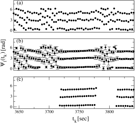
Our algorithm for the detection of phase synchronization episodes is based on the study of cardio-respiratory synchrograms tutorial ; Schaefer ; Toledo ; Stefanovska . For each record, the times of heartbeats are mapped on the continuous cumulative phase of the respiratory signal, which we obtain by unfolding the phase of the Hilbert transform of the normalized thermistor recording. Figure 1(a) shows a representative synchrogram, where is plotted versus . In case of :1 synchronization (i. e., if heartbeats fit to one breathing cycle) one observes parallel horizontal lines in the synchrogram [ in Fig. 1(a)]. In general, to find different ratios : of phase synchronization, we plot versus .
While most earlier work relies on a visual evaluation of the synchrograms Schaefer ; Stefanovska , we detect the episodes in a fully systematic way. For each synchronization ratio : we first replace the phase points in each respiratory cycles by the averages calculated over the corresponding points in the time windows from to [Fig. 1(b)]. In the second step, the algorithm deletes all phase points where the mean standard deviation of the points in each breathing cycles, , is larger than . In the third step, only the phase points in uninterrupted sequences of durations exceeding seconds are kept [Fig. 1(c)].

Figure 2(a) shows a comparison of the detected synchronization rates in real data and in surrogate data for several values of the parameter . The ratio of the mean percentage of synchronization in real data over the mean percentage in surrogate data increases from 1.6 for s to 3.4 for s. There is no generic limit for . However, we choose s in order to keep the number of arbitrary detection of synchronization at an acceptable low level (% in the surrogate data) while at the same time still detecting synchronization episodes during all kinds of sleep stages. In addition, for s we have only one effective time scale parameter, which is identical with the standardized time frame used for the detection of sleep stages rechtschaffen . We note that changing the parameter has a similar effect on the results as changing , and we chose based on similar considerations as for . Our results do hardly depend on the duration of the initial running average.
When studying the percentages of synchronized episodes in real data separately during wakefulness, REM sleep, and non-REM sleep we obtain a highly significant difference in the frequency of cardio-respiratory synchronization between the two major physiological states during sleep. We find 3.8% synchronization in non-REM sleep compared with just 0.6% in REM sleep – a difference by a factor of 6.3. Wakefulness is clearly intermediate, since we find 1.6% for it. Similar differences are observed for other values of and . Since our data base contains records of 112 subjects, we can also study synchronization separately for several age groups, several groups with different body mass index (BMI), and men and women. Figures 2(b,c) show that the results, for wakefulness, REM sleep, and non-REM sleep are practically the same for both genders, all BMI groups, and all age groups, although both, heart rate and breathing rate are known to depend on BMI and age. These results prove that our finding of significant differences between the cardio-respiratory synchronization in REM and non-REM sleep is very stable.
Similar stable differences between REM and non-REM sleep were found in the correlation properties of both heartbeat HBcor and breathing BRcor fluctuations, but hardly in the magnitude of these fluctuations fn1 . The differences were attributed to the influence of the central nervous system with its sleep stage regulation in higher brain regions on the autonomous nervous system. The similarity leads us to suggest that the diminished synchronization during REM sleep is also caused by influences of the central nervous system. As long as the heartbeat oscillator and the breathing oscillator (as parts of the autonomous nervous system) are only affected by uncorrelated noise from higher brain regions, they run like two weakly coupled oscillators – and they clearly show synchronization as expected, possibly enhanced by the noise noise . However, if the higher brain regions are more active and impose long-term correlated noise on the two oscillators, as is the case during REM sleep, the noise disturbs the emergence of synchronized patterns, leading to a drastic reduction of synchronization episodes. Hence we suggest from the experimental data that correlated noise is rather suppressing synchronization while uncorrelated noise might increase it.
Our interpretation is consistent with the result that cardio-respiratory synchronization is enhanced in heart transplanted patients, where correlated signals from the brain can hardly affect the heartbeat oscillator Toledo . Hence, it supports that any relation of the synchronization patterns with cardiac impairments can only be an indirect one as reported recently Hoyer . Diminished long-term correlated regulation activity might explain the increase of synchronization in well-trained athletes Schaefer , where fluctuations of heartbeat and breathing might be avoided to optimize the cardiovascular system for optimal performance.
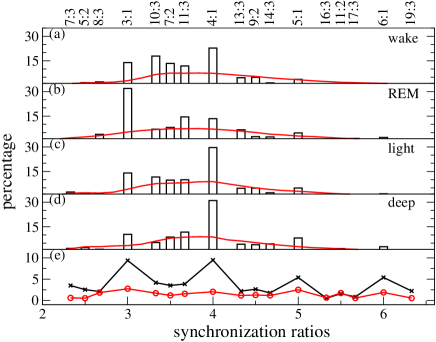
In order to gain insight into the mechanism of cardio-respiratory phase synchronization we have studied, again for all 112 subjects, the distribution of the synchronization ratios :, where cardiac cycles are synchronized with breathing cycles. Figure 3 shows the normalized histograms of the synchronization ratios during (a) wakefulness, (b) REM sleep, (c) light sleep, and (d) deep sleep. The underlying continuous curves in Fig. 3(a-d) show the distributions of the frequency ratios between heartbeat and breathing independent of synchronization.
To clarify the efficiency of the synchronization mechanism, we show the quotient of the synchronization rate histogram with the distribution of the frequency ratios (crosses) in Fig. 3(e). When comparing with the corresponding curve for surrogate data (circles) it becomes obvious that :1 synchronization is preferred. This involves not just the common ratios of 3:1, 4:1, and 5:1, but also clearly 6:1 synchronization. In particular, we do not find any indication of a suppression of 4:1 synchronization as has been reported recently based on modelling Kotani . This suggests that the feedback from baroreceptors to the respiratory centers introduced in the model is probably not very important in the healthy subjects we studied. The synchronization ratios :2 and :3 are weakly efficient if is low, but not efficient at all for large values of . Hence, Fig. 3(e) proves that cardio-respiratory synchronization is nearly limited to frequency ratios : with very small , but quite independent of the number of heartbeats .
¿From this behavior we suggest that the physiological synchronization mechanism is mainly based on an interaction of the respiratory cycle upon heartbeat and not vice versa. This assumption can explain the weak (or absent) efficiency of : synchronization with . If the heartbeat oscillator gets a synchronizing kick at a particular phase of each respiratory cycle and , only half or even less of the kicks coincide with a heartbeat and thus : synchronization cannot be effective. The assumption is consistent with the result obtained when studying the direction of synchronization in children and adults Rosenblum and in a recent model Smely . It is also coherent with the result that synchronization is enhanced under paced respiration Prokhorov .
In conclusion, we have studied cardio-respiratory phase synchronization during different well-defined physiological stages in sleep for a large data base of healthy subjects. We observed clearly reduced synchronization during REM sleep and enhanced synchronization during non-REM sleep. The result is stable for all studied subgroups of subjects; it is neither affected by gender, nor by age, nor by BMI. Since REM and non-REM sleep differ mainly in the type of activity of higher brain centers, it seems probable that the differences in cardio-respiratory synchronization are caused by the more and less long-term correlated regulation actions of the brain during REM and non-REM, respectively. Heart rate and breathing rhythm generators behave like two weakly coupled oscillators, where the coupling direction is from breathing to heartbeat. They become synchronized if uncorrelated noise is imposed from the brain while long-term correlated noise disturbs the emergence of the synchronized patterns. Hence, the experimental data suggests that correlated noise is suppressing synchronization while uncorrelated noise might increase it.
Acknowledgement: We thank Shay Moshel, Meir Plotnik, and Diego Rybski for discussions. This work has been supported by the Deutsche Forschungsgemeinschaft (grants KA 1676/3 and PE 628/3), by the Minerva Foundation, by the Israel Science Foundation, and by the EU project DAPHNet (grant 018474-2).
References
- (1) S.H. Strogatz and I. Stewart, Sci. Am. 269, 102 (1993); L. Glass, Nature (London) 410, 277 (2001); S.H. Strogatz, Sync: How Order Emerges from Chaos in the Universe, Nature, and Daily Life (Penguin Books, 2004).
- (2) M.G. Rosenblum et al., Phys. Rev. Lett. 76, 1804 (1996); E. Rosa, Jr. et al., ibid. 80, 1642 (1998); K. J. Lee et al., ibid. 81, 321 (1998); G. V. Osipov et al., ibid. 91, 024101 (2003); Z. Liu et al., Europhys. Lett. 71, 200 (2005); D.A. Smirnov and R.G. Andrzejak, Phys. Rev. E 71, 036207 (2005); Y.-Ch. Lai et al., ibid. 73, 026214 (2006); K. Wood et al., Phys. Rev. Lett. 96, 145701 (2006).
- (3) A. Maritan and J.R. Banavar, Phys. Rev. Lett. 72, 1451 (1994); Ch. Zhou and J. Kurths, ibid. 88, 230602 (2002); J. Teramae and D. Tanaka, ibid. 93, 204103 (2004); B. Blasius, Phys. Rev. E 72, 066216 (2005); Sh. Guan et al., Phys. Rev. E 73, 046210 (2006); S.F. Brandt et al., Phys. Rev. Lett. 96, 034104 (2006).
- (4) Ch. Zhou et al., Phys. Rev. Lett. 89, 014101 (2002); I.Z. Kiss et al., Phys. Rev. E 70, 026210 (2004).
- (5) S. Boccaletti et al., Phys. Rev. Lett. 89, 194101 (2002); C.S. Zhou et al., Phys. Rev. E 67, 066220 (2003).
- (6) D. Maraun and J. Kurths, Geophys. Res. Lett. 32, L15709 (2005).
- (7) P. Tass et al., Phys. Rev. Lett. 81, 3291 (1998); L. Angelini et al., ibid. 93, 038103 (2004).
- (8) P. Engel et al., Pflügers Arch. 298, 258 (1968); H. Passenhofer and T. Kenner, Pflügers Arch. 355, 77 (1975); F. Raschke, in: Temporal Disorder in Human Oscillatory System, edited by L. Rensing et al. (Springer, Berlin, 1987), pp. 152.
- (9) M.G. Rosenblum et al., in: Handbook of Biological Physics 4, ed. S. Gielen and F. Moss (Elsevier, New York, 2001); A.S. Pikovsky et al., Synchronization – A universal concept in nonlinear science (Cambridge University Press, 2001).
- (10) C. Schäfer et al., Nature 392, 239 (1998); Phys. Rev. E 60, 857 (1999).
- (11) E. Toledo et al., Med. Eng. Phys. 24, 45 (2002).
- (12) M.B. Lotric and A. Stefanovska, Physica A 283, 451 (2000); A. Stefanovska et al., Phys. Rev. Lett. 85, 4831 (2000); M.-Ch. Wu and Ch.-K. Hu, Phys. Rev. E 73, 051917 (2006).
- (13) M.D. Prokhorov et al., Phys. Rev. E 68, 041913 (2003).
- (14) K. Kotani et al., Phys. Rev. E 65, 051923 (2002).
- (15) V.N. Smelyanskiy et al., Phys. Rev. Lett. 94, 098101 (2005).
- (16) A. Rechtschaffen and A. Kales, A manual of standardized terminology, techniques, and scoring system for sleep stages of human subjects (U.S. Government Printing Office, Washington, 1968).
- (17) C.-K. Peng et al., Phys. Rev. Lett. 70, 1343 (1993); P.Ch. Ivanov et al., Europhys. Lett. 48, 594 (1999); Nature 399, 461 (1999).
- (18) A. Bunde et al., Phys. Rev. Lett. 85, 3736 (2000); J.W. Kantelhardt et al., Phys. Rev. E 65, 051908 (2002); Europhys. Lett. 62, 147 (2003); T. Penzel et al., IEEE Transact. Biomed. Eng. 50, 1143 (2003).
- (19) J.W. Kantelhardt et al., Physica A 319, 447 (2003); S. Rostig et al., Sleep 28, 411 (2005).
- (20) We note that there is very little (%) increase in the amplitude of heartbeat or breathing fluctuations during REM sleep when compared with non-REM sleep. The changes in the synchronization behavior can thus not be due to variations in the strength of the influences from the brain. Rather they must be due to the correlation structure imposed by these influences. Long-term correlations are nearly absent in both, heartbeat and breathing during non-REM sleep HBcor ; BRcor .
- (21) G. Klösch et al., IEEE Eng. in Med. and Biol. 20/3, 51 (2001); H. Danker-Hopfe et al., J. Sleep Res. 13, 63 (2004).
- (22) We note that the detection of phase synchronization in oscillators affected by the same uncorrelated noise might be spurious if band-pass filtering techniques (Fourier filtering) are applied [L. Xu et al., Phys. Rev. E 73, 065201(R) (2006)]. However, our synchrogram based algorithm works without any band-pass filtering.
- (23) D. Hoyer et al., Med. Eng. Phys. 24, 33 (2002).
- (24) M.G. Rosenblum et al., Phys. Rev. E 65, 041909 (2002); M. Palus and A. Stefanovska, Phys. Rev. E 67, 055201(R) (2003).