Demonstration of an erbium doped microdisk laser on a silicon chip
Abstract
An erbium doped micro-laser is demonstrated utilizing microdisk resonators on a silicon chip. Passive microdisk resonators exhibit whispering gallery type (WGM) modes with intrinsic optical quality factors of up to and were doped with trivalent erbium ions (peak concentration using MeV ion implantation. Coupling to the fundamental WGM of the microdisk resonator was achieved by using a tapered optical fiber. Upon pumping of the erbium transition at 1450 nm, a gradual transition from spontaneous to stimulated emission was observed in the 1550 nm band. Analysis of the pump-output power relation yielded a pump threshold of 43 W and allowed measuring the spontaneous emission coupling factor: .
The increasing demand in computing power and communication bandwidth has generated an increased interest in the field of silicon photonics which aims at creating photonic elements utilizing standard, complementary metal oxide semiconductor (CMOS) processing technology and materials, such as silica and silicon. As silica and silicon intrinsically lack direct optical transitions, alternative methods such as erbium doping or creating nanostructures Min1996 ; Zacharias2002 have been used to achieve optical emission. Despite these advances, achieving lasing in CMOS compatible structures has remained challenging and has only been observed recently via the Raman nonlinearity Kippenberg2004 ; Rong2005 and by Er doping of silicon chip-based silica toroid micro-cavities Polman2004 .
In this context, erbium is a particularly promising optical dopant as it provides gain in the 1.55 m telecommunication range, and can be incorporated into a by ion implantation Polman1997 , which is an inherently CMOS compatible process. However, due to the small emission cross section of Er at 1.55 ) in conjunction with the fact that Er concentrations are limited to ions due to clustering, the modal gain is limited to approximately 7 dB/cm. Lasing action under these Er gain conditions requires optical resonators with quality factors (assuming mode overlap and refractive index n=1.44). These quality factors are readily available in toroidal micro-cavities Armani2003 or silica microspheres Braginskii1990 , which use a laser assisted reflow process to create ultra-high Q cavities. Indeed, using these microcavity geometries rare-earth doped microcavity lasers were first demonstrated Polman2004 ; Yang2003 ; Sandoghdar1996 . However, the use of a laser reflow makes control through ion implantation difficult, since restructuring of the silica takes place. Increased control of the Er distribution relative to the optical mode is essential to achieve low lasing threshold or high gain. In addition, increased control is important in more complex materials systems such as e.g. Er-doped silica co-doped with Si nanocrystals that act as sensitizers for Er Kik2000 .
A more amenable geometry to these studies are planar microdisks McCall1992 ; Kippenberg2003 , which can be fabricated with small transverse dimensions on a Si chip. Fabrication of these disks does not rely on a laser reflow process and doping with rare earth ions or Si ions by ion implantation can be readily performed. Earlier work has already demonstrated that (at 1550 nm) can be achieved in silica microdisks Kippenberg2003 , which indicates the possibility to observe Er lasing in microdisks.
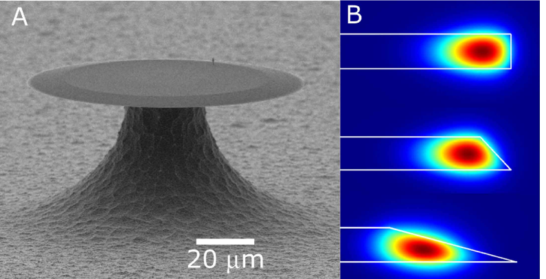
In this Letter we demonstrate lasing in an Er-doped microdisk on a silicon chip. These micro-lasers combine modal engineering of microdisk resonators with the nanoscale precise control of the Er ion distribution in the disk through ion implantation, yielding optimum overlap of the Er ions with the fundamental whispering gallery modes (WGM). By optical pumping of the Er ions at 1.48 m via a tapered optical fiber Spillane2003 , lasing at 1.55 m was observed to occur at a threshold power of less than 43 W. These results demonstrate Er lasing from a microdisk resonator for the first time, using CMOS compatible fabrication. Fabrication of Er-doped microdisk lasers proceeded in several steps. The substrate used in the present experiments was a Si(100) wafer covered with 1 m thick thermally-grown film. This oxide thickness represents a compromise between optical cavity design and ion distribution. The Er ions were incorporated in the by 2 MeV ion implantation at room temperature. The corresponding implantation range of 560 nm was chosen to obtain a good modal overlap with the fundamental whispering gallery modes (WGM)(cf. Fig. 1). A total Er fluence of ions/ ions was implanted, yielding a Gaussian depth distribution with a standard deviation . The average Er density within the implanted layer is ions/. (integrated over the full-width at half maximum of the distribution), which corresponds to a modal gain of 2.66 for . Upon implantation the oxidized wafer was annealed in Ar for 1 hour at 800 C, which yielded optimized photoluminescence intensity and lifetime. The lifetime found (14 ms) demonstrated successful passivation of implantation induced defects. In addition, a reference sample was fabricated in which the Er implantation at an energy of 4 MeV led to an implantation depth exceeding the oxide thickness of 1000 nm. While this implantation depth precludes the observation of lasing, it served as a reference to assess any deterioration of the Q factor due to ion implantation.
Following Er ion implantation, microcavity fabrication was carried out by first defining circular silica microdisks using optical lithography and hydrofluoric etching as detailed in Refs.Armani2003 ; Kippenberg2003 . The resulting microdisk had a diameter of 60 m as shown in the scanning electron micrograph (SEM) in Fig. 1. A key feature of the disk, as seen in Fig. 1, is the strong inclination of the cavity sidewalls that is inherent to the fabrication process, which employs an isotropic HF etch. The disks were then undercut using a gas to isotroptically etch the silicon and thereby create an air-clad whispering gallery mode structure. To optically test the microcavities, tapered optical fibers were used which provide high coupling ideality Spillane2003 .
First, the reference microdisk resonators were tested in which no lasing of Er is expected. In these experiments the cavity modes typically appeared as doublets in the transmission spectrum, which is well known to result from scattering-induced coupling of the clockwise and counterclockwise cavity modes Kippenberg2002 ; Weiss1995 . An example of such a transmission spectrum is plotted as an inset in Fig. 2. Upon fitting the data the inferred Q-factor for each of the two modes was . This is a very high value for a planar microdisk, but still one order of magnitude lower than in the case of toroid microcavities Armani2003 . Importantly, these Q-factors should readily allow for observation of lasing of the Er ions. The observed high Q is attributed to the wedged-shaped edge of the disk microcavity, which is believed to isolate modes from the disk perimeter and thereby reduce scattering losses Kippenberg2003 . This conjecture is further corroborated using numerical finite-element simulations as shown in Fig. 1, which demonstrate that an increased sidewall angle leads to an optical mode that is progressively more removed from the outer, lower cavity boundary (which can induce scattering losses). The Q value of also provides a lower bound on the effect of ion implantation induced defects.
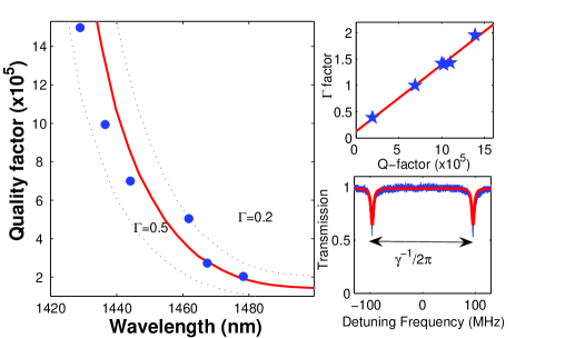
Next, the 2 MeV Er-implanted microdisks, with an active Er distribution peaking at a depth of 560 nm were analyzed. First, the Q-factors in the range from 1410-1480 nm were measured for a microdisk with a diameter of 60 m (and an equivalent free-spectral-range, FSR=9.1 nm) as shown in Fig. 2. To avoid variations in the overlap factor and population-dependent Q, care was taken to measure the Q at the same launched power for the same mode family (fundamental microdisk WGM). Upon approach of the absorption band an increase in loss is evidenced. Specifically, while at 1428 nm the quality factor was observed to be it gradually reduced to a value as low as at 1470 nm. Both the absolute value of the Q factor as well as its spectral dependence as shown in Fig. 2 are in good agreement with the theoretically predicted absorption-limited Q (solid red curve in Fig. 2) assuming =0.3, a typical value for the fundamental WGM modes of the microdisk (cf. Fig. 1). Calculations for =0.2 and =0.5 are shown for comparison (dotted lines). The inset of Fig. 2 shows the Q-dependence of the modal coupling parameter Kippenberg2002 () defined as the ratio of is the splitting frequency of the degeneracy-lifted cavity modes (), and the intrinsic cavity linewidth (), both derived from the transmission spectra as in the inset of Fig. 2. Data are taken for several fundamental WGM. The linear dependence that is observed demonstrates that the quality factors are dominated by absorption losses, in this case by (i.e. since the scattering rate is observed to be nearly constant for all pump modes the variation of must be caused by a change in absorption).
The excited erbium ions within the microcavity readily couple to the cavity modes. Due to the Stark broadening of transitions of in , the ions are coupled to many modes of the cavity. Indeed, as shown in Fig 3, upon pumping at 1450 nm the spectrum collected through the coupled fiber taper contains several peaks throughout the erbium emission band (separated by the FSR of 9.1 nm) clearly demonstrating that the erbium ions are coupled to the (fundamental) cavity modes. Weaker, subsidiary peaks observed in Fig. 3 are attributed to Er ions coupling to fundamental WGM’s of opposite polarization. Note that relative strength of the observed peak emission collected in the tapered fiber depends both on the emission spectrum, and the wavelength-dependent ratio of internal to external quality factor, and therefore does not correspond exactly to the emission cross section spectrum. Upon increasing the launched pump power, the erbium related luminescence in all modes increases linearly (data not shown), demonstrating that the emission observed in Fig. 3(a) is due to spontaneous emission.
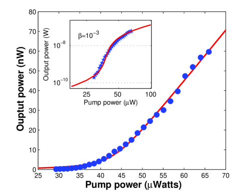
Upon further increase in launched pump power super-linear behavior in the spontaneous emission is observed, as plotted in Fig. 4, followed by a linear pump-output relationship of one of the modes at high power (cf. Fig. 3(b)), which we attribute to lasing. This change is accompanied by an increase in differential slope efficiency for the lasing mode. The remaining (non-lasing) modes did not show this transition. This behavior is well known in microcavity lasers, which due to a large spontaneous emission coupling factor exhibit a gradual transition from spontaneous emission to stimulated emission Yokoyama1989 . From linear interpolation the lasing threshold was estimated to be 43 W. This threshold value is consistent with the predicted valueMin2004 using V cm nm, nm, and assuming that the intrinsic Q-factor of the lasing mode is . The emission was observed to be single-mode for pump powers up to 400 W, and the highest output power observed was 10 W. As we have demonstrated for toroidal cavities Min2004 higher output powers can be achieved for overcoupled conditions, but at the expense of a higher pump threshold.
To determine the spontaneous emission coupling factor (), defined as the fraction of spontaneous emission coupled into the lasing mode with respect to the spontaneous emission into all modes, the input-output power relationship was modeled using a rate equation model Yokoyama1989 , which yields
| (1) |
Here is the cavity photon number, the pump rate (of Er ions) and the cavity loss rate. The solid line in Fig. 4 is a three parameter fit using in Eqn. (1), fitting s to the measured output power, , and . The pump coupling efficiency from fiber into the cavity is assumed unity. The fit exhibits satisfactory agreement with the data. It is noted however, that this model can deviate from the observed dependency pump-output relations close to the threshold, for several reasons. First, the evanescent waveguide coupling renders the coupled power sensitive to the intrinsic cavity Q, which due to the presence of Er is varying. Such a pump power-dependent Q (due to a pump-induced reduction of Er absorption or nonlinear effects), has already been observed in Raman micro-cavity lasers Kippenberg2004 ; Min2003 . Specifically the coupled power () is given by: where denotes the launched power and is the total quality factor at the pump wavelength, which contains a contribution from Erbium absorption ( ) and other cavity loss mechanisms ( ). The former is given by where are the Gilles factors describing gain and loss at the pump wavelength[18] and the normalized upper state population is given (below threshold) by (where is the pump photon flux and the erbium upper state lifetime).For low pump powers , whereas for high pump powers the Q increases to (which can be close to transparency[18]). Since the Er absorption is observed to influence the total cavity Q (cf. Fig, 2), it is clear the pumping dependent Q will lead to loading effects. As the observed Q factors in the Er absorption band are limited by Er absorption for low pump powers, this effect will thus lead to an increased intrinsic Q. Effects of varying Q are most prominent around the threshold, since clamping of the population occurs above threshold, leading to constant Q as is the case for low pump powers. Thus the low and high power laser dynamics are well captured by the above model. A second effect that is not taken into account by the above model is a pump power-dependent Er excited state lifetime, due to up cooperative upconversion between excited Er ions Polman1997 . To obtain an improved fit in the region of low pump and high pump power which determines the value of (and in which the aforementioned effects are negligible), the pump-output relationship was fitted on a double logarithmic scale as shown in the inset of Fig. 4. From the model we derive the spontaneous emission coupling factor, which describes the fraction of spontaneous emission coupled into cavity modes: .
We ascribe this relatively low value of , to a number of effects. First of all, ions in glass exhibit large homogeneous broadening. As a consequence the erbium population can decay via a large number of cavity modes. From the spontaneous emission spectrum in Fig. 2(a), we can estimate that the total number of cavity modes (N) to which the Er ions couple is 20 ( 10 modes at each TE and TM polarization ). Consequently, even in the case of spontaneous emission taking place only in cavity modes the which can be expected is (=1/N). The yet lower experimentally observed value is ascribed to that fact that spontaneous emission also occurs into free-space modes (non-cavity modes).
In summary, we have realized an erbium-implanted microlaser using a silica microdisk on a silicon wafer. A pump threshold as low as 43 W is observed, and the spontaneous emission coupling factor is determined to be for the lasing mode, in fair agreement with theory. The disk geometry presents several advantages over previous toroidal geometries as it enables the direct use of ion implantation, a planar technology, for doping optical microcavities. The CMOS compatibility of all fabrication step, including ion implantation, may enable the use of these microdisk lasers in photonic and opto-electronic components on a Si chip. Furthermore, these cavities are ideal microlaboratories to study fundamental effects of a broad range of ion beam doped optical materials.
.1 Acknowledgements
This work was supported by DARPA and the Caltech Lee Center for Advanced Networking. TJK acknowledges a postdoctoral fellowship from the Caltech Center of the Physics of Information. The Dutch part of this work is part of the research program of FOM which is financially supported by NWO. The authors thank Ali Dabirian from the MPQ for the finite element numerical modeling.
References
- (1) D. K. Armani, T. J. Kippenberg, S. M. Spillane, and K. J. Vahala. Ultra-high-q toroid microcavity on a chip. Nature, 421(6926):925–928, 2003.
- (2) V. B. Braginskii, V. S. Ilchenko, and M. L. Gorodetskii. Optical microresonators with the modes of the whispering gallery type. Uspekhi Fizicheskikh Nauk, 160(1):157–159, 1990.
- (3) P. G. Kik, M. L. Brongersma, and A. Polman. Strong exciton-erbium coupling in si nanocrystal-doped sio2. Applied Physics Letters, 76(17):2325–2327, 2000.
- (4) T. J. Kippenberg, S. M. Spillane, D. K. Armani, and K. J. Vahala. Fabrication and coupling to planar high-q silica disk microcavities. Applied Physics Letters, 83(4):797–799, 2003.
- (5) T. J. Kippenberg, S. M. Spillane, D. K. Armani, and K. J. Vahala. Ultralow threshold microcavity raman laser on a microelectronic chip. Optics Letters, 2004.
- (6) T. J. Kippenberg, S. M. Spillane, and K. J. Vahala. Modal coupling in traveling-wave resonators. Optics Letters, 27(19):1669–1671, 2002.
- (7) S. L. McCall, A. F. J. Levi, R. E. Slusher, S. J. Pearton, and R. A. Logan. Whispering-gallery mode microdisk lasers. Applied Physics Letters, 60(3):289–291, 1992.
- (8) B. K. Min, T. J. Kippenberg, and K. J. Vahala. Compact, fiber-compatible, cascaded raman laser. Optics Letters, 28(17):1507–1509, 2003.
- (9) B. K. Min, T. J. Kippenberg, L. Yang, K. J. Vahala, J. Kalkman, and A. Polman. Erbium-implanted high-q silica toroidal microcavity laser on a silicon chip. Physical Review A, 70(3), 2004.
- (10) K. S. Min, K. V. Shcheglov, C. M. Yang, H. A. Atwater, M. L. Brongersma, and A. Polman. Defect-related versus excitonic visible light emission from ion beam synthesized si nanocrystals in sio2. Applied Physics Letters, 69(14):2033–2035, 1996.
- (11) A. Polman. Erbium implanted thin film photonic materials. Journal of Applied Physics, 82(1):1–39, 1997.
- (12) A. Polman, B. Min, J. Kalkman, T. J. Kippenberg, and K. J. Vahala. Ultralow-threshold erbium-implanted toroidal microlaser on silicon. Applied Physics Letters, 84(7):1037–1039, 2004.
- (13) H. S. Rong, A. S. Liu, R. Jones, O. Cohen, D. Hak, R. Nicolaescu, A. Fang, and M. Paniccia. An all-silicon raman laser. Nature, 433(7023):292–294, 2005.
- (14) V. Sandoghdar, F. Treussart, J. Hare, V. LefevreSeguin, J. M. Raimond, and S. Haroche. Very low threshold whispering-gallery-mode microsphere laser. Physical Review A, 54(3):R1777–R1780, 1996.
- (15) S. M. Spillane, T. J. Kippenberg, O. J. Painter, and K. J. Vahala. Ideality in a fiber-taper-coupled microresonator system for application to cavity quantum electrodynamics. Physical Review Letters, 91(4):art. no.–043902, 2003.
- (16) D. S. Weiss, V. Sandoghdar, J. Hare, V. Lefevreseguin, J. M. Raimond, and S. Haroche. Splitting of high-q mie modes induced by light backscattering in silica microspheres. Optics Letters, 20(18):1835–1837, 1995.
- (17) L. Yang, D. K. Armani, and K. J. Vahala. Fiber-coupled erbium microlasers on a chip. Applied Physics Letters, 83(5):825–826, 2003.
- (18) H. Yokoyama and S. D. Brorson. Rate-equation analysis of microcavity lasers. Journal of Applied Physics, 66(10):4801–4805, 1989.
- (19) M. Zacharias, J. Heitmann, R. Scholz, U. Kahler, M. Schmidt, and J. Blasing. Size-controlled highly luminescent silicon nanocrystals: A sio/sio2 superlattice approach. Applied Physics Letters, 80(4):661–663, 2002.