\ethnote
{Authlist} F. Nessi-Tedaldi \InstfootethSwiss Federal Institute of Technology (ETH), CH-8093 Zürich, Switzerland
Crystals for High-Energy Physics calorimeters in extreme environments
1 Introduction
This report addresses the performance of scintillating crystals used for high-energy physics calorimetry, when operation implies high radiation levels and intense particle fluxes.
The effect of high levels of ionising radiation on crystals has been studied in depth and reported upon by many authors, as crystals were used e.g. in collider experiments, and their growth parameters were optimised for best performance in such environments. They are briefly summarised herein. Hadron collider detectors today share the same concern, but add to it the need to ensure adequate performance when crystals are exposed to large particle fluxes. Such running conditions are namely expected in several experiments under construction or designed. Some new results are thus presented here, together with a fresh look at existing, older ones, to provide, as far as possible, a complete picture.
2 Performance under high ionising radiation levels
Ionising radiation is known to produce absorption bands through formation of colour centres, which reduce the Light Transmission (LT) and thus the Light Output (LO), due to oxygen contamination in alkali halides like and CsI, and to oxygen vacancies and impurities in oxides like BGO and [1]. Phosphorescence or afterglow appear sometimes[2], which increase the noise levels in the detected light signal, possibly worsening the energy resolution (in a negligible way for in LHC experiments[3]), while the scintillation mechanism is generally not damaged. Recovery of damage at room temperature can occur depending on crystal type and growth parameters, giving rise to a dose-rate dependence of damage equilibrium levels[1, 4] and to a recovery speed dependent on the depth of traps. That ionising radiation only affects LT, means the damage can be monitored through light injection and corrected for, as it is done in the CMS Electromagnetic Calorimeter (ECAL)[5].
3 Performance in large particle fluxes
The way hadron fluxes affect crystals has become a crucial question while detectors making use of this calorimetry technique are being constructed. In particular, it had to be ascertained whether such fluxes cause a specific, possibly cumulative damage, and if so, what its quantitative importance is, whether it only affects LT or also the scintillation mechanism. Extensive studies have been recently performed on at IHEP Protvino[6] and, for the CMS ECAL, at CERN and ETH-Zürich[7]. Their main results are quoted and discussed herein.
Crystal tests at Protvino were using and beams and sources up to a few krad at 1 to 60 rad/h at one end, and a very intense mixed beam of charged hadrons, neutrons and up to 3 Mrad at 1 krad/h and 100 krad/h equivalent fluxes at the other end. In individual , and irradiations, the signal loss behaviour is found to be qualitatively similar between electrons and pions, and the damage appears to reach equilibrium at a dose-rate dependent level. Furthermore, no indication of damage to the scintillation mechanism from irradiation is found[8]. A concern remains however, that an additional, specific, possibly cumulative damage from hadrons cannot be excluded and could appear when a high total integrated dose is reached. This concern is partially confirmed by irradiations in the very intense, mixed beam. Under the constant flux used, the damage appears in fact to be steadily increasing with accumulated dose. This is unlike pure ionising radiation damage, which reaches equilibrium at a level depending on dose rate, not beyond what saturation of all colour centres can yield. Therefore, this constitutes an indication for a cumulative, hadron-specific damage.
For CMS, hadron fluences have been calculated[9] for (10 y running at LHC), yielding in the ECAL barrel (end caps) charged hadrons/ . A hadron-specific damage could arise from the production, above a MeV threshold, of heavy fragments (“stars”), with up to 10 m range and energies up to 100 MeV, causing a displacement of lattice atoms and energy losses along their path up to 50000 times the one of minimum-ionising particles. The damage caused by these processes is likely different from the one of ionising radiation, thus possibly cumulative. The primarily investigated quantity was the damage to Light Transmission measured longitudinally through the length (L) of the crystal and quantified as the induced absorption coefficient at peak-of-emission wavelength, , with and the longitudinal Light Transmission at 420 nm before and after irradiation. The Perkin Elmer spectrophotometer used, allows in fact to measure LT very accurately, to better than 1%. Transmission is furthermore related to LO changes, provided scintillation is not affected.
Eight CMS production crystals of consistent quality were irradiated at the IRRAD1 facility of the CERN PS accelerator T7 beam line[10] in a 20 GeV/c proton flux of


(crystals a”, b, c, d, e, h) or of (crystals E, F’, G) 111Prime (respectively ”) indicates a second (or third) irradiation of the same crystal.. To disentangle the contribution to damage from the associated ionising dose, complementary -irradiations were performed at a dose rate of 1 kGy/h on seven further crystals (t, u, v, w, x, y, z) at the ENEA Casaccia Calliope plant[11]. In fact, a flux of has an associated ionising dose rate in of 1 kGy/h. The LT data in Fig. 1(a) show a smooth worsening of LT with increasing proton fluence over the entire range of wavelengths, and a clear shift of the Transmission band-edge. In -irradiated crystals (Fig. 1(b), where also the emission spectrum[12] is indicated), the band-edge does not shift at all, even after the highest cumulated dose reached: just the usual absorption band appears around 420 nm. These data thus give prominence to the qualitatively different fundamental nature of proton-induced and -induced damage.
The correlation in Fig. 2(a), between and fluence, is consistent with a linear


behaviour over two orders of magnitude, showing that proton-induced damage in is predominantly cumulative, unlike -induced damage, which reaches equilibrium[1, 4]. Figure 2(b) shows plotted versus light wavelength for the proton-irradiated crystal a” and for the two - irradiated crystals v and y. The dot-dashed line shows fitted to the data of the proton-damaged crystal a”. The good agreement is an indication of Rayleigh scattering from small centres of severe damage. This is consistent with an origin of damage due to the high energy deposition of heavily ionising fragment along their path, that changes locally the crystal structure. Taking into account the difference in composition and energy spectra between 20 GeV/c protons and CMS, simulations indicate that the test


results cover the CMS running conditions up to . An experimental confirmation is expected in the future from a pion-irradiation of , closely approximating the CMS particle spectrum and energies.
The evolution of Light Output was also monitored on the same set of irradiated crystals, using cosmic muons, traversing the crystals transversely and thus leaving approximately 30 MeV of energy deposit, to excite scintillation. The correlation[13] between and Light Output loss is shown in the top part of Fig. 3 for all proton-irradiated crystals, and at the bottom for all -irradiated ones. The vertical bars indicate the systematic scale uncertainty affecting the data for a’ and F’. For both, proton-irradiated and -irradiated crystals, the measured Light Output loss correlates well with . Furthermore, within the precision of the measurements, no difference can be observed in this correlation between the two sets of crystals and thus no hadron-specific alteration of the scintillation properties can be claimed.
Proton and data are also compared in a study performed on BGO[14]. The changes in band-edge are similar to what is seen in , and long enough after irradiation, when the ionising-radiation damage contribution has recovered, one can extract a remaining proton-induced damage that behaves linearly with fluence, as visible in Fig. 4. The same exercise is not possible on CsI data from the same authors[15] because the damage
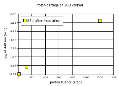
caused by ionising radiation gives a contribution which is too important to allow disentangling the proton-specific one.
In conclusion, one can say that for all crystals commonly used in calorimetry, beyond the well-studied damage from ionising radiation, the understanding of additional contributions to the damage, when crystals experience a substantial hadron flux, has become important since experiments are being built having to cope with such running conditions. A hadron-specific, cumulative contribution, likely due to the intense local energy deposition from heavy fragments, has been observed in and BGO. Over the explored flux and fluence ranges and within the accuracy of the measurements, this contribution is observed to only affect Light Transmission, and thus can be monitored through light injection. Additional studies are expected to consolidate the present understanding of hadron damage.
References
- [1] R.Y. Zhu et al., Nucl. Instr. Meth. A413 (1998) 297-311.
- [2] H. Hofer, P. Lecomte, F. Nessi-Tedaldi, Nucl. Instr. Meth. A433 (1999) 630-636.
- [3] R.Y.Zhu et al. Nucl. Instr. Meth. A376 (1996) 319.
- [4] H.F. Chen, K. Deiters, H. Hofer, P. Lecomte, F. Nessi-Tedaldi, Nucl. Instr. Meth. A414 (1998) 149-155.
- [5] L. Zhang et al., IEEE Trans. Nucl. Sci. 52 (2005) 1123-1130.
-
[6]
V Batarin et al.,
Nucl. Instr. Meth. A512 (2003) 488-505;
V. Batarin et al., Nucl. Instr. Meth. A530 (2004) 286-292. - [7] M. Huhtinen, P. Lecomte, D. Luckey, F. Nessi-Tedaldi, F. Pauss, Nucl. Instr. Meth. A545 (2005) 63-87.
- [8] V. Batarin et al., Nucl. Instr. Meth. A540 (2005) 131-139.
- [9] M. Huhtinen, P. Lecomte, D. Luckey, F. Nessi-Tedaldi, First Results on radiation damage in crystals exposed to a 20 GeV/c proton beam, Talk presented at the 8th ICATPP conference, Como, October 2003.
- [10] M. Glaser et al., Nucl. Instr. Meth. A 426 (1999) 72.
- [11] S. Baccaro, A. Festinesi, B. Borgia, CERN CMS TN-1995/192, Geneva, 1995.
- [12] R.Y. Zhu, IEEE Trans. Nucl. Sci. 51 (2004) 1560-1567.
- [13] P. Lecomte, D. Luckey, F. Nessi-Tedaldi, F. Pauss, to be published.
- [14] M. Kobayashi et al., Nucl. Instr. Meth. 206 (1983) 107-117.
- [15] M. Kobayashi et al., Nucl. Instr. Meth. A328 (1993) 501-505.