,
Coherent control with shaped femtosecond laser pulses applied to ultracold molecules
Abstract
We report on coherent control of excitation processes of translationally ultracold rubidium dimers in a magneto-optical trap by using shaped femtosecond laser pulses. Evolution strategies are applied in a feedback loop in order to optimize the photoexcitation of the Rb2 molecules, which subsequently undergo ionization or fragmentation. A superior performance of the resulting pulses compared to unshaped pulses of the same pulse energy is obtained by distributing the energy among specific spectral components. The demonstration of coherent control to ultracold ensembles opens a path to actively influence fundamental photo-induced processes in molecular quantum gases.
pacs:
32.80.Qk, 33.80.-b, 82.53.-kI Introduction
The interest to study interactions in ultracold molecular gases Doyle et al. (2004) is propelling various research programs to produce and trap dense ultracold molecular ensembles Fioretti et al. (1998); Weinstein et al. (1998); Bethlem et al. (1999); Jochim et al. (2003); Greiner et al. (2003); Zwierlein et al. (2003). The goals and perspectives in this field range from quantum computation DeMille (2002) and quantum scattering Chin et al. (2005) to ultrahigh precision spectroscopy Hudson et al. (2002) and coherent ultracold chemistry Heinzen et al. (2000). The manipulation of molecular wavepacket dynamics with shaped femtosecond pulses may open exciting possibilities to this field. In recent years, the control of molecular processes by shaped fs-laser pulses through application of evolution strategies in a feedback loop has attained considerable success. Since it was proposed Judson and Rabitz (1992), coherent control with shaped femtosecond laser pulses has been applied to a large variety of experiments, i.e. selective fragmentation Assion et al. (1998), population transfer Bardeen et al. (1997), ionization Vajda et al. (2001), and high harmonic generation Bartels et al. (2000). By using an iterative process, the method enables the discovery of an optimal laser pulse shape which drives the system towards a desired target state. It even became possible to decipher the molecular dynamics underlying the laser-molecule interaction by analyzing the optimal pulse shapes Vajda et al. (2001); Daniel et al. (2003).
Theoretical work on photoassociation with pulsed lasers has started several years ago Machholm et al. (1994) and first experiments with thermal mercury atoms in a gas cell Marvet and Dantos (1995) and with sodium atoms in a magneto-optical trap Fatemi et al. (2001) have been performed. More recently, optimal control calculations were done proposing efficient ultracold molecule formation Vala et al. (2000) and vibrational cooling Koch et al. (2004) by appropriately shaped laser pulses. However, up to now there are no experimental results on the application of feedback-controlled optimization to photoassociation or manipulation of ultracold molecules. In this article we present first experimental steps towards this goal by investigating the interaction of femtosecond laser pulses with ultracold ground state molecules in view of a pulsed photoassociation experiment. The low temperature of the atomic and molecular sample, while not directly influencing the interaction with the femtosecond pulses, leads to molecules in very high lying vibrational levels and allows us to study molecules under the same conditions of typical photoassociation experiments. In this work we demonstrate in particular the enhancement of the electronic excitation and subsequent fragmentation of ultracold molecules from a magneto-optical trap using shaped femtosecond laser pulses, which were adaptively optimized by an evolutionary algorithm.
II Experimental setup
In our experiment, schematically depicted in Fig. 1, about 85Rb atoms are captured in a magneto-optical trap (MOT) at a density of atoms/cm3 and a temperature of K. Diatomic rubidium molecules continuously form in the magneto-optical trap due to either three body collisions or light assisted two-body collisions of trapped Rb atoms. They populate predominantly the highest vibrational states below the dissociation limit in the triplet electronic ground state Gabbanini et al. (2000); Kemmann et al. (2004). These Rb2 molecules, which are not trapped by the trap magnetic field, are detected in this experiment via resonant two photon ionization and time-of-flight mass analysis. The ionization laser is a 15 Hz Nd:YAG pumped dye laser at wavelengths between 600 and 610 nm, a spectral width of 0.5 cm-1 and a pulse energy of 20 mJ. In the steady state of molecule formation and loss a maximum count rate of 0.5 Rb molecular ions per laser pulse is observed, measured at a wavelength of 602.5 nm.
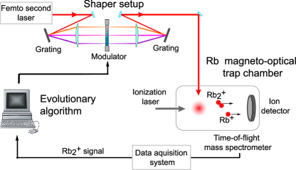
The femtosecond (fs) laser pulses are generated in a Ti:Sapphire oscillator (Tsunami; Spectra Physics) that provides pulses of 120 fs duration (FWHM) at a rate of 80 MHz, a spectral width of nm and an energy of up to 18 nJ per pulse. Low pulse energies ensure that the laser-molecule interaction is well described in a perturbative picture and no high intensity effects have to be considered. To modify the spectral components of the pulses we use a pulse shaper that allows independent phase and amplitude modulation Wefers and Nelson (1995). It consists of a liquid crystal modulator (CRI; SLM-256) Weiner et al. (1992) with 2x128 pixels, placed in the Fourier plane of a double grating zero dispersion compressor. A lens focuses the beam to a spot of 150 m diameter at the center of the trap, illuminating about 10% of the cloud volume. For the experiments the fs-laser is tuned in the range between 780 and 820 nm.
III Laser-induced loss
Transform-limited fs-pulses with wavelengths near the Rb resonance lines and focused into the MOT are found to strongly interact with the trapped atoms, as observed through a significant decrease of MOT fluorescence. At small pulse energies, the trap loading rate outside the femtosecond beam can partly compensate for the losses in the focus, resulting in an area of reduced fluorescence predominantly where the beam is passing, whereas for high energies the trap is completely depleted. We attribute this to photon scattering from the femtosecond pulses, which causes an outward directed light force on the atoms, leading to trap loss. The effect is significantly stronger when the D2 atomic resonance at 780 nm was part of the pulse spectrum than for the D1 resonance at 795 nm, reflecting the higher transition dipole moment of the D2 line. A narrow slit, representing a bandpass of 0.3 nm width, is scanned through the Fourier plane of the pulse shaper, which showed that only components resonant with the D1 and D2 atomic transitions are responsible for this effect. Singly-charged rubidium ions have been detected when the central pulse frequency was resonant with one of the atomic transitions, indicating that resonant three-photon ionization contributes to the loss of atoms from the MOT. To study the laser pulse interaction with rubidium molecules, the atomic resonance components were removed from the pulse spectrum by a notch filter, realized by a physical block in the shaper’s Fourier plane. In this way atomic losses from the MOT could be reduced below the detection threshold.
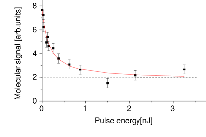
The rubidium dimers interact with the fs-laser pulses over the entire accessible range of central wavelengths from 780 nm to 820 nm. As shown in Fig. 2, the molecular signal decreases rapidly at small pulse energies and levels off to 25% at a pulse energy of 0.6 nJ. As only molecules in the electronic ground state are detected, the signal reduction can be attributed to excitation by the fs-pulses. The process can be modeled by a simple rate equation for the number of detected ground state molecules:
| (1) |
where is the production rate of molecules from trapped atoms, is the rate of molecular loss induced by interaction with the femtosecond laser, and the molecular loss rate induced by other processes such as rest gas collisions or gravitation accelerating the molecules out of the detection volume. In the steady state the number of molecules is given by
| (2) |
The curve in fig. 2 represents a fit to the data assuming a linear dependence of on the pulse energy of the laser, i.e. . The assumption of a quadratic or higher order dependence of the excitation rate on pulse energy does not fit the data. This indicates that, in the regime of pulse energies employed in the experiment, the interaction with the molecules has the character of an effective one-photon excitation Ban et al. (2005).
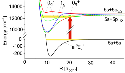
According to Gabbanini et al. (2000), the molecules in the MOT initially populate the highest levels in the a state. Due to selection rules and Franck-Condon factors, they are preferably excited to the 0 and 1g 5s5p1/2 states (see Fig. 3). Emission back into the electronic ground state could only lead to signal reduction if the vibrational level population moves out of the excitation window of the ionization laser. However, scans of the detection laser with and without fs-beam show reduced but qualitatively similar spectra which should not be the case for a vibrational redistribution. Instead it can be expected that excited molecules absorb further photons, so the whole process of molecular loss can be regarded as a resonance enhanced multi-photon excitation, followed by dissociation, predissociation or ionization. This happens either within one pulse, or, as the laser repetition rate is comparable to the lifetime of the first excited state, it occurs in the subsequent pulse. At high energies all molecules in the laser focus are excited or dissociated and the residual signal in Fig. 2 is due to molecules which did not interact with the femtosecond laser. This shows that most of the molecules are produced within a small volume inside the MOT which is consistent with the picture that they form at the MOT center where the atom number density is at its maximum Townsend et al. (1995). At high energies all molecules in the laser focus are excited or dissociated and the residual signal in Fig. 2 is due to molecules which did not interact with the femtosecond laser, indicated by the dashed line. This shows that most of the molecules are produced within a small volume inside the MOT which is consistent with the picture that they form at the MOT center where the atom number density is at its maximum Townsend et al. (1995).
IV Application of coherent control
In order to demonstrate the practical applicability of coherent control concepts to ultracold molecules, the Rb signal acts as an input for a self-learning optimization algorithm which autonomously programs the pulse shaper in a closed loop experiment. The algorithm is based on evolution strategies and is described in detail in Bartelt et al. (2001). Because of the small molecular ion count rate and hence the low signal-to-noise ratio the signal is averaged over 128 dye laser pulses for each individual of the algorithm. To reduce the search space for the learning algorithm we chose a mixed scheme of parametric amplitude and free phase optimization where the algorithm tries to find the optimal pulse shape under the restriction that only a few sharp spectral peaks contribute to the pulse shape. During an optimization the parameters of these peaks, their spectral positions and amplitudes, are altered to find the most efficient excitation pulse. Moreover, the phase was optimized freely in order to allow a temporal modulation of the pulse. The evolutionary algorithm administrates 31 individuals, each representing the pulse shaper parameters to produce pulses consisting of up to eleven Gaussian peaks of 7 cm-1 FWHM.
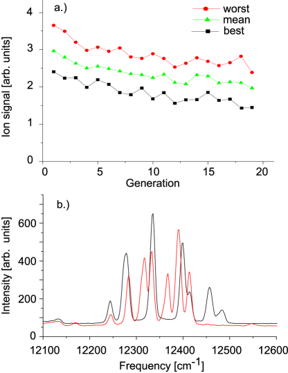
The adaptive algorithm was applied to manipulate the excitation pulses with the aim to minimize the molecular signal from the MOT. For each iteration the ion signals corresponding to the best and worst individuals are protocoled together with the mean fitness of the whole generation. As depicted in Fig. 4(a), all three signals decrease during the particulate optimization to about 70% of the initial value after 20 iterations. The spectra of the final best individuals of two successive runs shown in Fig. 4(b) display several peaks which coincide in some but not all spectral positions. The frequency span of the fs-pulse supports our assignment of excitation to the 0 and 1g 5s5p1/2 states (see Fig. 3). By comparing the excitation yield of the best individuals with transform-limited pulses of the same energy it is observed that the optimized pulse excites the molecules on average 25% more efficiently, which demonstrates the feasibility and potential of adaptive control.
We attribute the observed excitation enhancement to an increased spectral intensity at particular molecular resonances found by the evolutionary algorithm. Starting from a narrow band in the a state Gabbanini et al. (2000); Kemmann et al. (2004), molecules are excited into bound states below the D1 resonance. By shifting the peak positions, the algorithm finds transition frequencies from this band to certain vibrational states, thereby sharing the pulse energy more efficiently than a broad Gaussian pulse. The algorithm therefore has a large number of possible solutions to choose from and so the final pulse shapes after an optimization are not identical. In the spectral region between 12000 and 12500 cm-1, the vibrational level separation is about 10 cm-1 in the 0 and 1g 5s5p1/2 states, respectively. The high density of states also explains the limited potential of the optimization because the optimization factor depends on the chosen peakwidth which is limited by the shaper resolution. The Franck-Condon factors may also be relevant for the excitation process since they differ for different vibronic transitions and favor particular frequencies which are enhanced in the experimentally acquired spectra. Yet, as the initial ground state population distribution in the vibrational states is not known accurately, no quantitative treatment or assignment can be made.
V Conclusions
In this article we demonstrate the application of iterative adaptive control techniques to the manipulation of ultracold molecules. The minimization of molecular signal from a rubidium magneto-optical trap results in pulse shapes that significantly excite more Rb2 molecules than transform-limited pulses of the same energy. The resulting pulse spectra are not unambiguous because of the large number of possible optimal solutions within the experimental accuracy. For future applications of shaped fs-pulses in photoassociation experiments, the dissociative effect of the pulses has to be suppressed. On the one hand, photoassociated cold molecules may be dissociated by subsequent femtosecond pulses due to the high repetition rate of the femtosecond laser system. Therefore, photoassociated molecules have to be detected directly after their formation by increasing the repetition rate of the detection system, ideally matching the rate of the photoassociating beam. On the other hand, the excitation of ground state molecules may be coherently suppressed while enhancing the molecule formation through actively shaping the pulses. For this purpose, we currently theoretically investigate realistic scenarios for femtosecond photoassociation of cold molecules by numerically simulating the dynamical behavior of an atom pair interacting with an arbitrarily shaped electromagnetic field. It is also envisioned to use specifically shaped femtosecond laser pulses for the cooling of vibrational molecular excitations by pump-dump processes via an intermediate excited state. This approach could be applied to produce quantum-degenerate molecular gases in arbitrary vibrational states or superpositions of states.
Acknowledgements.
We thank C. Koch, R. Kosloff, B. Schäfer-Bung and V. Bonačić-Koutecký for theoretical support and many stimulating discussions. This work was supported by the Deutsche Forschungsgemeinschaft in the frame of the Sonderforschungsbereich 450 and the Schwerpunktprogramm 1116. F. Sauer acknowledges the Studienstiftung des deutschen Volkes, A. Merli thanks the Cusanuswerk.References
- Doyle et al. (2004) J. Doyle, B. Friedrich, R. V. Krems, and F. Masnou-Seeuws, Eur. Phys. J. D 31, 149 (2004).
- Fioretti et al. (1998) A. Fioretti, D. Comparat, A. Crubellier, O. Dulieu, F. Masnou-Seeuws, and P. Pillet, Phys. Rev. Lett. 80, 4402 (1998).
- Weinstein et al. (1998) J. D. Weinstein, R. deCarvalho, T. Guillet, B. Friedrich, and J. M. Doyle, Nature 395, 148 (1998).
- Bethlem et al. (1999) H. L. Bethlem, G. Berden, and G. Meijer, Phys. Rev. Lett. 83, 1558 (1999).
- Jochim et al. (2003) S. Jochim, M. Bartenstein, A. Altmeyer, G. Hendl, S. Riedl, C. Chin, J. Hecker-Denschlag, and R. Grimm, Science 302, 2101 (2003).
- Greiner et al. (2003) M. Greiner, C. A. Regal, and D. S. Jin, Nature 426, 537 (2003).
- Zwierlein et al. (2003) M. W. Zwierlein, C. A. Stan, C. H. Schunck, S. M. F. Raupach, S. Gupta, Z. Hadzibabic, and W. Ketterle, Phys. Rev. Lett. 91, 250401 (2003).
- DeMille (2002) D. DeMille, Phys. Rev. Lett. 88, 067901 (2002).
- Chin et al. (2005) C. Chin, T. Kraemer, M. Mark, J. Herbig, P. Waldburger, H.-C. Nägerl, and R. Grimm, Phys. Rev. Lett. 94, 123201 (2005).
- Hudson et al. (2002) J. J. Hudson, B. E. Sauer, M. R. Tarbutt, and E. A. Hinds, Phys. Rev. Lett. 89, 023003 (2002).
- Heinzen et al. (2000) D. J. Heinzen, R. Wynar, P. D. Drummond, and K. V. Kheruntsyan, Phys. Rev. Lett. 84, 5029 (2000).
- Judson and Rabitz (1992) R. S. Judson and H. Rabitz, Phys. Rev. Lett. 68, 1500 (1992).
- Assion et al. (1998) A. Assion, T. Baumert, M. Bergt, T. Brixner, B. Kiefer, V. Seyfried, M. Strehle, and G. Gerber, Science 282, 919 (1998).
- Bardeen et al. (1997) C. J. Bardeen, V. V. Yakovlev, K. R. Wilson, S. D. Carpenter, P. M. Weber, and W. S. Warren, Chem. Phys. Lett. 280, 151 (1997).
- Vajda et al. (2001) S. Vajda, A. Bartelt, E.-C. Kaposta, T. Leisner, C. Lupulescu, S. Minemoto, P. Rosendo-Francisco, and L. Wöste, Chem. Phys. 267, 231 (2001).
- Bartels et al. (2000) R. Bartels, S. Backus, E. Zeek, L. Misoguti, G. Vdovin, I. P. Christov, M. M. Murnane, and H. C. Kapteyn, Nature 406, 164 (2000).
- Daniel et al. (2003) C. Daniel, J. Full, L. González, C. Lupulescu, J. Manz, A. Merli, S. Vajda, and L. Wöste, Science 299, 536 (2003).
- Machholm et al. (1994) M. Machholm, A. Giusti-Suzor, and F. H. Mies, Phys. Rev. A 50, 5025 (1994).
- Marvet and Dantos (1995) U. Marvet and M. Dantos, Chem. Phys. Lett. 245, 393 (1995).
- Fatemi et al. (2001) F. Fatemi, K. M. Jones, H. Wang, I. Walmsley, and P. D. Lett, Phys. Rev. A 64, 033421 (2001).
- Vala et al. (2000) J. Vala, O. Dulieu, F. Masnou-Seeuws, P. Pillet, and R. Kosloff, Phys. Rev. A 63, 013412 (2000).
- Koch et al. (2004) C. P. Koch, J. P. Palao, R. Kosloff, and F. Masnou-Seeuws, Phys. Rev. A 70, 013402 (2004).
- Gabbanini et al. (2000) C. Gabbanini, A. Fioretti, A. Lucchesini, S. Gozzini, and M. Mazzoni, Phys. Rev. Lett. 84, 2814 (2000).
- Kemmann et al. (2004) M. Kemmann, I. Mistrik, S. Nussmann, H. Helm, C. J. Williams, and P. S. Julienne, Phys. Rev. A 69, 022715 (2004).
- Wefers and Nelson (1995) M. M. Wefers and K. A. Nelson, J. Opt. Soc. Am. B 12, 1343 (1995).
- Weiner et al. (1992) A. M. Weiner, D. E. Leaird, J. S. Patel, and J. R. Wullert, IEEE J. Quant. Elect. 28, 908 (1992).
- Ban et al. (2005) T. Ban, D. Aumiler, and G. Pichler, Phys. Rev. A 71, 022711 (2005).
- Townsend et al. (1995) C. G. Townsend, N. H. Edwards, C. J. Cooper, K. P. Zetie, C. J. Foot, A. M. Steane, P. Szriftgiser, H. Perrin, and J. Dalibard, Phys. Rev. A 52, 1423 (1995).
- Bartelt et al. (2001) A. Bartelt, S. Minemoto, C. Lupulescu, S. Vajda, and L. Wöste, Eur. Phys. J. D 16, 127 (2001).