EXPERIMENTS WITH PARAMETRIC X-RAY RADIATION (PXR) FROM NON-RELATIVISTIC ELECTRONS
Abstract
Interaction of non-relativistic electrons with single crystal target may produce coherent x-rays. That is the result of interference between two known x-ray generation mechanisms having orientational behavior, namely parametric x-rays and coherent Bremsstrahlung. Experiments aimed to PXR research were performed with 50-100 keV electrons and its distinctive features were observed. Requirements to the experimental set-up, detector instrumental response, and targets as well as experiment geometry are discussed in detail. Series of PXR spectra in various conditions were recorded and their distinctive features were observed. Tuning of radiation frequency with crystal-target rotation was observed for the first time for low energy electrons. Dependence of the x-ray frequency on the beam energy was detected. Soft PXR peak with energy below 1 keV was observed for the first time. Possible applications of PXR for structure analysis and crystallography are discussed. These results are obtained in the framework of ISTC project #B626.
I INTRODUCTION
Parametric x-rays produced by non-relativistic electrons passing through a single crystal target were described in 1 on the basis of general theory of PXR 2 . Due to the fact that the angular distributions of x-rays originated from all radiation mechanisms are almost isotropic in this case, peaks produced by coherent effects, i.e. parametric x-rays and coherent Bremsstrahlung (CBS), are the result of PXR and CBS interference and may be observed on the intensive uniform background. Their shape and intensity depend on the target thickness and the detector spectral resolution. Some experiments with non-relativistic electrons are currently known 3 -5 , but their authors outlined the effect taking into consideration only CBS mechanism. Model 1 presumably describes x-rays from non-relativistic electrons in crystal targets more adequately.
To study x-ray properties followed from the theory 1 , we have performed experiments focused on the PXR research with 50100 keV electrons of the electron microscope. Let us briefly consider requirements of the PXR experimental observation. In general case a charged particle passing though matter undergoes elastic and inelastic collisions with the atoms of a medium. As a result, the angular and energy distributions of electrons change during beam passing through a medium. Influence of electron scattering on the spectral-angular distribution becomes significant if either the width of velocity distribution or the width of angular distribution becomes larger or equal to the width of the spectral line: or , where is the wave-vector of the radiated photon, is the electron path in the crystal.
Angles of multiple scattering and influence of inelastic scattering have been estimated for several crystals at low electron energy. It was obtained, that to observe coherent x-radiation in wide frequency range the rigid requirements for target thickness and electron beam parameters should be fulfilled. They are: the target thickness should not exceed 0.5 m and the energy of electron beam should be above 50-60 keV (in this energy range angle of multiple scattering . Initial energy and angular dispersion of the electron beam should be less than 10-1.
We propose to detect x-ray photons, which are radiated at small sliding angle with respect to the target surface in the direction, which is opposite to electron velocity. In this case the detector would register the photons from the electron pass (if ), here is approximately equal to the absorption length of radiation in a crystal, is the imaginary part of crystal susceptibility. Therefore, a part of electron beams with less dispersion () radiates. This experiment can be realized for such target and radiation frequency, which correspond to small absorption length. For example, absorption length for LiH crystal is 1 m for 10 radiation wavelength. Then, at the angle 0.1 () one can observe radiation from the 0.1 m pass.
The PXR photons can be radiated at all directions. The frequency of PXR photons depends not only on the angle between the electron velocity and crystallographic plane H but also on the angle between the electron velocity and direction of registration
| (1) |
where is the interplanar interval, is the electron velocity, is the speed of light. When the observation angle is fixed, the PXR spectral width is defined by the length of the coherent interaction of the electron in crystal, that is before the multiple scattering becomes essential. So, the effective observation of PXR with the non-relativistic electrons is possible for the single crystal films with the thickness lower than effective scattering length, . In this case the relative spectral width of the PXR ”harmonic” is defined by the coherent interaction of the electrons with crystal and can be calculated as follows:
| (2) |
II EXPERIMENT
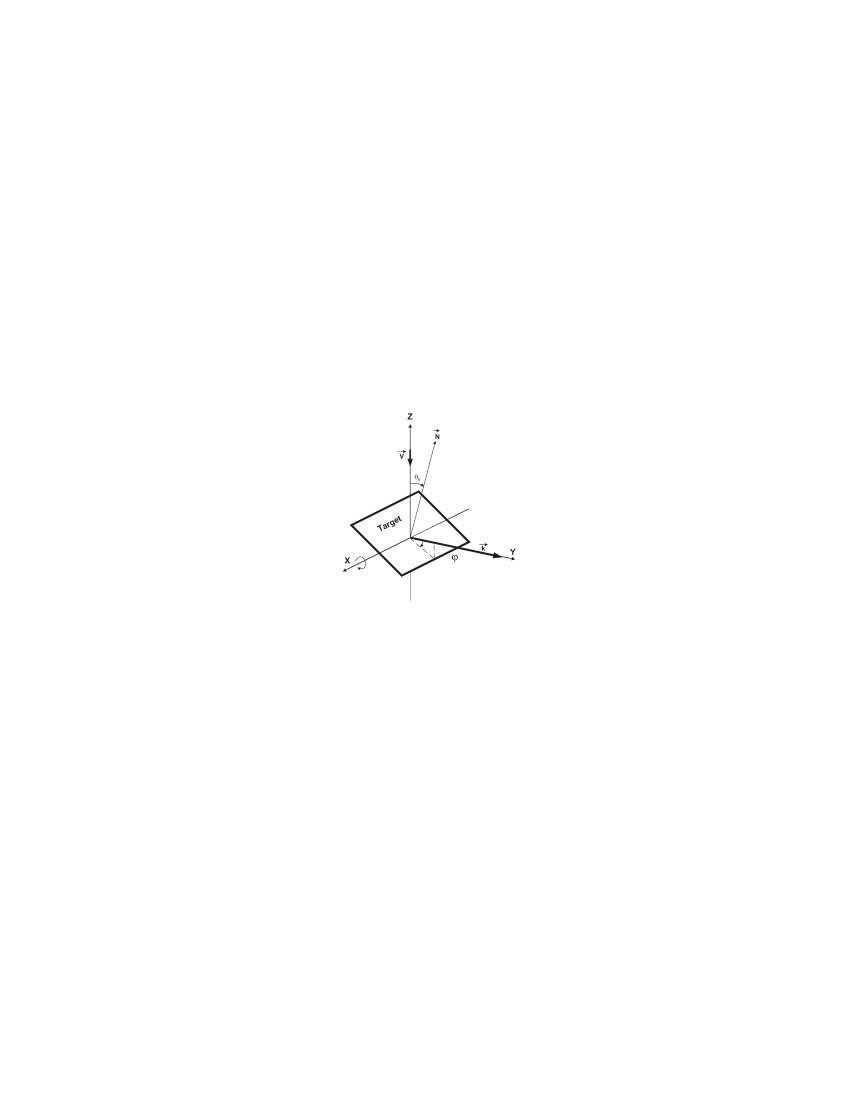
Taking into account conditions described above, we have performed the experiment outlined in Fig. 1. Narrow electron beam falls on the surface of the single crystal target, is the velocity of electron beam (). Axis is coplanar the target surface, is orthogonal to the crystal surface. Angle is the angle between and , which determines crystal rotation around the axis . At the target surface is orthogonal to the velocity of electron beam. At crystal rotation around the axis , moves in the plane . Detector window is placed at the angle to the velocity of electrons in the direction .
To provide requirements to the beam, we have applied electron beam of an electron microscope. Electron beam injector was thoroughly tested for stability and reliability of beam shaping systems and to optimize beam parameters for CXR observation. Some measured beam parameters are as follows:
-
•
electron energy 50-100 keV
-
•
relative instability of accelerating voltage 2*10-5
-
•
brightness 7*104 /m2 sr.
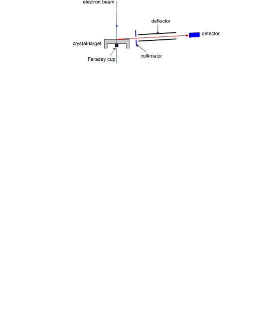
Careful selection of optimal currents of lenses and voltage between Venelt cylinder and anode minimized parasitic scattering inside the microscope and provide minimal x-ray background inside the experimental area. To reduce background caused by electron scattered at the target, we have applied electrostatic deflector made of two brass plates of the 50 mm length positioned in the input window of the detector. The six kilovolts voltage was applied between to them that decrease background rate significantly.
We have used Si(Li) detector with thermoelectric cooling and Si(Li) detector with cryostat both with 20 mm2 sensitive areas and thin polymer windows for soft x-ray detection supplied by BSI (Riga, Latvia). Energy resolution of detectors was evaluated as 170 eV. Detector angular aperture was 0.2 mrad. ORTEC 2056-C 4096-channel analyzer collected detector output data. Off-line spectra processing was performed after spectra transferring to the computer. It involved calibration, smoothing, baseline subtraction, and fitting by the set of Gauss peaks.
Analysis above shows that optimal target thickness should be below half of micron. Applied target (Fig.2) is the silicon crystal substrate of 2x2 mm dimensions and 200 m thickness with 0.5 m thickness membrane of 1.0 mm diameter. Basic plane has (100) or (111) orientation. Membrane material is layer of pure epitaxial Si of 0.9–1.0 m thickness deposited on substrate of heavily doped p+ Si of KDB 0.01 100 grade. Choice of such structure was determined by electrochemical etching technique, in which pure epitaxial Si serves as termination layer. For membrane of other thickness one should take structures with epitaxial layer thickness close to desired one. Precise membrane thickness adjustment can be performed by ion-beam etching with 10-15 nm/min rate.

We have used relatively simple technique to measure thickness of such ultra-thin Si targets. As membranes with thickness of about micron and below are going to be semi-transparent in visible light range, one can record their optical transmittance spectra. At this spectra interference fringes can be observed and than thickness may be calculated. Dispersion of measured thickness values normally is below 5%.
III PRELIMINARY RESULTS AND DISCUSSION
Experiments described below were performed with (111) Si target of 410 nm thickness. Working current of 150 nA was chosen to provide count rate at detector below 3 kHz and avoid peak distortion. Data acquisition time was 5,000 seconds for the majority of spectra. Some spectra were recorded at 10,000 seconds, but due to equipment instabilities obtained spectra did not have considerable improvement.
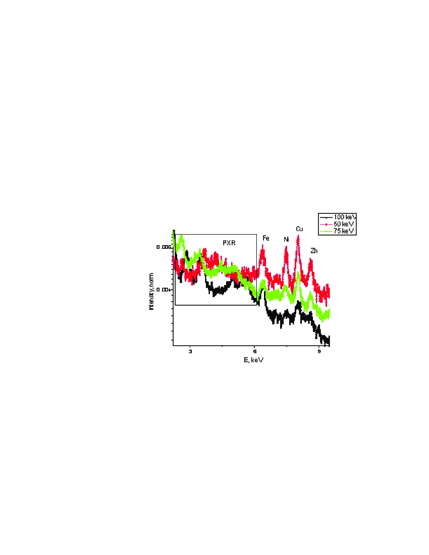
Fragments of raw (just smoothed by Savitsky-Golay algorithm) normalized PXR spectra are shown in Fig.3. One can see dependence of PXR peaks frequency on beam energy. PXR frequency is increasing with beam energy increase in conformity with formula (1). Group of characteristic x-ray peaks in right part of spectra correspond to the microscope constructional materials excited by electrons scattered by the target. Characteristic peak of Si locates at 1.73 keV and it is not shown in picture because it is very intensive in comparison with other peaks.
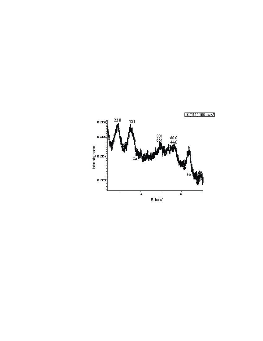
After more detailed PXR spectra examinations we can attribute peaks to corresponding crystallographic reflexes, Fig.4. Most intensive peak in 2.5-2.9 keV region is related to (220) reflexes with various indexes permutations. Reflex (131) can correspond to peak at 3.5 keV. Region 4.9-5.2 keV can be attributed to (331) and (511) including permutations. Finally, region 5.4-6.0 keV is corresponding to (800) and (440) reflexes.
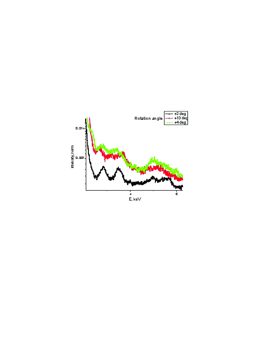
As followed from Eq.(1), PXR frequency must depend on beam incidence angle. Experimentally observed frequency sifts are shown in Fig.5. Basically, peaks should be splitted and distance must increase depending on incidence angle. At 3 degrees incidence angle split should be about 0.2 keV, at 10 degrees it may be as high as 0.7 keV. One can see this split in some spectra. Changes in spectra connected with incidence angle change are close to calculated values.
IV CONCLUSION
Series of PXR spectra in various conditions were recorded and their distinctive features were observed. Tuning of radiation frequency with crystal-target rotation was observed for the first time for low energy electrons. Dependence of the x-ray frequency on the beam energy was detected. Despite of their relatively low quantum yield, they can be considered as prospective source for structure analysis and crystallography.
We have obtained and used big amount of targets made of various materials. Spectra of acceptable quality were measured with only a few thin Si membranes. Expecting application of PXR of non-relativistic electrons as base for a source of tunable x-rays, problem of thin single crystal membrane target must be a matter of high-tech challenge.
These results are obtained in the framework of ISTC project #B626.
References
- (1) I.D. Feranchuk, A. Ulyanenkov, J. Harada, J.C.H. Spence // Phys. Rev. E, v. 62, No3 (2000) 4225.
- (2) V.G.Baryshevsky, I.D. Feranchuk // J. de Phys. (Paris), v. 44, (1983) 913.
- (3) Yu.S. Korobochko, V.F. Kosmach, V.I. Mineev // Sov. JETP, v. 21 (1965) 834.
- (4) G.M. Reese, J.C.H. Spence, N. Yamamoto // Philos. Mag. A, v. 49, No5 (1984) 697.
- (5) K.S. Vecchio, D.B. Williams // Journ. of Microscopy, v. 147, pt. 1 (1987) 15.