Nanostructures and enhanced absorption in intense laser interaction with matter: effect of laser prepulses
Abstract
Hard x-ray emission (20 - 200 keV) from plasmas produced by intense laser pulses on nanoparticle coated targets is compared with that from optically polished targets. The yield enhancement offered by nanoparticles is studied under different prepulse conditions. It is observed that the enhancement reduces when the nanoparticle coated target is irradiated with a prepulse with intensity greater than Wcm-2. When the prepulse intensity exceeds Wcm-2, the enhancement vanishes completely. This is attributed to preplasma formation on nanoparticles their subsequent structural modification before the arrival of the main pulse. It is suggested that high-contrast ultrashort pulses are essential for nanoparticles to function as yield enhancers.
pacs:
52.25.Nr, 52.40.Nk, 52.50.Jm, 42.65.ReIn recent years, plasmas generated by intense, ultrashort lasers have attracted multifaceted research to explore basic physics as well as applications. One of the major reasons for this attention is their high brightness as x-ray sources Gibbon . Radiation and particle emissions from these plasmas are inherently ultrashort in nature Murnane . The radiation pulses are potentially useful in lithography, time-resolved mapping of ultrafast atomic and molecular processes, precision imaging etc. Teubner ; Tinten ; Westneat . Since a practical realization of such sources demands high flux levels, there is a great deal of interest in methods to enhance the x-ray yield and the influence of various laser and target conditions has been the subject of many recent studies. For example, laser prepulses Pelletier and modulated surfaces Murnane2 ; Nishikawa ; Kulcsar enhance laser absorption considerably and subsequently increase the x-ray yields (mostly in soft and moderately hard x-ray regimes). Methods of enhancing emission in the very hard x-ray spectral region are being explored only recently. Such studies are interesting not only from the point of view of the enhanced radiation, but also to understand the role of surface structures or ‘roughness’ in enhancing the production of hot electrons in plasma responsible for the emission. Enhanced x-ray yield is a signature of enhanced hot electron production, a central issue in inertial fusion research Tabak and high energy particle generation and acceleration Wilks .
Recently we have reported that metal nanoparticles can be used as excellent sources of hard x-ray pulses Rajeev . A simple, yet general and quantitative model for the enhanced emission was also recently presented Rajeev ; OL . We had proposed that the non-planar geometry of these nanostructures modifies the local electric fields around them, resulting in enhanced absorption and hard x-ray emission. However, to make this method generally applicable, we need to examine the behaviour of nanoparticles during their exposure to intense, ultrashort light pulses. Since intense femtosecond laser pulses invariably possess prepulses as well as picosecond pedestals - particularly, if sufficient care is not taken to specifically avoid them - it is important to examine how nanoparticles respond to prepulses.
The effect of strong prepulses on flat targets and the resulting particle emissions has been investigated in detail before Pelletier . A prepulse could be intentional or inherent (the latter could be due to leakage from the cavity dumping in a regenerative amplifier). Further, the pedestal of an intense pulse resulting from incomplete pulse compression or amplified spontaneous emission, could also serve as a prepulse. If the prepulse intensity is above the plasma formation threshold, it would form a plasma layer before the main pulse arrives, thus crucially altering the interaction process. Depending on the prepulse-main pulse delay, plasma length-scale could increase and instabilities can build-up in the plasma, resulting in ripples in the critical density layer. The unevenness of critical layer as well as a long density profile favor resonance absorption and thus, laser absorption and x-ray production increase with preplasma formation Pelletier ; Sandhu_JAP . However, the effect of preplasma formation on nanostructured targets has not yet been explored. Such a study is very interesting as it can establish the validity or otherwise of the basic assumptions Murnane2 ; Kulcsar ; Rajeev ; OL in the modelling and understanding of the phenomenon of enhanced absorption.
In this paper, we address this problem in detail by monitoring x-ray emission from optically polished copper surfaces as well as those coated with spherical copper nanoparticles.. We observe that nanoparticle-coated targets offer 3-4 fold enhancement in hard x-ray production as compared to uncoated copper targets, when irradiated with high-contrast laser pulses (main pulse-prepulse contrast :1) at light field intensities Wcm-2. This enhancement is examined under different pre-pulse conditions realized in a standard two-pulse (prepulse-main pulse) set-up. It is observed that the enhancement offered by nanoparticles reduces and even vanishes completely when exposed to prepulses with intensity levels above a certain threshold. The preplasma formed on nanoparticles modifies them before the main pulse. This results in a reduction and complete removal of yield enhancements. This study therefore provides crucial information on the exact mechanism of nanostructure-induced absorption enhancement.
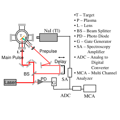
The laser used for our experiments (Fig. 1) is a custom-designed chirped pulse amplification Ti: Sapphire system, producing pulses of 100 fs duration at 10 Hz, centered at 806 nm. The linearly polarized laser beam is split into two and both beams are focused on the targets housed in a vacuum chamber at Torr, with precise overlap of the focal regions. The target is continuously translated such that each laser shot irradiates a fresh area on the target. The weak pre-pulse and strong main pulse are focussed respectively by 30 cm and 20 cm focal length plano-convex lenses. The maximum main beam energy is limited to 4.5 mJ in the present series of experiments, yielding a light intensity of about 4.5 Wcm-2 at a focal spot of 20 microns diameter. A variable optical delay is introduced in the weaker beam path using a motorized precision translation stage. The delay between the two beams can be continuously adjusted from -0.5 ns to +0.5 ns - negative delay implies that the prepulse arrives before the main pulse. We keep the prepulse intensity sufficiently above the plasma formation threshold ( Wcm-2 for Copper APB ) such that a pre-plasma is formed before the main pulse is incident. The intensity of the prepulse is varied using a half-wave plate-polarizer combination. The spatial overlap of main pulse and prepulse is checked through a CCD imaging system which observes the plasma formation region. To determine the temporal overlap, the prepulse reflectivity (at very low prepulse levels, such that the prepulse does not produce plasma) is monitored as the delay is changed. A sharp drop in the prepulse reflectivity, which is indicative of plasma formation by the main pulse, establishes the ”zero” of temporal overlap Sandhu_PRL . X-ray emission from the plasma is monitored using a time-gated NaI (Tl) scintillation detector kept in the plane of incidence at to the target normal. A detailed description of this diagnostic system can be found elsewhere OC .
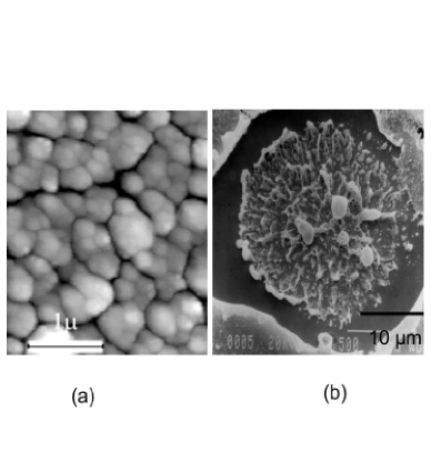
Copper nanoparticles are deposited by high-pressure dc-magnetron (Atom Tech 320-O) sputtering Pushan ; APB on optically polished copper discs held at C. The nanocrystalline thin films typically consist of a collection of densely packed, spherical nanoparticles, as shown in Figure 2 (a). The resulting nanocrystalline Cu films are optically flat and 1 m in thickness. The crystallographic domain size () is obtained from x-ray diffraction line broadening. For a film deposited in 180 mTorr Ar environment at a sputtering power of 200W, we obtain = 15nm. Since the thickness of the nanoparticle layer is greater than the skin depth of the laser light, the laser essentially interacts only with the film and not the substrate behind it. However, during the interaction, the film is locally destroyed in the focal spot, if the laser intensity exceeds the plasma formation threshold. Figure 2 (b) is a Scanning Electron Micrograph of one such focal spot. The micrograph, which represents the surface after the laser irradiation, further evidences that the laser interacts just with the nanoparticle coating leaving the optically flat copper surface behind it unaffected.
Figure 3 (inset) presents a typical comparison of bremsstrahlung emission spectrum, measured from an optically polished copper surface and such a surface coated with spherical nanoparticles, irradiated at at Wcm2 with a single pulse. The total energy emitted per pulse from a polished target is J while the spherical nanoparticles yield J, giving about 3 - fold enhancement in the range 20-200 keV. This is quantitatively substantiated by a simple model, which ascribes observed yield enhancements to the electric field enhancements near the nanostructures Rajeev ; OL . The model assumes that the integrity of the nanostructures is preserved during the interaction time . This assumption is fairly reasonable as the plasma does not expand significantly to alter the shape of the structure before the peak of the pulse is reached. Thus, one can consider the system as a ’nanoplasma’, with just a different dielectric constant; the change in the dielectric function does not hamper the predictions of the model as the field enhancement is shown not to depend critically on dielectric functions, under our experimental conditions Rajeev ; OL .

We now examine the effect of prepulses on the nanostructured targets. Figure 3 shows the result of irradiating a spherical nanoparticle-coated target with a prepulse of intensity Wcm-2 at normal incidence. The intensity of the main pulse was around Wcm-2. A constant 4-fold enhancement was obtained from the nanoparticle-coated surface, when irradiated with just the main pulse, without the prepulse, similar to the result shown in the inset. This is obviously not a function of the prepulse delay and the plotted data points (top curve) indicate just the values obtained with different measurements. As is clear from the figure, there is a drastic drop in the enhancement on irradiation of a prepulse, even at small ( 10 ps) delays. The enhancement vanishes completely as the delay between the prepulse and the main pulse increases.
This observation is very significant as it proves that the nanostructure has to be intact before the intense pulse, to observe any yield enhancements. It becomes clear that the preformed plasma on nanoparticles is not the reason for the observed enhancements on nanostructured surfaces. Thus the observed enhancements in single pulse experiments at these intensities, themselves substantiate the integrity of the surface structures in single pulse interaction. Apart from ruling out the prepulse/pedestal levels in single pulse experiments, this result also suggests that it is necessary to irradiate with high-contrast pulses for observation of yield enhancement using nanostructured surfaces.
This is especially important in the context of low contrast pulses. Typically ultrashort pulses ride over a long pedestal and wings of duration from picoseconds to nanoseconds. Most high-power laser systems have prepulses as well. This is mainly caused by the non-ideal behaviour of the Pockels cell-polarizer combination in the regenerative amplifier The prepulse just before the cavity dumping is normally the strongest of all.
Both prepulse and pedestal can form preplasma provided their intensity levels are above the plasma formation threshold. In flat targets, as mentioned before, this causes the plasma length-scale to exceed the wavelength, which is never the case under our experimental conditions with clean pulses. Further, instabilities can build-up in a long-lived plasma, resulting in ripples in the critical density layer. Both of these effects favor resonance absorption, the major mechanism of light coupling in plasma in our experimental conditions. However, in the case of nanostructured surfaces, preplasma formation affects the interaction detrimentally, as discussed above. The enhanced absorption- otherwise present- could be reduced or eliminated completely.

It is thus important to parameterize conditions at which nanostructures provide enhancements in x-ray yield. We have studied the x-ray production from laser produced plasma on nanoparticle-coated targets and flat copper targets under various prepulse levels. Fig. 4 summarizes the results of such a study. Yield enhancement from nanoparticles at main pulse intensities Wcm-2 and under different prepulse levels is presented. Notice that even at small delays, the enhancement starts reducing once the prepulse levels exceed Wcm-2. Prepulse levels less than Wcm-2 do not affect the enhancement. This is understandable as the nanoparticle coating is about a micron thick and contains several layers of nanoparticles. Even after some layers are destroyed or modified as a result of the preplasma formation, some fresh layers can be preserved for the further interaction if the prepulse intensity is low enough. Thus, some field enhancement and subsequent enhanced absorption is still possible with the main pulse. However, it has to be noted that the enhancement reduces monotonically with prepulse intensity. Though, the preplasma formed would be mostly underdense at these intensities, it may not be produced in a uniform fashion all around the nanostructure. It is well known that any non-planar geometry severely alters the local field distribution. It can be seen that most of the field enhancement is concentrated near the maximum curvature point - at the tip of the ellipsoidal particle. Thus, plasma is presumably produced first near the tip of these structures. This spatially inhomogeneous production of plasma can in turn lead to alterations in the surface structure, which can reduce enhancement, as observed. However, this effect may not be strong enough to completely nullify the enhancements at low prepulse levels. The fact that the shape of the critical layer in nanostructures is not seriously altered even at intensities beyond the plasma formation threshold is further evidenced in our reflectivity studies reported elsewhere; the strong dependence of polarization in absorption was found to be present at intensity levels immediately beyond the plasma formation threshold APB . It takes a prepulse intensity Wcm-2 to completely nullify the effect of nanoparticles. Lower intensities do not seem to alter or destroy the nanostructures to an extent that their effects is removed completely in the interaction with the following ultrashort pulse.
In conclusion, the hard x-ray emission from plasmas produced on optically polished copper surfaces and those coated with spherical copper nanoparticles is examined with femtosecond laser pulses. The emission is studied under different prepulse levels irradiated at different times before the main ultrashort pulse. It is observed that with significant prepulse levels, the enhancement in x-ray emission from nanoparticle targets gets adversely affected and even nullified. These observations are especially important in studies to find efficient laser plasma sources of short wavelength radiation. The prepulses and pedestals invariably associated with high power lasers can affect the enhanced absorption induced by the nanostructures detrimentally. Extra pulse cleaning devices such as a Pockels cell or saturable absorbers should be used to increase the contrast of the temporal profile of the pulse in order to use nanostructured surfaces to facilitate enhanced x-ray production.
References
- (1) See, for instance, P. Gibbon, and E. Forster, Plasma Phys. Control. Fusion , 769 (1996).
- (2) M. Murnane, H. Kapteyn, M. D. Rosen, and R. Falcone, Science , 531 (1991).
- (3) U. Teubner, T. Missala, I. Uschman, E. F rster, W. Theobald and C. Wulker, Appl.Phys.B , 213 (1996).
- (4) K. Sokolowski-Tinten, C. Blome, J. Blums, A. Cavalleri, C. Dietrich, A. Tarasevitch, I. Uschmann, E. F rster, M. Kammler, M. Horn-von-Hoegen and D. von der Linde, Nature, , 287 (2003).
- (5) M. W. Westneat, O. Betz, R. W. Blob, K. Fezzaa, W. J. Cooper, and W. Lee, Science , 558 (2003).
- (6) J. F. Pelletier, M. Chaker, and J.C. Kieffer, J. Appl. Phys. , 5980 (1997); H. Nakano, T. Nishikawa, H. Ahn, and N. Uesugi, Appl. Phys. Lett. , 2992 (1996).
- (7) M. M. Murnane, H. C. Kapteyn, S. P. Gordon, J. Bokor, E. N. Glytsis, and R. W. Falcone, Appl. Phys. Lett. , 1068 (1993).
- (8) T. Nishikawa, H. Nakano, Naoshi Uesugi, M. Nakao, and H. Masuda, Appl. Phys. Lett. , 4079 (1999); T. Nishikawa, H. Nakano, H. Ahn, and N. Uesugi, Appl. Phys. Lett. , 1653 (1997).
- (9) G. Kulcsar, D. AlMawlawi, F. W. Budnik, P.R. Herman, M. Moskovits, L. Zhao, and R. S. Marjoribanks, Phys. Rev. Lett. , 5149 (2000).
- (10) M. Tabak, J. Hammer, M. E. Glinsky, W. L. Kruer, S.C. Wilks, J.Woodworth, E. M. Campbell, and M. D. Perry, Phys. Plasmas , 1626 (1994).
- (11) S. C. Wilks, A. B. Langdon, T. E. Cowan, M. Roth, M. Singh, S. Hatchett, M. H. Key, D. Pennington, A. MacKinnon, and R. A. Snavely, Phys. Plasmas , 542 (2001).
- (12) P. P. Rajeev, P. Taneja, P. Ayyub, A. S. Sandhu and G. R. Kumar, Phys. Rev. Lett. , 115002 (2003).
- (13) P. P. Rajeev, P. Ayyub, S. Bagchi and G. R. Kumar, Opt.Lett. , 2662 (2004).
- (14) A.S. Sandhu, A.K. Dharmadhikari and G. R. Kumar, J. Appl. Phys. , 023526 (2005).
- (15) P.P. Rajeev et al., Appl. Phys. B (in press)
- (16) A. S. Sandhu, A. K. Dharmadhikari, P. P. Rajeev, S. Sengupta, A. Das, P. K. Kaw and G. R. Kumar, Phys. Rev. Lett , 225002 (2002)
- (17) P. P. Rajeev and G. R. Kumar, Opt. Commun. , 9 (2003)
- (18) P. Ayyub, R. Chandra, P. Taneja, A. K. Sharma, and R. Pinto, Appl. Phys. A , 67 (2001).