Coupled Dynamics of Voltage and Calcium in Paced Cardiac Cells
Abstract
We investigate numerically and analytically the coupled dynamics of transmembrane voltage and intracellular calcium cycling in paced cardiac cells using a detailed physiological model and its reduction to a three-dimensional discrete map. The results provide a theoretical framework to interpret various experimentally observed modes of instability ranging from electromechanically concordant and discordant alternans to quasiperiodic oscillations of voltage and calcium.
Over the last decade, there has been a growing recognition that dynamic instability of the cardiac action potential can play a crucial role in the initiation of life-threatening arrhythmias karma ; garfinkel ; modalt ; expalt ; hall ; foxmap . Most studies to date have focused on the dynamics of the transmembrane voltage governed by the standard equation
| (1) |
where is the membrane capacitance, is the total membrane current, which is the sum of the individual currents for Na+, K+, and Ca2+ ions depicted schematically in Fig. 1, and is the external current representing a sequence of suprathreshold stimuli equally spaced in time by , the pacing period. A widely used approach to model the nonlinear dynamics of voltage is the one-dimensional discrete map which relates the action potential duration (APD) at two subsequent beats via the restitution curve, , where is the interval between the end of the previous action potential and the next karma ; garfinkel ; modalt ; expalt ; hall ; foxmap . The periodic fixed point of this map corresponding to the stable 1:1 rhythm undergoes a period-doubling instability to alternans, a sequence LSLS… of long (L) and short (S) APD, when the slope of the restitution curve is .
Even though this map has been successful to model the unstable dynamics of voltage in some ionic models modalt and experiments expalt , its predictions are inconsistent with a wide range of observations hall ; foxmap ; pruvot ; lab . For example, Hall et al. hall found that alternans can be absent even when the slope of the restitution curve is significantly larger than one, and conversely alternans are observed under ischemic conditions in which the restitution curve is flat lab . An important limitation of the restitution relationship is highlighted by recent experimental pruvot ; chudin ; eisner-sr and theoretical studies shiferaw which suggest that alternans may result from an instability of intracellular calcium cycling. The coupled nonlinear dynamics of voltage and calcium, however, remains largely unexplored.

In this letter, we investigate this dynamics by a numerical study of a detailed physiological model and an analysis of the dynamics based on iterated maps. The model consists of Eq. 1, with membrane currents (Fig. 1) modeled based on modifications by Fox et al. fox of the Luo-Rudy currents rudy , coupled to equations from a recent model of calcium cycling shiferaw
| (2) | |||
| (3) | |||
| (4) | |||
| (5) | |||
| (6) |
where , , and are the concentrations of free Ca2+in a thin layer just below the cell membrane (submembrane space), in the bulk myoplasm, and the sarcoplasmic recticulum (SR), with volumes , , and , respectively, where the SR volume includes both the junctional SR (JSR) and the network SR (NSR); is the average JSR concentration in the whole cell as defined in Ref. shiferaw . The concentrations and are in units of M, whereas and are in units of M. All Ca2+ fluxes are divided by and have units of M/s. Instantaneous buffering of calcium to SR and calmodulin sites in and is accounted for by the functions and , and the currents describe time-dependent buffering to troponin C shiferaw .
Calcium release from the SR is triggered by calcium entry into the cell via calcium-induced-calcium-release (CICR) fabiato . Release occurs at a very large number of junctions where several L-type Ca channels () and a few release channels (ryanodine receptors; RyRs) face each other in close proximity. Only one of these junctions is shown in Fig. 1 for clarity. The total release current for the whole cell is the sum , of local currents at each junction where release channels are activated. Active junctions appear as bright localized spots, or “sparks”, in confocal microscope imaging of calcium activity bers . The number of sparks varies in time since sparks are recruited stochastically and extinguish. The model takes into account this spatially localized nature of release and the dynamical equation for the release current (Eq. 6) captures phenomenologically three key experimental observations: (i) sparks are recruited at a rate proportional to the whole cell , or collier , which insures that calcium release is graded with respect to calcium entry bers ; wier , (ii) the spark life-time is approximately constant, and (iii) the amount of calcium released increases with SR concentration (SR-load) shannon .
Instability mechanisms. Ca2+ alternans, a period-doubling sequence … of large () and small () calcium transient ( peak ), can occur independently of voltage alternans in experiments with a single cell paced with a periodic voltage waveform chudin . Both theoretical analyses shiferaw ; eisner and recent experiments eisner-sr support that a steep dependence of release on SR-load is the underlying mechanism of these alternans. The sensitivity of release to SR-load is controlled in the model by the slope of the function at high load
| (7) |
For a large enough slope, the model produces Ca2+ alternans when paced with a periodic voltage waveform shiferaw as in the experiments of Ref. chudin .
Steep APD-restitution in the absence of Ca2+ alternans can also induce APD alternans. This steepness is especially sensitive to the recovery from inactivation of the calcium current fox ; rudy
| (8) |
where is the single channel current and () is a fast (slow) voltage-dependent activation (inactivation) gate. For the intermediate range of pacing rates studied in the present work, increasing the time constant of the gate in the equation steepens APD-restitution and promotes voltage alternans.
Voltage-calcium coupling. The mutual influence of voltage and calcium during the action potential is controlled by the membrane currents that depend on intracellular calcium concentration. These include and the sodium-calcium exchanger . A crucial property is that a change in the magnitude of the calcium transient has opposite effects on these currents with respect to prolonging or shortening the APD. A larger calcium transient following a larger release enhances inactivation of via the calcium-dependent gate , and hence shortens the APD, but increases the chemical driving force for Ca2+ extrusion from the cell via the exchanger. Since 3 Na+ enter the cell for every Ca2+ extruded, this increase in driving force increases the inward membrane current which prolongs the APD. Therefore, depending on the relative contributions of and , increasing the magnitude of the calcium transient can either prolong (positive coupling) or shorten (negative coupling) the APD, as illustrated in Fig. 2. The sign of this coupling can be changed in the model by varying the exponent in the phenomenological expression for the steady-state value of , where the constant sets the concentration range for inactivation. Increasing enhances calcium-dependent inactivation of and tends to make the coupling negative.
Numerical results. The dynamics of the model was studied numerically as a function of the two instability parameters and which promote Ca2+ and voltage alternans, respectively, and for two values of that were found to yield a positive () and a negative () coupling between voltage and calcium. All the other parameters are the same as in Ref. shiferaw ; fox and the pacing period is fixed to ms.
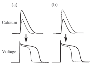
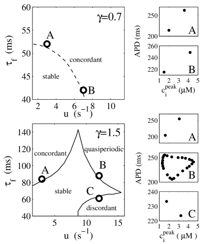
The results plotted in Fig. 3 highlight the crucial role of the coupling between voltage and calcium in the dynamics. For positive coupling, the instability of the 1:1 periodic state always occurs through a period-doubling bifurcation to electromechanically concordant alternans with the long (short) APD corresponding to a large (small) calcium transient, independently of whether voltage or calcium is the dominant instability mechanism. In contrast, for negative coupling, three distinct modes of instability are found that correspond to (i) concordant alternans, as for positive coupling, but only when the instability is dominated by voltage (large and small ), (ii) discordant alternans with the long (short) APD corresponding to a small (large) calcium transient when the instability is dominated by calcium (small and large ), and (iii) quasiperiodic oscillations of APD and calcium transient amplitude with a phase and a Hopf frequency that vary with and for the in between case where the instability is driven by both voltage and calcium. Both electromechanically concordant and discordant alternans have been widely observed experimentally under various conditions discordant-papers . In addition, there is experimental evidence for quasiperiodicity in recordings of voltage gilmour1 and, more recently, calcium entcheva .
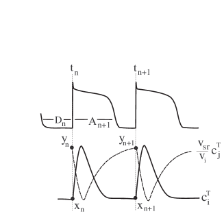
Iterated map of voltage-calcium dynamics. To interpret our results, we extend the two-dimensional iterated map developed in Ref. shiferaw for calcium cycling when the cell is paced with a fixed periodic voltage waveform, to the present case where the voltage is unclamped. To a good approximation, and preceding a stimulus shiferaw , such that we only need to track beat-to-beat changes of and . Furthermore, we assume for simplicity that buffering of calcium is instantaneous such that there exists a unique nonlinear relationship between the concentration of free calcium () and total calcium (free plus bound) (). The basic variables of the map (Fig. 4) are then and at time of the stimulus, defined by and where both and are in units of M, and the APD corresponding to this stimulus, .
The map is obtained by extending the restitution map to include the effect of calcium on the APD and by integrating the calcium flux equations
| (9) | |||||
| (10) |
from time to time . This yields
| (11) | |||||
| (12) | |||||
| (13) |
respectively, where , , and are the integrals of , , and over the time interval , respectively, and are functions of for a fixed pacing period; and are the total amount of calcium released from and pumped into the SR over one beat, respectively, and is the net total calcium entry into the cell over one beat which can be positive (negative) if the exchanger extrudes more (less) calcium from the cell than brings into the cell.
To study the stability of the fixed point of the map , we exploit the fact that the total amount of calcium inside the cell is approximately constant during steady-state pacing. Hence, we can approximate the 3-dimensional (3-d) map (Eqs. 11-13) by a 2-d map by assuming that , where is the total calcium in the cell at time . This 2-d map is given by Eqs. 11 and 12 with , , and . A straightforward linear stability analysis of this 2-d map yields the eigenvalues
| (14) |
where we have defined the quantities
| (15) | |||||
| (16) | |||||
| (17) |
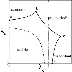
which are evaluated at the fixed point of the map. Here, and govern the degree of instability of the voltage and calcium systems, respectively, while determines the sign of the coupling between the two systems. Making APD-restitution () or the relationship between release and SR-load () steeper by increasing and in the ionic model is equivalent to increasing and , respectively. Graded release implies that is positive for high pacing rates where depends on , such that the sign of is governed by where the latter reflects the effect of the magnitude of the calcium transient on APD via and (Fig. 2). The periodic fixed point undergoes a period doubling bifurcation when and a Hopf bifurcation for when the pair of complex eigenvalues , with and , crosses the unit circle . For the latter case, the beat-to-beat oscillations of voltage and calcium are modulated with a period . Examination of the eigenvectors for reveals that alternans are discordant when is real and . We plot in Fig. 5 the corresponding stability boundaries for positive and negative coupling in the plane which are remarkably isomorphic to the stability boundaries obtained by simulations of the ionic model in the plane of Fig. 3. This agreement shows that this simple map captures the main robust features of the instability of the voltage-calcium system observed in the ionic model and experimentally.
The numerical study of both the ionic model and the map in a nonlinear regime reveals the existence of a rich dynamical behavior including higher order periodicities (3:3, 4:4, etc) as well as transitions to chaos mediated by a period-doubling cascade or intermittency depending on the parameters. Moreover, this model naturally contains memory gilmour1 ; Watanabe due to the slow change of total calcium concentration over several beats. Both of these aspects will be discussed in more details elsewhere.
In conclusion, we have outlined the essential three-dimensional parameter space that controls dynamic instability of membrane voltage coupled to calcium cycling and we have presented a theoretical framework in which to interpret experiments beyond the limitations of the one-dimensional restitution relationship. The main axes of this parameter space are the degree of instability of the voltage and calcium systems, and the sign of the coupling between the two systems, which is an important new parameter to emerge from this work. These results offer new concepts to help identify the mechanisms which underly various heart rhythm disorders. This research is supported by NIH SCOR P50-HL52319.
References
- (1) A. Karma, Chaos 4, 461 (1994).
- (2) A. Garfinkel et al., Proc. Natl. Acad. Sci. USA 97, 6061 (2000).
- (3) M. Courtemanche, L. Glass, and J. P. Keener Phys. Rev. Lett. 70, 2182 (1993); B. Echebarria and A. Karma, Phys. Rev. Lett. 88, 208101 (2002).
- (4) J. B. Nolasco and R. W. Dahlen, J. App. Physiol. 25, 191 (1968); M.R. Guevara et al., IEEE Comp. Cardiol. 562, 167 (1984).
- (5) G. M. Hall, S. Bahar, and D.J. Gauthier, Phys. Rev. Lett. 82, 2995 (1999); G. M. Hall and D.J. Gauthier, Phys. Rev. Lett. 88, 198102 (2002).
- (6) J. J. Fox, E. Bodenschatz, and R. F. Gilmour Jr., Phys. Rev. Lett. 89, 138101 (2002).
- (7) E.J. Pruvot et al., Circ. Res. 94, 1083 (2004).
- (8) S. G. Dilly and M. J. Lab, J. Physiol. 402, 315 (1988).
- (9) E. J. Chudin et al. Biophys. J. 77, 2930 (1999).
- (10) M. E. Díaz, S. C. O Neill, and D. A. Eisner, Circ. Res. 94, 650 (2004).
- (11) Y. Shiferaw et al., Biophys. J. 85, 3666 (2004).
- (12) J. J. Fox, J. L. McHarg, and R. F. Gilmour, Am. J. Physiol. 282, H1534 (2002).
- (13) C. H. Luo and Y. Rudy, Circ. Res. 74, 1071 (1994).
- (14) A. Fabiato, J. Gen. Physiol. 85, 189 (1985).
- (15) D. M. Bers, Excitation-contraction coupling and cardiac contractile force, (Kluwer, Boston, 2001).
- (16) M. L. Collier, A. P. Thomas, and J. R. Berlin, J. Physiol. 516, 117 (1999).
- (17) W. G. Wier et al., J. Physiol. 474, 463 (1994).
- (18) T. R. Shannon, K. S. Ginsburg, and D. M. Bers, Biophys. J. 78, 334 (2000).
- (19) D. A. Eisner et al., Circ. Res. 87, 1087 (2000).
- (20) D. S. Rubenstein and S. L. Lipsius, Circulation 91, 201 (1995); M. L. Walker and D. S. Rosenbaum, Cardiovascular Res. 57, 599 (2003); and earlier references therein.
- (21) R. F. Gilmour, N. F. Otani, and M. A. Watanabe, Am. J. Physiol, 272, H1826 (1997); N.F. Otani and R.F. Gilmour, J. Theor. Biol. 187, 409 (1997).
- (22) L. Yin, H. Bien, and E. Entcheva, “Calcium Instabilities in Cardiomyocyte Networks,” (preprint, 2004).
- (23) M. A. Watanabe and M. L. Koller, Am. J. Physiol. 282, H1534 (2002).