Hyperpolarized xenon nuclear spins detected by optical atomic magnetometry
Abstract
We report the use of an atomic magnetometer based on nonlinear magneto-optical rotation with frequency modulated light (FM NMOR) to detect nuclear magnetization of xenon gas. The magnetization of a spin-exchange-polarized xenon sample (cm3 at a pressure of bar, natural isotopic abundance, polarization ), prepared remotely to the detection apparatus, is measured with an atomic sensor (which is insensitive to the leading field of 0.45 G applied to the sample; an independent bias field at the sensor is G). An average magnetic field of nG induced by the xenon sample on the 10-cm diameter atomic sensor is detected with signal-to-noise ratio , limited by residual noise in the magnetic environment. The possibility of using modern atomic magnetometers as detectors of nuclear magnetic resonance and in magnetic resonance imaging is discussed. Atomic magnetometers appear to be ideally suited for emerging low-field and remote-detection magnetic resonance applications.
pacs:
07.55.Ge,82.56.Dj,76.60.PcNuclear magnetic resonance (NMR) is a versatile technique for the study of structure and dynamics on both molecular and macroscopic scales, and on time scales from nanoseconds to hours. Spin-polarized 129Xe (nuclear spin-1/2, magnetic moment , where is the nuclear magneton) is particularly well suited for NMR and magnetic-resonance imaging (MRI) studies for several reasons. It is possible to polarize it using the laser-optical-pumping techniques Walker and Happer (1997) to a degree that is orders of magnitude higher than what is possible via thermal polarization in high-field magnets. In contact with various analytes, xenon displays a wide range of relative chemical shifts of up to several hundred ppm Goodson (2002), which makes it an ideal probe of its local physiochemical environment. Finally, it has a long longitudinal relaxation time of several minutes or longer, even at low fields. Xenon can also be used in solution, and is especially soluble in organic solvents.
An important recent development in NMR/MRI is the technique of remote detection Moulé et al. (2003); Seeley et al. (2004), in which information about an analyte is transferred onto a mobile spin-polarized substance, and is then read out at a different location. This technique allows the separate optimization of the encoding and the detection environment. As the signal in this case is due to the net magnetization of the spin-polarized sample, the task becomes to read out this information efficiently and with high sensitivity. Detection using superconducting quantum interference devices (SQUIDs) Clarke (1996) and atomic magnetometers Budker et al. (2002a) provides an alternative to the traditional techniques involving inductive detection. In addition to eliminating the need for a strong magnetic field for the detector, another advantage of these methods is that the time constant of the measurement in this case is limited by the longitudinal relaxation, which could be significantly slower than transverse relaxation limiting induction detection. SQUID detection has already proven useful in NMR experiments Greenberg (1998); McDermott et al. (2002).
Atomic magnetometers (the essential components of which consist only of a diode laser, an atomic vapor cell, and the necessary optics and electronics) are also attractive for NMR applications as they have the potential to be very cheap and compact and—in contrast to SQUIDs, which require cryogenic temperatures—they operate at room temperature. The first use of an atomic magnetometer for detection of the static magnetic field produced by a sample of gaseous nuclear-polarized atoms was reported nearly 35 years ago in the pioneering work of Cohen-Tannoudji et al. Cohen-Tannoudji et al. (1969). In that work, a 6-cm diameter vapor cell containing 5-optically-polarized 3He gas at a pressure of 3 torr was placed next to a cell of similar dimensions containing 87Rb that served as a sensor of an optical-pumping magnetometer. The field produced by the nuclear spins was detected with a sensitivity . A similar setup was also used in Ref. Newbury et al. (1993).
Modern atomic magnetometers utilizing alkali-metal vapors in anti-relaxation coated cells can achieve sensitivities to magnetic field better than G, see, for example, Ref. Budker et al. (2000) and review Budker et al. (2002a). Recently, a sensitivity of was demonstrated Kominis et al. (2003) with an atomic sensor of a volume of only cm3 where, instead of anti-relaxation coating, buffer gas was used to reduce relaxation in wall collisions, and operation at a high alkali-atom density ensured the rapid spin-exchange-collision regime Happer and Tam (1977), where spin-relaxation in collisions between alkali atoms is also reduced. Due to such advances, the use of atomic magnetometers for low-field NMR experiments becomes attractive.
With a view toward future NMR applications, particularly remote detection, we have carried out exploratory measurements of samples of gaseous spin-exchange-polarized xenon with a modified version of the atomic magnetometry apparatus previously described in Refs. Budker et al. (2000, 2002b); Malakyan et al. (2004) (Fig. 1).
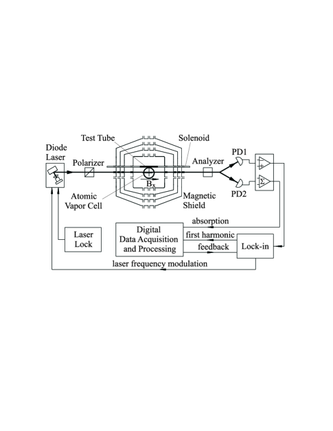
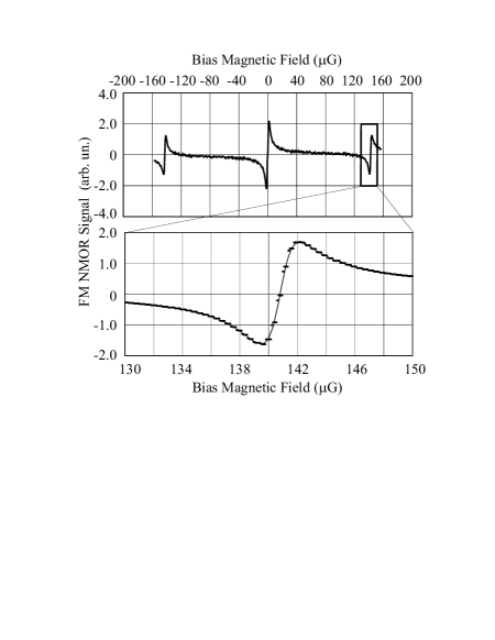
The apparatus incorporates a 10-cm diameter spherical 87Rb-vapor cell at room temperature with no buffer gas, whose inner walls are coated Alexandrov et al. (2002) with paraffin to reduce spin relaxation in collisions of Rb atoms with the wall. The magnetic-field measurement is based on the technique of nonlinear magneto-optical rotation with frequency-modulated light (FM NMOR) Budker et al. (2002b). The key feature of the method is the use of an ultra-narrow resonance arising when the frequency of the light is modulated at twice the Larmor-precession frequency of the Rb atoms (Fig. 2). The magnetometer operates in a closed feedback loop involving digital signal processing, locking to an FM NMOR resonance by adjusting the diode-laser-modulation frequency. The mean frequency of the W laser light delivered to the sensor is locked to the D1-resonance using a technique Yashchuk and Budker (2004) involving an auxiliary Rb-vapor cell (not shown). The atomic sensor cell is placed inside a multi-layer magnetic shield (shielding factor Yashchuk et al. (1999)) equipped with internal magnetic-field coils. The FM NMOR magnetometer is intrinsically a scalar device; however, a bias field G applied along the direction of light propagation renders the sensor linearly sensitive only to the average component of the magnetic field produced by the sample in that direction.
The xenon samples of natural isotopic abundance containing about 26 of 129Xe were prepared using a commercial spin-exchange polarizer [MITI IGI 9800 Xe, Magnetic Imaging Technologies, Inc. (Polarean), Durham, NC]. A gas mixture of 1% Xe, 10% N2, and 89% He was used. After polarization, pure xenon was frozen out of the gas mixture into a cold finger that was immersed in liquid nitrogen. At the end of the polarization process, the Xe batch was thawed into a custom sapphire sample tube equipped with a miniature titanium spring-loaded valve mechanism with overall outer diameter of 6.4 mm and sample volume dimensions of 4.8 mm inner diameter and mm length Yashchuk et al. (2004). These materials were chosen to ensure a long spin-polarization relaxation time Haake et al. (1998). The outer diameter of the tube was dictated by the tight constraints given by the geometry of the atomic magnetometer’s magnetic shield Yashchuk et al. (1999). The tube was designed for operation with gas pressures of up to 30 bar. The xenon polarization was measured by the polarizer’s onboard NMR spectrometer and calibrated using thermally-polarized xenon dissolved in pentane on a Varian Unity Inova NMR spectrometer equipped with an Oxford 7-T magnet. The longitudinal relaxation time constant in the tube was found to be min within the 7-T magnet, and typically min in the earth and laboratory fields. For the experimental runs described here, we used samples with total xenon pressure of bar with initial 129Xe polarization.
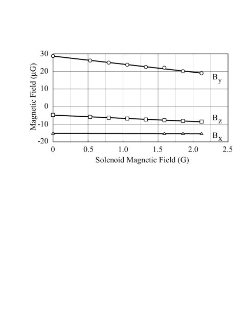
A piercing solenoid (see Fig. 1) was used to apply a leading field of 0.45 G to the xenon sample. The setup is designed so that the leakage field outside of the solenoid has negligible effect on the atomic sensor; the field due to the piercing solenoid is a factor of smaller at the vapor cell, as determined by an auxiliary measurement with the atomic magnetometer (Fig. 3). During the xenon measurement, this leakage field provides a constant magnetic field offset that is much smaller than and transverse to the G bias field applied to the atomic sensor. Thus, the FM NMOR measurement is largely insensitive to the solenoid leakage field.
After introducing spin-exchange-polarized xenon into the tube, the sample is hand-delivered to the atomic magnetometry laboratory located in a different building and loaded into the magnetometer apparatus inside the piercing solenoid (Fig. 1). Typically, it takes about 5 min between the completion of xenon gas preparation and the beginning of the atomic magnetometer measurements. Differential measurement is achieved by moving the sample within the piercing solenoid in and out of the position next to the Rb cell where maximal sensitivity to the xenon magnetic field is obtained. This modulation is effective in discriminating between the xenon magnetization signal and the slow drift (typically, nG/min) of the magnetic field within the shield. When the sample tube is in the position where the atomic sensor is most sensitive to its field, the average magnetic field from the xenon magnetization over the volume of the magnetometer sensor cell (equal to the field in the center of the cell) is a factor of 5000 smaller than the field in the sample tube. This suppression factor was calculated from the experimental geometry and verified experimentally by replacing the sample cell with a calibrated solenoid of the same dimensions, and could be greatly reduced with a sensor geometry designed specifically for this application. When the sample tube is moved away to the “out” position, the suppression factor is about two orders of magnitude larger. An example of experimental data is shown in Fig. 4.
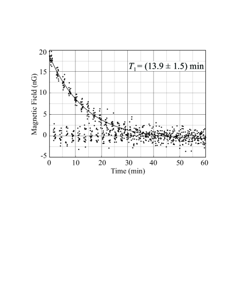
With a measurement time of about 3 s per point, we detect the decay of xenon magnetization corresponding to an initial average field at the atomic sensor of nG with a signal-to-noise ratio (S/N) of about 10, clearly demonstrating the ability of the atomic sensor to detect dc magnetization of a small gaseous sample. It should be emphasized that the S/N obtained in this work is many orders of magnitude lower than what one could obtain with straightforward modifications of this technique. Perhaps most significant would be the improvement of the geometrical suppression factor from 5000 to about 10 with optimized geometry. In addition, the magnetic noise in this experiment is dominated by the fluctuations of the magnetic field within the magnetic shield and exceeds the projected intrinsic sensor noise of our apparatus Budker et al. (2000, 2002b) of G by about two orders of magnitude. This noise can be effectively suppressed (as was done, for example, in Ref. Kominis et al. (2003)) by employing a gradiometric arrangement of magnetic sensors.
With a system consisting of two compact, high-precision atomic magnetometers operating in gradiometric mode, it should be possible to measure the magnetization of about fully-polarized nuclear (e.g., 129Xe) spins in less than a second with a signal-to-noise ratio of 10. The open geometry of an atomic magnetometer equipped with a piercing solenoid would allow measurements in which polarized samples can be continuously transported through the magnetometer, an important feature for remote detection experiments Moulé et al. (2003). Commercial xenon hyperpolarization systems are capable of producing over a liter of xenon gas at 1 bar with typical polarization of . Using such a system and an optimized atomic magnetometer, it will be possible to make point-by-point low-field measurements, in which a single-point sample will constitute mL of xenon gas whose magnetization will be determined with S/N in s, allowing ten thousand single-point measurements to be taken in less than an hour. Faster and/or higher resolution scans can, in principle, be obtained by using multiple atomic sensors in parallel, and/or by sacrificing the S/N.
In principle, it is also possible to perform manipulations on the nuclear spins within the magnetometer, including adiabatic spin-flips, spin-echoes, etc. This capability may be of interest for ultra-low field NMR studies (leading fields can be as low as G limited by the magnetic-shielding system; if necessary, much larger leading fields than those used in this work may be applied as well). Ultra-low field NMR with SQUID detection has been recently demonstrated as a powerful tool for analytical chemistry McDermott et al. (2002). A useful feature of the present approach is the ability, due to the presence of the piercing solenoid, to apply a leading field to the sample under investigation and an independent bias field to the atomic-magnetometer sensor cell.
In conclusion, we have demonstrated reliable detection of nuclear spin polarization of gaseous samples of spin-exchange-polarized xenon using an atomic magnetometer based on nonlinear magneto-optical rotation with frequency-modulated light. The present apparatus is not optimized for NMR/MRI work, and there is a large () geometrical suppression factor that will be reduced in a future dedicated setup. That setup will also employ a gradiometric arrangement of atomic sensors, thus reducing the presently dominant source of noise resulting from fluctuations and drift of the magnetic field within the magnetic shield. Estimates of the sensitivity of such a device show its great promise as a detector for NMR and MRI studies.
The authors are grateful to Dr. Song-I Han for useful discussions and to A. Vaynberg, T. Millet, and M. Solarz for making crucial parts of the apparatus and technical assistance. This work was supported by the Office of Naval Research (grant N00014-97-1-0214), by NSF, and by the Director, Office of Science, Office of Basic Energy Sciences, Materials Sciences and Nuclear Science Divisions, of the U.S. Department of Energy under contract DE-AC03-76SF00098. J.G. gratefully acknowledges the Swiss National Science Foundation for support through a postdoctoral fellowship.
References
- Walker and Happer (1997) T. G. Walker and W. Happer, Rev. Mod. Phys. 69, 629 (1997).
- Goodson (2002) B. M. Goodson, J. Mag. Reson. 155, 157 (2002).
- Moulé et al. (2003) A. J. Moulé, M. M. Spence, S. Han, J. A. Seeley, K. L. Pierce, S. Saxena, and A. Pines, Proc. Natl. Acad. Sci. U. S. A 100, 9122 (2003).
- Seeley et al. (2004) J. A. Seeley, S. Han, and A. Pines, J. Mag. Reson. 167, 282 (2004).
- Clarke (1996) J. Clarke, in SQUID Sensors: Fundamentals, Fabrication, and Applications, edited by H. Weinstock (Kluwer Academic, The Netherlands, 1996), pp. 1–62.
- Budker et al. (2002a) D. Budker, W. Gawlik, D. F. Kimball, S. M. Rochester, V. V. Yashchuk, and A. Weis, Rev. Mod. Phys. 74, 1153 (2002a).
- McDermott et al. (2002) R. McDermott, A. H. Trabesinger, M. Muck, E. L. Hahn, A. Pines, and J. Clarke, Science 295, 2247 (2002).
- Greenberg (1998) Y. S. Greenberg, Rev. Mod. Phys. 70, 175 (1998).
- Cohen-Tannoudji et al. (1969) C. Cohen-Tannoudji, J. DuPont-Roc, S. Haroche, and F. Laloë, Phys. Rev. Lett. 22, 758 (1969).
- Newbury et al. (1993) N. R. Newbury, A. S. Barton, P. Bogorad, G. D. Cates, M. Gatzke, H. Mabuchi, and B. Saam, Phys. Rev. A 48, 558 (1993).
- Budker et al. (2000) D. Budker, D. F. Kimball, S. M. Rochester, V. V. Yashchuk, and M. Zolotorev, Phys. Rev. A 62, 043403 (2000).
- Kominis et al. (2003) I. K. Kominis, T. W. Kornack, J. C. Allred, and M. V. Romalis, Nature 422, 596 (2003).
- Happer and Tam (1977) W. Happer and A. C. Tam, Phys. Rev. A 16, 1877 (1977).
- Budker et al. (2002b) D. Budker, D. F. Kimball, V. V. Yashchuk, and M. Zolotorev, Phys. Rev. A 65, 055403 (2002b).
- Malakyan et al. (2004) Y. P. Malakyan, S. M. Rochester, D. Budker, D. F. Kimball, and V. V. Yashchuk, Phys. Rev. A 6901, 3817 (2004).
- Alexandrov et al. (2002) E. B. Alexandrov, M. V. Balabas, D. Budker, D. English, D. F. Kimball, C. H. Li, and V. V. Yashchuk, Phys. Rev. A 66, 042903/1 (2002).
- Yashchuk and Budker (2004) V. V. Yashchuk and D. Budker, To be published (2004).
- Yashchuk et al. (1999) V. Yashchuk, D. Budker, and M. Zolotorev, in Trapped Charged Particles and Fundamental Physics (Asilomar, CA, USA, 1999), vol. 457 of AIP Conf. Proc., pp. 177–81.
- Yashchuk et al. (2004) V. V. Yashchuk, J. Granwehr, A. H. Trabesinger, and D. Budker, To be published (2004).
- Haake et al. (1998) M. Haake, B. M. Goodson, D. D. Laws, E. Brunner, M. C. Cyrier, R. H. Havlin, and A. Pines, Chem. Phys. Lett. 292, 686 (1998).