Observation of Large Atomic-Recoil Induced Asymmetries in Cold Atom Spectroscopy
Abstract
The atomic recoil effect leads to large (25 %) asymmetries in simple spectroscopic investigations of Ca atoms that have been laser-cooled to K. Starting with spectra from the more familiar Doppler-broadened domain, we show how the fundamental asymmetry between absorption and stimulated emission of light manifests itself when shorter spectroscopic pulses lead to the Fourier transform regime. These effects occur on frequency scales much larger than the size of the recoil shift itself, and have not been observed before in saturation spectroscopy. These results are relevant to state-of-the-art optical atomic clocks based on freely expanding neutral atoms.
pacs:
32.80.-t, 42.62.FiWhen we recently started using much colder Ca atoms for our ultra-high resolution optical clock spectroscopy, we discovered unexpectedly large asymmetries in the resulting spectra, which we found to be the direct result of atomic recoil.Curtis et al. (2003) Most surprising was that these effects appeared on a frequency scale more than ten times larger than the size of the recoil effect itself. In this letter we use a simple saturation spectroscopic configuration to show how the small effect of atomic recoil can cause these large asymmetries in the spectra of samples of freely expanding cold atoms. Not only are these effects of interest from the point of view of basic physics, they also have important implications for future optical clocks based on laser-cooled neutral atoms.Curtis et al. (2003); Wilpers et al. (2002)
The effects of atomic recoil have been of considerable interest to laser spectroscopists for more than 30 years Kol’chenko et al. (1969) and are the foundation of laser cooling of atoms. The first experimental evidence that saturation absorption spectroscopy produced a recoil splitting in the Doppler-free spectra of atomic and molecular lines was demonstrated by Hall, Bordé, and Uehara using a high-resolution laser spectrometer.Hall et al. (1976) Since then there have been several beautiful demonstrations of recoil splitting via various forms of saturation spectroscopy (see for example refs. Barger et al. (1979); Sterr et al. (1992); Riehle et al. (1988); Bagayev et al. (1989)). In all of these studies the closely spaced recoil components were superimposed on a broad Doppler background, thereby obscuring the recoil-induced asymmetries inherent in light-atom interactions. In the work presented here, the width of the Doppler background is only seven times that of the recoil splitting, thereby clearly exposing the asymmetric aspect of the recoil components in the Doppler-broadened regime (see Fig 1).
Moreover, by gradually changing the resolution, we show how this leads to large spectral asymmetries in the Fourier-transform limit. These effects differ from asymmetries described in earlier papers, which resulted from spontaneous emission or collisional quenching.Kol’chenko et al. (1969); Hall et al. (1976)
As described elsewhere, the recoil doublet can be readily understood from conservation of momentum and energy.Kol’chenko et al. (1969); Hall et al. (1976) When an atom initially at rest absorbs a photon of frequency , it recoils with a velocity, , where is the atomic mass. For a two-level atom with an energy level spacing of , this absorption resonance occurs at a frequency,
| (1) |
Eq. 1 shows that the incident photon needs to supply energy for both the atomic excitation and recoil. For an atom at rest but starting in the excited state, a similar analysis shows that the resonance for stimulated emission occurs at a frequency . This resonance is red-shifted relative to that for absorption by , the splitting of the recoil doublet, which is typically tens of kilohertz for optical transitions. This frequency splitting ensures that the alternating absorption and stimulated emission cycles in Rabi flopping are simultaneously resonant even when the Doppler shift associated with the atomic recoil is included. Thus, one cannot observe the recoil splitting with a singly-passed laser beam. Instead, one must use two counter-propagating laser beams, the usual configuration for saturation spectroscopy. Then the atomic recoil due to photon absorption from one beam pushes an atom toward the counter-propagating beam, effectively reversing the sign of the recoil shift. In this way it is possible to see two distinct sub-Doppler features split by , although this small splitting can only be resolved in ultra-high-resolution experiments.
In this investigation, we take advantage of the capabilities of our optical clock apparatus Curtis et al. (2003) to perform the simplest form of saturation spectroscopy. We excite a laser-cooled (K) sample of neutral Ca atoms using the closed 657 nm transition between the 1S0 ground state and the meta-stable 3P1 excited state, which has a lifetime of s. With transit-time broadening and spontaneous emission negligible, we can change the spectroscopic resolution just by changing the duration of the square probe pulses. Finally, we can excite the atoms sequentially with laser pulses of equal intensity and frequency detuning. Sequential excitation greatly simplifies the analysis, since it removes the possibility of events containing more than two photons from different directions that can distort the lineshapes Hall et al. (1976); Barger et al. (1979); Ishikawa et al. (1994).
We realize these experimental conditions with the following measurement cycle.Curtis et al. (2003) We first load Ca into a magneto-optic trap using the strongly-allowed 423 nm cooling transition. We then turn off the 423 nm light and use a 3-dimensional quenched narrow-line cooling scheme based on the clock transition to reduce the temperature of the atomic sample ( atoms) to less than 10 K.Curtis et al. (2003); Binnewies et al. (2001) When the atoms are cold, we switch off the trap and turn on a greater than 2 G magnetic bias field to perform the spectroscopy on the narrow clock transition. We then probe the 657 nm clock transition with pulses derived from a cw diode laser, which is locked tightly to a narrow Fabry-Perot cavity fringe (producing a laser linewidth less than 70 Hz). Some light from this stabilized master laser is used to injection-lock a slave laser whose output is sent through two acousto-optic modulators to generate the pulses for the opposing directions. The deflected light is steered into optical fibers to spatially filter the beams. The beams coupled out of the fibers expand and are collimated to about 6 mm, so that the atoms see flat wavefronts (radius of curvature greater than 50 m). As much as 13 mW can be coupled into these beams, although we adjust the power to yield unit excitation on resonance (commonly called a -pulse in Rabi flopping parlance). For these measurements we illuminate the atoms with a pulse from one direction, wait 6 s (to make sure the first beam is completely turned off), and then illuminate the atoms with a pulse from the opposite direction. Finally, the fraction of atoms in the excited state is measured using a normalized shelving fluorescence detection technique.Curtis et al. (2003) We scan the probe frequency () slowly (4 s sweep time) while continuously repeating the measurement cycle (duration 35 ms) to generate our spectra as a function of the laser detuning ().
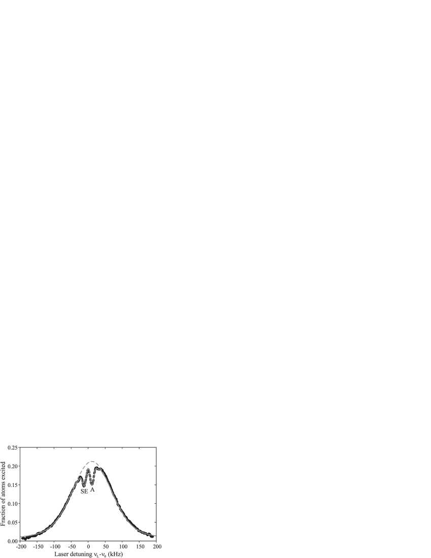
To make a connection with previous experiments, we first consider the Doppler-broadened regime, for which we choose a pulse duration (s) such that the spectroscopic resolution is about 21 kHz. This is less than the 150 kHz (FWHM) Doppler width of the 10 K atoms and slightly less than the 23.1 kHz recoil splitting of the 657 nm clock transition. In Fig. 1 we show the resulting excitation spectrum, whose envelope is primarily determined by the Doppler background. In fact this envelope is simply twice the height of the curve seen with a single probe pulse, because for most laser frequencies the two counter-propagating laser beams excite non-overlapping velocity classes. However, at the absorption resonance frequency, (‘A’ in Fig. 1), there is not this doubling of the Doppler background, since the first laser pulse has already excited the majority of these atoms to the long-lived excited state. Note that the second pulse cannot de-excite many of these atoms either, since due to atomic recoil they have been shifted out of resonance. The second resonance (‘SE’ in Fig. 1), at , results from atoms that started with a velocity , and were thus excited by the first pulse (in the atoms’ rest frame, ); now at rest in the lab frame due to atomic recoil, they are resonant with the second pulse for stimulated emission, thereby reducing the net fraction of atoms excited.
The two dips resulting from these resonances are analogous to those seen in earlier experiments but with one important distinction: due to the small width of the Doppler background, we can readily see that the dips are asymmetrically located about the center of the Doppler background. Since the Doppler background results from absorption, it is naturally centered around the resonance at , coincident with the absorption recoil dip. The dip associated with stimulated emission is located one recoil frequency () below the absorption resonance, on the red side of the Doppler curve. It is important to emphasize that this asymmetry is not a result of the order of the laser pulses. If we reverse the temporal order, so that the pulse directions are reversed, we observe the identical lineshape, not its mirror image. Rather, this asymmetry is a fundamental feature of saturation spectroscopy, though one that is easily overlooked in experiments with broad Doppler backgrounds. As has been noted by other observers,Ishikawa et al. (1994); Dingler et al. (1994) this asymmetry can lead to undesired offsets in realizing optical frequency standards based on saturation absorption since the unperturbed line center of the transition (midway between the recoil components) is not centered on the background.
We now consider what happens to this spectrum as we broaden the Fourier spectrum of the probe pulse, not just beyond the recoil splitting but well beyond the width of the Doppler distribution itself. This regime has not previously been investigated experimentally, but is becoming important in state-of-the-art optical atomic clocks based on neutral atoms.Curtis et al. (2003); Wilpers et al. (2002) We access this regime by reducing the duration of the probe pulses (while maintaining the pulse area to keep the excitation probability constant). In Fig. 2, we show a set of spectroscopic lineshapes taken over probe durations ranging from 40 s down to 2.6 s.
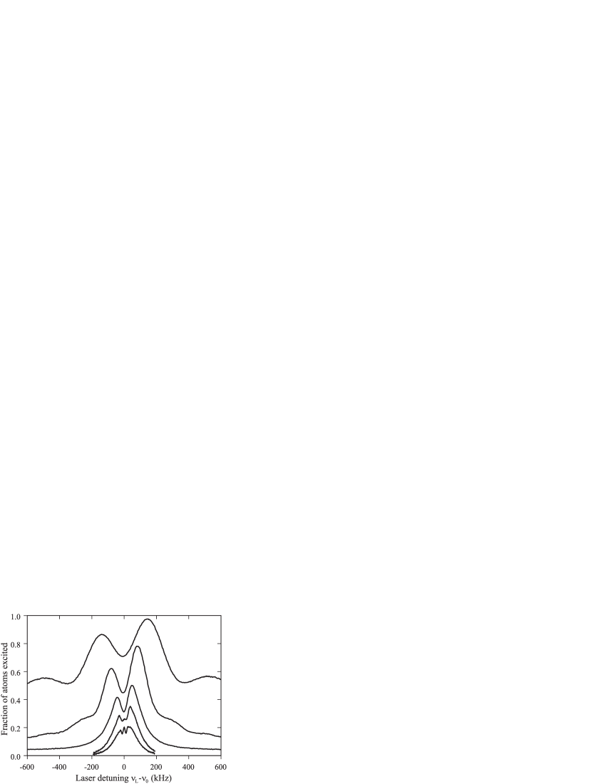
Note that the fraction of atoms excited grows with decreasing resolution as our probe spectrum covers a larger fraction of the velocity distribution. For the shortest pulse length, the Fourier transform of the probe pulse has a spectral width approximately 2.5 times that of the Doppler distribution. As we see, changing the resolution from 21 kHz to 42 kHz (20 s pulse duration) begins to obscure the recoil splitting, but the large asymmetry persists. As we move to 85 kHz resolution (10 s pulse duration), the recoil splitting is no longer visible as the two dips merge into a single dip centered at , but the spectrum now appears to consist of two peaks whose separation is determined by the spectroscopic resolution and whose amplitudes differ by more than 25 % ! The asymmetry persists for even the lowest resolution, where the maxima are separated by 280 kHz, more than 10 times that of the recoil splitting itself.
Examination of Fig. 2 provides an intuitive picture of how such a small effect can cause such large asymmetries. In the Fourier-transform regime we find the unusual spectroscopic condition where the widths of both the envelope and the dip are determined almost solely by the probe time, but are slightly offset from one another. This small offset leads to the envelope asymmetry by causing the dip to intersect the Doppler envelope at different heights on the two sides. Thus, the size of the asymmetry is related to the slope of the envelope multiplied by the size of the recoil effect.
Alternatively, we can think of this spectrum as resulting from two contributions: one symmetric (due solely to absorption) and one asymmetric (due to stimulated emission by the second pulse). This framework allows a simple model to describe our spectra well (an exact theoretical treatment of coherent saturation spectroscopy including multi-photon effects has been developed by Bordé and co-workersIshikawa et al. (1994); Bordé et al. (1984). We start by considering the excitation spectrum resulting from the first (idealized) square -pulse of duration and Rabi frequency illuminating a sample of ground-state atoms initially at rest. This yields the well-known Rabi spectrum Rabi et al. (1939) for the excitation probability:
| (2) |
where is the laser detuning from the absorption resonance. Illuminating the atoms with a square -pulse from the opposite direction gives two sets of atoms to consider. First, there are atoms that remained in the ground state after the first pulse (a fraction equal to ); they will be resonant with a second pulse at frequency , so the above equation applies to these atoms as well. Second, the atoms that were excited by the first pulse (a fraction equal to ) now have a velocity toward the second counter-propagating laser beam. The associated Doppler effect will shift the stimulated emission resonance down by one recoil, so these atoms will be resonant with light at frequency , or . The fraction of these atoms transferred back to the ground state can then be derived from the same probability function , but with the argument .
To find the total fraction in the excited state after two pulses, we simply add the two contributions:
| (3) |
The first product in this expression gives the ground-state contribution and is symmetric (both pulses are resonant at the same frequency). The second term gives the excited-state contribution, but it is asymmetric due to the offset of between arguments of the multiplicands, thus yielding a net asymmetry for . We can easily connect this model with experiment by including the initial velocity distribution (via the detuning) and the unequal laser intensities seen by the atoms due to the spatial distribution of the atoms in the laser mode (via the Rabi frequency). The free parameters in our simulation were overall signal amplitude (imperfect normalization required a 10 - 15 % reduction in signal size) and atomic cloud size (which we fixed at the same value for all simulations). We measured the velocity distributions and the laser mode size separately and used the resulting values for the simulations. We see good agreement over a variety of probe resolutions (simulations are shown as solid lines in Figs. 1 and 3), although we see small differences in the wings at the lowest resolutions,
most likely resulting from the non-ideal square pulses used in the experiment.
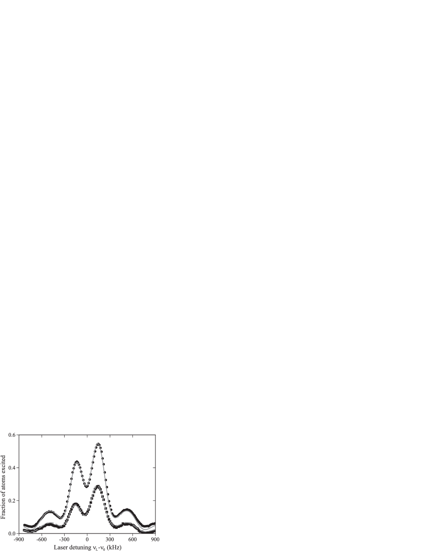
Interestingly, we can isolate the second product in the expression for experimentally by using recoil suppression.Riehle et al. (1988); Dingler et al. (1994) In this case we suppress the ground-state (absorption) contribution by illuminating the atoms with a resonant 423 nm pulse (duration 20 s) after the first red probe pulse but before the second red pulse. This heats the atoms in the ground state to the point where excitation by the second pulse produces a nearly flat Doppler background, upon which sits the contribution from the atoms shelved in the excited state. Fig. 3 shows that the feature associated with stimulated emission is fully responsible for the lineshape asymmetry, as the absolute peak height differences are virtually identical. We emphasize that this asymmetric envelope persists in four-pulse Bordé-Ramsey saturation spectroscopy Bordé et al. (1984) in the Fourier-transform regime, which is used in ultra-high resolution studies and for cold atom optical clocks.Curtis et al. (2003); Wilpers et al. (2002) However, the good agreement between theory and experiments give us good confidence that these effects will not limit the accuracy of these clocks.
In summary, we have used ultra-cold two-level atoms and ultra-high resolution spectroscopy to probe the atomic recoil structure unique to saturated absorption spectroscopy at an unprecedented level. The resulting spectra clearly reveal the fundamental asymmetry in the location of recoil components on the Doppler background. In addition we have shown how this asymmetry in the recoil frequencies leads to large amplitude asymmetries in the Fourier transform limit, a regime that has not previously been investigated but is important for state-of-the-art precision metrology. Extending these studies to a regime with Ca atoms laser cooled to sub-recoil temperatures, as demonstrated by Curtis et al. Curtis et al. (2003), would allow us to individually address the recoil components via laser detuning. This could enhance the sensitivity of precision measurements such as that of the photon recoil or other atom interferometry experiments Weiss et al. (1993); Snadden et al. (1998); Gupta et al. (2002).
Acknowledgements.
We thank J. Bergquist and J. Kitching for their insightful comments and J. Hall for illuminating discussions. This work was supported in part by NASA and ONR-MURI. G. W. acknowledges support from the Alexander von Humboldt Foundation. Work of a US government agency; not subject to copyright.References
- Curtis et al. (2003) E. A. Curtis, C. W. Oates, and L. Hollberg, J. Opt. Soc. Am. B 20, 977 (2003).
- Wilpers et al. (2002) G. Wilpers, T. Binnewies, C. Degenhardt, U. Sterr, J. Helmcke, and F. Riehle, Phys. Rev. Lett. 89, 230801 (2002).
- Kol’chenko et al. (1969) A. P. Kol’chenko, S. G. Rautian, and R. I. Sokolovskiĭ, Soviet Physics JETP 28, 986 (1969).
- Hall et al. (1976) J. L. Hall, C. J. Bordé, and K. Uehara, Phys. Rev. Lett. 37, 1339 (1976).
- Barger et al. (1979) R. L. Barger, J. C. Bergquist, T. C. English, and D. J. Glaze, Appl. Phys. Lett. 34, 850 (1979).
- Sterr et al. (1992) U. Sterr, K. Sengstock, J. H. Müller, D. Bettermann, and W. Ertmer, Appl. Phys. B 54, 341 (1992).
- Riehle et al. (1988) F. Riehle, J. Ishikawa, and J. Helmcke, Phys. Rev. Lett. 61, 2092 (1988).
- Bagayev et al. (1989) S. N. Bagayev, A. E. Baklanov, V. P. Chebotayev, and A. S. Dychkov, Appl. Phys. B 48, 31 (1989).
- Ishikawa et al. (1994) J. Ishikawa, F. Riehle, J. Helmcke, and C. J. Bordé, Phys. Rev. A 49, 4794 (1994).
- Binnewies et al. (2001) T. Binnewies, G. Wilpers, U. Sterr, F. Riehle, J. Helmcke, T. E. Mehlstäubler, E. M. Rasel, and W. Ertmer, Phys. Rev. Lett. 87, 123002 (2001).
- Dingler et al. (1994) F. E. Dingler, V. Rieger, K. Sengstock, U. Sterr, and W. Ertmer, Opt. Commun. 110, 99 (1994).
- Bordé et al. (1984) C. J. Bordé, C. Salomon, S. Avrillier, A. Van Lerberghe, C. Bréant, D. Bassi, and G. Scoles, Phys. Rev. A 30, 1836 (1984).
- Rabi et al. (1939) I. I. Rabi, S. Millman, P. Kusch, and J. R. Zacharias, Phys. Rev. 55, 526 (1939).
- Weiss et al. (1993) D. S. Weiss, B. C. Young, and S. Chu, Phys. Rev. Lett. 70, 2706 (1993).
- Snadden et al. (1998) M. J. Snadden, J. M. McGuirk, P. Bouyer, K. G. Haritos, and M. A. Kasevich, Phys. Rev. Lett. 81, 971 (1998).
- Gupta et al. (2002) S. Gupta, K. Dieckmann, Z. Hadzibabic, and D. E. Pritchard, Phys. Rev. Lett. 89, 140401 (2002).