Wavelet analysis of epileptic spikes
Abstract
Interictal spikes and sharp waves in human EEG are characteristic signatures of epilepsy. These potentials originate as a result of synchronous, pathological discharge of many neurons. The reliable detection of such potentials has been the long standing problem in EEG analysis, especially after long-term monitoring became common in investigation of epileptic patients. The traditional definition of a spike is based on its amplitude, duration, sharpness, and emergence from its background. However, spike detection systems built solely around this definition are not reliable due to the presence of numerous transients and artifacts. We use wavelet transform to analyze the properties of EEG manifestations of epilepsy. We demonstrate that the behavior of wavelet transform of epileptic spikes across scales can constitute the foundation of a relatively simple yet effective detection algorithm.
pacs:
87.10. +e, 05.45.-aRecordings of human brain electrical activity (EEG) have been the fundamental tool for studying the dynamics of cortical neurons since 1929. Even though the gist of this technique has essentially remained the same, the methods of EEG data analysis have profoundly evolved during the last two decades. In 1985 Babloyantz et al. demonstrated that certain nonlinear measures, first introduced in the context of chaotic dynamical systems, changed during slow-wave sleep Babloyantz et al. (1985). The flurry of research work that followed this discovery focused on the application of nonlinear dynamics in quantifying brain electrical activity during different mental states, sleep stages, and under the influence of the epileptic process (for a review see for example West et al. (1995); Pritchard and Duke (1992)). It must be emphasized that a straightforward interpretation of neural dynamics in terms of such nonlinear measures as the largest Lyapunov exponent or the correlation dimension is not possible since most biological time series, such as EEG, are nonstationary and consequently do not satisfy the assumptions of the underlying theory. On the other hand, traditional power spectral methods are also based on quite restrictive assumptions but nevertheless have turned out to be successful in some areas of EEG analysis. Despite these technical difficulties, the number of applications of nonlinear time series analysis has been growing steadily and now includes the characterization of encephalopaties Stam et al. (1999), monitoring of anesthesia depth Widman et al. (2000), characteristics of seizure activity Casdagli et al. (1997), and prediction of epileptic seizures Lehnertz and Elger (1998). Several other approaches are also used to elucidate the nature of electrical activity of the human brain ranging from coherence measures Nunez et al. (1997, 1999) and methods of nonequilibrium statistical mechanics Ingber (1982) to complexity measures Rezek and Roberts (1998); van Putten and Stam (2001).
One of the most important challenges of EEG analysis has been the quantification of the manifestations of epilepsy. The main goal is to establish a correlation between the EEG and clinical or pharmacological conditions. One of the possible approaches is based on the properties of the interictal EEG (electrical activity measured between seizures) which typically consists of linear stochastic background fluctuations interspersed with transient nonlinear spikes or sharp waves. These transient potentials originate as a result of a simultaneous pathological discharge of neurons within a volume of at least several mm3.
The traditional definition of a spike is based on its amplitude, duration, sharpness and emergence from its background Gotman and Gloor (1976); Gotman and Wang (1982). However, automatic epileptic spike detection systems based on this direct approach suffer from false detections in the presence of numerous types of artifacts and non-epileptic transients. This shortcoming is particularly acute for long-term EEG monitoring of epileptic patients which became common in 1980s. To reduce false detections Gotman and Wang Gotman and Wang (1991) made the process of spike identification dependent upon the state of EEG (active wakefulness, quiet wakefulness, desynchronized EEG, phasic EEG, and slow EEG). This modification leads to significant overall improvement provided that state classification is correct.
Diambra and Malta Diambra and Malta (1999) adopted nonlinear prediction for epileptic spike detection. They demonstrated that when the model’s parameters are adjusted during the “learning”phase to assure good predictive performance for stochastic background fluctuations, the appearance of an interictal spike is marked by a very large forecasting error. This novel approach is appealing because it makes use of changes in EEG dynamics. One expects good nonlinear predictive performance when the dynamics of the EEG interval used for building up the model is similar to the dynamics of the interval used for testing. However, it is uncertain at this point whether it is possible to develop a robust spike detection algorithm based solely on this idea.
As Clark et al. put it succinctly, automatic EEG analysis is a formidable task because of the lack of “…features that reflect the relevant information ”Clark et al. (1995). Another difficulty is the nonstationary nature of the spikes and the background in which they are embedded. One technique developed for the treatment of such nonstationary time series is wavelet analysis Mallat (1998); Unser and Aldroubi (1996). The goal of this paper is to characterize the epileptic spikes and sharp waves in terms of the properties of their wavelet transforms. In particular, we search for features which could be important in the detection of epileptic events.
The wavelet transform is an integral transform for which the set of basis functions, known as wavelets, are well localized both in time and frequency. Moreover, the wavelet basis can be constructed from a single function by means of translation and dilation:
| (1) |
is commonly referred to as the mother function or analyzing wavelet. The wavelet transform of function is defined as
| (2) |
where denotes the complex conjugate of . The continuous wavelet transform of a discrete time series of length and equal spacing is defined as
| (3) |
The above convolution can be evaluated for any of values of the time index . However, by choosing all successive time index values, the convolution theorem allows us to calculate all convolutions simultaneously in Fourier space using a discrete Fourier transform (DFT). The DFT of is
| (4) |
where is the frequency index. If one notes that the Fourier transform of a function is then by the convolution theorem
| (5) |
frequencies are defined in the conventional way. Using (5) and a standard fast Fourier transform (FFT) routine it is possible to efficiently calculate the continuous wavelet transform (for a given scale ) at all simultaneously Torrence and Compo (1998). It should be emphasized that formally equation (5) does not yield the discrete linear convolution corresponding to (3) but rather a discrete circular convolution in which the shift is taken modulo . However, in the context of this work, this problem does not give rise to any numerical difficulties. This is because, for purely practical reasons, the beginning and the end of the analyzed part of data stream are not taken into account during the EEG spike detection.
From a plethora of available mother wavelets, we employ the Mexican hat
| (6) |
which is particularly suitable for studying epileptic events.
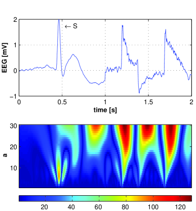
In the top panel of Fig. 1 we present two pieces of the EEG recording joined at approximately . The digital 19 channel recording sampled at 240 Hz was obtained from a juvenile epileptic patient according to the international 10-20 standard with the reference average electrode. The epileptic spike in this figure (marked by the arrow) is followed by two artifacts. The bottom panel of Fig. 1 displays the contour map of the absolute value of Mexican hat wavelet coefficients . It is apparent that the red prominent ridges correspond to the position of either spike or the motion artifacts. What is most important, for small scales, , the values of the wavelet coefficients for the spike’s ridge are much larger than those for the artifacts. The peak value along the spike ridge corresponds to . In sharp contrast, for the range of scales used in Fig. 1 the absolute value of coefficients for the artifacts grow monotonically with .
The question arises as to whether the behavior of the wavelet transform as a function of scale can be used to develop a reliable detection algorithm. The first step in this direction is to use the normalized wavelet power
| (7) |
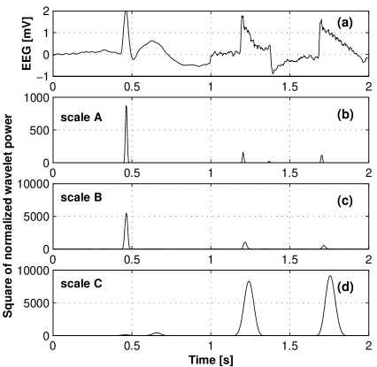
instead of the wavelet coefficients to reduce the dependence on the amplitude of the EEG recording. In the above formula is the variance of the portion of the signal being analyzed (typically we use pieces of length 1024 for EEG tracings sampled at 240 Hz). In actual numerical calculations we prefer to use the square of to merely increase the range of values analyzed during the spike detection process. In Fig. 2 for the signal used in Fig. 1 is plotted for three scales , and .
In the most straightforward approach, we identify an EEG transient potential as a simple or isolated epileptic spike if and only if:
the value of at is greater than a predetermined threshold value ,
the square of normalized wavelet power decreases from scale to ,
the value of at is greater than a predetermined threshold value .
The threshold values and may be considered as the model’s parameters which can be adjusted to achieve the desired sensitivity (the ratio of detected epileptic events to the total number of epileptic events present in the analyzed EEG tracing) and selectivity (the ratio of epileptic events to the total number of events marked by the algorithm as epileptic spikes).
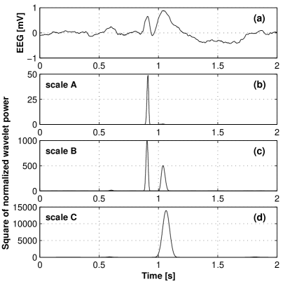
While this simple algorithm is quite effective for simple spikes such as one shown in Fig. 1 it fails for the common case of an epileptic spike accompanied by a slow wave with comparable amplitude. The example of such complex is given in Fig. 3(a). The overlap of the negative tail of the Mexican hat with the slow wave yields the inherently low values of at scale (panel (b)) and scale (panel (c)) as compared to those characteristic of the “isolated”spike. Nevertheless, the normalized wavelet power does decrease from scale to . Consequently, in the same vein as the argument we presented above, we can develop an algorithm which detects the epileptic spike in the vicinity of a slow wave by calculating the following linear combination of wavelet transforms:
| (8) |
and checking whether the square of corresponding normalized power at scales and exceeds the threshold value and , respectively. The second term in (8) allows us to detect the slow wave which follows the spike. The parameters and are chosen to maximize the overlap of the wavelet with the slow wave. For the Mexican hat we use and . By varying the values of coefficients and , it is possible to control the relative contribution of the spike and the slow wave to the linear combination (8).
For testing purposes, we built up the database of artifacts and spikes. We made available some of these EEG tracings at hom along with the examples of the numerical calculations. While the analysis of the pieces of EEG recordings such as those shown in Fig. 2 and 3 is essential in determining the generic properties of epileptic events, it can hardly reflect the difficulties one can encounter in interpretation of clinical EEG. Therefore we selected four challenging EEG tracings with 340 epileptic events. The algorithm described in this work marked 356 events out of which 239 turned out to be the epileptic events. Thus the sensitivity of the algorithm was 70% and its selectivity was equal to 67%. We then analyzed the same tracings with the leading commercial spike detector developed by the Persyst Development Corporation (Insight 2001.07.12). This software marked 654 events out of which 268 were epileptic events. Thus slightly better sensitivity of 79% was achieved at the expense of the low 41% selectivity. The performance of preliminary numerical implementation of the detection algorithm presented in this work is excellent and allows to process 24 hour EEG recording (19 channels sampled at 240 Hz) in a matter of minutes on the average personal computer.
The goal of wavelet analysis of the two types of spikes, presented in this paper, was to elucidate the approach to epileptic events detection which explicitly hinges on the behavior of wavelet power spectrum of EEG signal across scales and not merely on its values. Thus, this approach is distinct not only from the detection algorithms based upon discrete multiresolution representations of EEG recordings Blanco et al. (1996); D’Attellis et al. (1997); Sartoretto and Ermani (1999); Calvagno et al. (2000); Gutiérrez et al. (2001) but also from the method developed by Senhadji and Wendling which employs continuous wavelet transform Senhadji and Wendling (2002).
Epilepsy is a common disease which affects 1-2% of the population and about 4% of children Jallon (2002). In some epilepsy syndromes interictal paroxysmal discharges of cerebral neurons reflect the severity of the epileptic disorder and themselves are believed to contribute to the progressive disturbances in cerebral functions (eg. speech impairment, behavioral disturbances) Engel (2001). In such cases precise quantitative spike analysis would be extremely important. The epileptic event detector described in this paper was developed with this particular goal in mind and its application to the studies of the Landau-Kleffner syndrome will be presented elsewhere.
References
- Babloyantz et al. (1985) A. Babloyantz, J. M. Salazar, and C. Nicolis, Physics Letters A 111, 152 (1985).
- West et al. (1995) B. J. West, M. N. Novaes, and V. Kovcic, in Fractal Geometry in Biological Systems, edited by P. M. Iannoccone and M. Khokha (CRC Press, Boca Raton, FL, 1995), pp. 267–316.
- Pritchard and Duke (1992) W. S. Pritchard and D. W. Duke, Intern. J. Neuroscience 67, 31 (1992).
- Stam et al. (1999) C. J. Stam, E. M. H. van der Leij R. W. M. Keunen, and D. L. J. Tavy, Theory Biosci. 118, 209 (1999).
- Widman et al. (2000) G. Widman, T. Schreiber, B. Rehberg, A. Hoeft, and C. E. Elger, Phys. Rev. E 62, 4898 (2000).
- Casdagli et al. (1997) M. C. Casdagli, L. D. Iasemidis, R. S. Savit, R. L. Gilmore, S. N. Roper, and J. C. Sackellares, Electroencephalogr. Clin. Neurophysiol. 102, 98 (1997).
- Lehnertz and Elger (1998) K. Lehnertz and C. E. Elger, Phys. Rev. Lett. 80, 5019 (1998).
- Nunez et al. (1997) P. L. Nunez, R. Srinivasan, A. F. Westdorp, R. S. Wijesinghe, D. M. Tucker, R. B. Silberstein, and P. J. Cadusch, Electroencephalogr. Clin. Neurophysiol. 103, 499 (1997).
- Nunez et al. (1999) P. L. Nunez, R. B. Silberstein, Z. P. Shi, M. R. Carpenter, R. Srinivasan, D. M. Tucker, S. M. Doran, P. J. Cadusch, and R. S. Wijesinghe, Clin. Neurophysiol. 110, 469 (1999).
- Ingber (1982) L. Ingber, Physica D 5, 83 (1982).
- Rezek and Roberts (1998) I. A. Rezek and S. J. Roberts, IEEE Trans. Biomed. Eng. 45, 1186 (1998).
- van Putten and Stam (2001) M. J. A. M. van Putten and C. J. Stam, Phys. Letters A 281, 131 (2001).
- Gotman and Gloor (1976) J. Gotman and P. Gloor, Electroencephalogr. Clin. Neurophysiol. 41, 513 (1976).
- Gotman and Wang (1982) J. Gotman and L. Y. Wang, Electroencephalogr. Clin. Neurophysiol. 54, 530 (1982).
- Gotman and Wang (1991) J. Gotman and L. Y. Wang, Electroencephalogr. Clin. Neurophysiol. 79, 11 (1991).
- Diambra and Malta (1999) L. Diambra and C. P. Malta, Phys. Rev. E 59, 929 (1999).
- Clark et al. (1995) I. Clark, R. Biscay, M. Echeverria, and T. Virues, Comput. Biol. Med. 25, 373 (1995).
- Mallat (1998) S. Mallat, A Wavelet Tour of Signal Processing (Academic Press, San Diego, 1998).
- Unser and Aldroubi (1996) M. Unser and A. Aldroubi, Proc. IEEE 84, 626 (1996).
- Torrence and Compo (1998) C. Torrence and G. P. Compo, Bull. Amer. Meteor. Soc. 79, 61 (1998).
- (21) URL http://republika.pl/eegspike/.
- Blanco et al. (1996) S. Blanco, C. E. D’Attellis, S. I. Isaacson, O. A. Rosso, and R. O. Sirne, PRE 54, 6661 (1996).
- D’Attellis et al. (1997) C. E. D’Attellis, S. Isaacson, and R. O. Sirne, Ann. Biomed. Eng. 25, 286 (1997).
- Sartoretto and Ermani (1999) F. Sartoretto and M. Ermani, Clin. Neurophysiol. 110, 239 (1999).
- Calvagno et al. (2000) M. Calvagno, M. Ermani, R. Rinaldo, and F. Sartoretto, in Proceedings of the 2000 IEEE International Conference on Acoustics, Speech, and Signal Processing (2000), vol. 6.
- Gutiérrez et al. (2001) J. Gutiérrez, R. Alcántara, and V. Medina, Medical Engineering & Physics 23, 623 (2001).
- Senhadji and Wendling (2002) L. Senhadji and F. Wendling, Neurophysiol. Clin. 32, 175 (2002).
- Jallon (2002) P. Jallon, Epileptic Disord. 4, 1 (2002).
- Engel (2001) J. Engel, Epilepsia 42, 796 (2001).