Hydration of Methanol in Water
A DFT-based Molecular Dynamics Study
Abstract
We studied the hydration of a single methanol molecule in aqueous solution by first-principle DFT-based molecular dynamics simulation. The calculations show that the local structural and short-time dynamical properties of the water molecules remain almost unchanged by the presence of the methanol, confirming the observation from recent experimental structural data for dilute solutions. We also see, in accordance with this experimental work, a distinct shell of water molecules that consists of about 15 molecules. We found no evidence for a strong tangential ordering of the water molecules in the first hydration shell.
INTRODUCTION
The solvation of alcohols in water has been studied extensively.[1] It is of fundamental interest in physics, chemistry and biology, but also of importance in technical applications. The characteristic hydroxyl group allows alcohols to form hydrogen bonds and is responsible for the good solubility of the smaller alcohols. In contrast, the alkyl group is hydrophobic and does not participate in the hydrogen bonding network of water. The presence of both hydrophobic and hydrophilic groups make the microscopic picture of solvation of alcohol in water a non-trivial and therefore interesting matter.
Understanding the solvation of methanol in water is a prerequisite for the study of chemistry of alcohols in aqueous solution. Important examples of such reactions are the conversion of ethanol into acetaldehyde in biological systems or the industrial ethanol production by acid-catalysed hydration of ethylene. An accurate microscopic understanding of the mechanism and kinetics of such reactions is of fundamental interest. However, presently, this picture is still far from complete. Density Functional Theory (DFT) based Molecular Dynamics simulation has proved to be a promising tool provide such an insight. An accurate calculation of the chemical bonding is incorporated via a DFT-based electronic structure calculations. The effect of temperature and solvent on the reactive events is implicitly accounted for via the Molecular Dynamics technique. The implementation of DFT-based MD as proposed by Car and Parrinello[2] has proven to be extremely efficient. It has successfully been applied to study of a large variety of condensed-phase systems at finite temperature. Applications to chemical reactions include the cat-ionic polymerization of 1,2,5-trioxane[3], or the acid-catalysed hydration of formaldehyde[4].
As a first step towards the study of chemical reactions involving alcohols we present in this paper a Car-Parrinello Molecular Dynamics (CPMD) study of the hydration of the simplest alcohol (methanol) in aqueous solution. Recent experimental work[5] has provided detailed structural information on the solvation shell. Various molecular simulation studies (e.g. Ref. [6, 7, 8, 9, 10] have addressed structure and dynamics of both the solute and the solvent. This experimental and numerical work has revealed that there is a distinct solvation shell around the methanol, and that the water structure is little affected by the presence of a methanol molecule. In this paper we will address these structural properties and in addition consider the dynamics of the methanol and the water molecules in the solvation shell.
This paper is organized as follows. First we outline the computational approach and its validation. Then we present the results for the structure and dynamics of a single solvated methanol in water. We conclude the paper with a summary and discussion.
METHODS AND VALIDATION
Electronic structure calculations are performed using the Kohn-Sham formulation[11] of DFT.[12] We employed the BLYP functional, that combines a gradient-corrected term for the correlation energy as proposed by Lee, Yang and Parr[14] with the gradient correction for the exchange energy due to Becke[13]. Among the available functionals, the BLYP functional has proven to give the best description of the structure and dynamics of water.[15, 16] All calculations[17] were performed using the CPMD package.[18]
The pseudopotential method is used to restrict the number of electronic states to those of the valence electrons. The interaction with the core electrons is taken into account using semi-local norm-conserving Martins-Troullier pseudopotentials.[19] The pseudopotential cutoff radius for the H was chosen 0.50 au. For O and C the radii are taken 1.11 and 1.23 a.u. for both the l=s and l=p term. The Kohn-Sham states are expanded in a plane-wave basis set matching the periodicity of the periodic box with waves up to a kinetic energy of 70 Ry. Test calculations showed that for this structural and energetic properties were converged within Å and kJ/mol, respectively. Frequencies are converged within 1 %, expect for CO and OH stretch modes that are underestimated by 3 % and 5 % compared to basis-set limit values.
To validate the computational methods outlined above we performed a series of reference calculations of relevant gas-phase compounds with the CPMD package. Energetics and geometry were calculated for methanol, water, two mono-hydrate configurations, and the di-hydrate configuration shown in Fig. 1. These calculations were performed using a a large periodic box of size 10x10x10 Å3. The interactions among the periodic images were eliminated by a screening technique similar to that of Ref. [20]. In addition we determined for the methanol molecule both the harmonic vibrational frequencies and the frequencies at finite temperature (T= 200 K). The latter includes the anharmonic contributions, and were obtained from the spectrum of the velocity auto correlation function (VACF) of a 3 ps CPMD calculation at E= 200 K. The calculated peak positions can be compared with experimental spectra. Results of the gas-phase calculations were compared with results obtained with a state-of-the-art atomic-orbital based DFT package (ADF[21]), and with results from MP2 calculations of Ref. [22]. In the comparison of the energies zero-point energies were not taken into account.
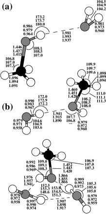
Complexation energies and geometries of the methanol hydrates are given in Tab. I and Fig. 1. Deviations among CPMD and ADF are within 1 kcal/mole for the energies, smaller than Å for the inter-molecular bonds and within Å for the weaker intra-molecular bonds. This indicates a state-of-the art accuracy for electronic structure methods employed in CPMD. Differences among BLYP and MP2 are within acceptable limits, with BLYP complexation energies smaller by 4 kJ/mole (dimer) and 10 kJ/mole (trimer). These deviations are similar to the comparison of BLYP and MP2 for the water dimer binding energy,[15, 23] where BLYP is 4 kJ/mole smaller, with the MP2 energy only kJ/mol below the experimental value. Assuming similar differences for the complexation energies bonds in the methanol hydrates would suggest that BLYP underestimates the methanol-water binding energy by approximately kJ/mol. Inter- and intra-molecular BLYP bond lengths are up to and 0.06 Å longer compared to the MP2 results, respectively.
Vibrational frequencies are listed in Tab. II. Again comparison of CPMD and ADF is excellent, consistent with the results for the energetics and geometries. Comparing the calculated finite-temperature frequencies against the experimental values shows that BLYP tends to underestimate the frequencies of almost all modes by 10 %. This trend is a known feature of BLYP. For example similar deviations are observed for BLYP calculation of water.[15]
Overall we conclude that the reference calculations of gas-phase
provides confidence that DFT-BLYP performs with a sufficient accuracy
for a quantitative study of methanol hydration.
| Harmonic | Anharmonic | |||
| (cm-1) | (cm-1) | |||
| mode | CPMD-BLYP | ADF-BLYPa | CPMD-BLYP | Exp.b |
| (T=200 K) | ||||
| (OH) | 280 | 380 | 280 | 270 |
| (CO) | 940 | 950 | 880 | 1034 |
| (CH3) | 1040 | 1050 | 980 | 1075 |
| (CH3) | 1130 | 1130 | 1070 | 1145 |
| (OH) | 1330 | 1340 | 1270 | 1340 |
| (CH3) | 1430 | 1430 | 1320-1430c | 1454 |
| (CH3) | 1460 | 1460 | 1320-1430c | 1465 |
| (CH3) | 1470 | 1470 | 1320-1430c | 1480 |
| (CH3) | 2940 | 2910 | 2640 | 2844 |
| (CH3) | 2990 | 2950 | 2740 | 2970 |
| (CH3) | 3060 | 3020 | 2830 | 2999 |
| (OH) | 3550 | 3590 | 3310 | 3682 |
SOLVATION
We performed Car-Parrinello Molecular Dynamics simulations of the solvation of a single methanol molecule. We considered two systems: one with 31 water molecules and the other with 63 water molecules, yielding methanol-water solutions with mole ratios of 1:31 and 1:63. In the following they are referred to as the small and large system, respectively. For reference we also performed a simulation of a pure water sample of 32 molecules. The molecules are placed in a periodic cubic box with edges of 9.98 Å (small solvated methanol system), 12.50 Å (large solvated methanol system), and (pure water) corresponding to the experimental densities at ambient conditions. The temperature of the ions is fixed at 300 K using a Nosé-Hoover thermostat [24, 25, 26]. The fictitious mass associated with the plane-wave coefficients is chosen at 900 a.u., which allowed for a time step in the numerical integration of the equations-of-motion of 0.145 fs. The two systems were equilibrated for 1 ps from an initial configuration obtained by a force-field simulation. Subsequently we gathered statistical averages from a 10 ps trajectory of the 31+1 molecule system, from a 7 ps trajectory of the 63+1 molecule system, and from a 10 ps trajectory of the pure water system.
Structure
In Fig. 2 we have plotted the radial distribution functions (RDF) of the water oxygen atoms. The minor variations among the RDF’s of the small methanol system, the large methanol system, and the pure water system is an indication that the local water structure, as measured by this RDF, is at only marginally changed by the solvation of a methanol molecule. Note, in this respect, that for the 32 molecule the first solvation shell constitutes a significant fraction of the total number of water molecules (see below).

Fig. 2 also shows the RDF of the methanol carbon and water oxygens for the small and large methanol system. A pronounced first peak clearly indicates the existence of shell of water molecules at a distance of 3.7 Å. Comparing the RDF’s of the small and large system shows a noticeable difference. This should be attributed to the limited size of the small system. It suggests that a proper description of the solvation structure of a single methanol in a cubic periodic simulation box requires at least 50 water molecules. Integrating the RDF for the large system up to the minimum at Å yields water molecules in the first solvation shell. The definite solvation shell observed in our simulations is consistent with the neutron diffraction data of Soper and Finney[5] who studied a 1:9 molar methanol-water system. Differences in molarity limits a quantitative comparison of the carbon-oxygen RDF, but a qualitative comparison learns that peak positions match with the peak values slightly more pronounced in the simulation results.
To analyze the orientational ordering of the water molecules around the methanol we computed the distribution function of the angle between the C-O bond vector and the normal to the plane of the water molecules in the first solvation shell. The results show that angle distribution is relatively uniform with a small tendency towards the tangential orientation, a feature occurs for all solvation shell radii in the range of 3.7-5.0 Å. Over the range of 0o-90o the distribution gradually decays, with the value at the tangential orientation (0o) about a factor of 2 larger than at the perpendicular orientation (90o). Qualitatively, this seems consistent with data for the orientational distribution obtained from neutron-diffraction data [5]. However from this experimental data it is concluded that the water molecules prefer to lie tangential and form a cage around the methanol. Our data do not give clear evidence for a cage-like structure. However, this might be a different interpretation from similar data. Note, in this respect, also that the experimental data cannot be quantitatively compared to our data, as different orientational distribution functions are employed.
To analyze the hydrogen bonding we adopted the definition of Ref. [7]: two molecules are hydrogen bonded if simultaneously the inter-oxygen distance is less than Åand the OHO angle is smaller than . From the simulation of the large system we found that the methanol hydroxyl group donates and accepts on average 0.9 and 1.5 hydrogen bonds, respectively. For a water molecule these numbers are equal and measured to be 1.7 in the simulation of the pure water sample. These results indicate that the methanol hydroxyl group participates strongly in the hydrogen bonding network with the a donating behavior similar to water hydrogen and a accepting character somewhat smaller than a water oxygen.
Dynamics
The time scale (7-10 ps) of the present simulations allows for a reliable analysis of dynamical properties occurring on the picosecond time scale.
The velocity auto correlation function (VACF) of the hydrogen atoms provides an important measure of hydrogen bonding. Fig. 3 shows the Fourier spectrum of the calculated VACF of hydrogen atoms of the water molecules in the small and large methanol sample. The three distinct peaks correspond to the vibrational (3100 cm-1), bending (1600 cm-1), and librational-translational (500 cm-1) modes of the water molecules. The most important observation is that mutual comparison of the two methanol samples and the comparison of these with the spectrum of the pure water sample (also plotted) shows no significant difference, not even for the small methanol sample where the solvation shell constitutes half of the water molecules in the system. This demonstrates that also the short-time dynamics of the water molecules is hardly affected by the solvation of a methanol molecule.

An indication for the average residence time of a water molecule in the first solvation shell is obtained by monitoring the trajectories of the individual water molecules. We found that in the large methanol system over 7 ps 10 water molecules left the region within Å from the methanol carbon. From this we estimate the average residence time to be of the order of a few picoseconds.
Fig. 3 shows the Fourier spectrum of the VACF of the hydroxyl H of methanol obtained from the trajectory of the large system. The spectrum is of limited accuracy due to the relatively short trajectories (7 ps). For comparison, the calculated spectrum for a single methanol molecule at is also plotted. In solution the OH stretch () peak, with a calculated gas-phase position of about 3300 cm-1, has shifted by 200 cm-1 to lower frequencies and has a relatively large width. The shift and width are both typical characteristics of a hydrogen bond and are also observed in the water spectrum (Fig. 3). In contrast to the OH stretch mode, we see that the OH-bending mode ( at 1300 cm-1) is blue-shifted by an amount of 50-100-1. A comparison with experimental frequency shifts in infrared spectra is limited as, to our knowledge, no experimental data for dilute methanol-water solutions are reported. However, a comparison with measured shifts in liquid methanol[27] shows similar trends for the shift of infrared stretch (-354 cm-1) and bend (+78 cm-1) peaks. The torsional mode (), expected to be shifted upward to around 600 cm-1, is not visible in our calculated spectra due to the large statistical errors.
DISCUSSION
We have studied the solvation of a single methanol molecule in water using DFT-based Car-Parrinello molecular dynamics simulation. Validation of the approach showed that energetics, structural, and dynamical properties of reference gas-phase compounds were sufficient to expect a quantitative accuracy of calculated properties.
The calculated solvation structure supports the experimental observation[5] that a shell of about 15 water molecules is formed around the methanol. Structural analysis also learns that the hydrogen bonded network of water is only minimally distorted by the presence of the methanol molecule. This confirms the proposition of Soper et al. [5] that speculations that the normal water structure is significantly enhanced by the hydrophobic alkyl group is groundless. The calculations showed that methanol OH group is strongly involved in hydrogen bonding, both as acceptor and as donor. Analysis of the dynamics learns that the average residence time of a water molecule in the first solvation shell is of the order of a few picoseconds. The vibrational spectrum of the water molecules is hardly changed by the presence of the methanol, indicating that the short-time dynamics is hardly affected by the presence of the methanol molecule. Vibrational analysis shows that methanol OH-stretch peak is a broad feature that is significantly red-shifted upon solvation, confirming its hydrogen-bonding character.
In conclusion, from comparison with available experimental data we have shown that first-principle DFT-based molecular dynamics simulation provides a reasonable accurate description of the structure and dynamics of a dilute aqueous methanol solution. This opens the way towards the study of chemistry involving methanol and larger alcohols in water.
Acknowledgements
The Netherlands Organization for Scientific Research is acknowledged financial support. E. J. M. acknowledges the ”Royal Netherlands Academy of Arts and Sciences” for financial support.
REFERENCES
- [1] F. Franks, in: F. Franks (Ed.), Water: A comprehensive treatised, Vol. 2, Plenum, 1973.
- [2] R. Car, M. Parrinello, Phys. Rev. Lett. 55 (1985) 2471.
- [3] A. Curioni, W. Andreoni, J. Hutter, H. Schiffer, M. Parrinello, J. Am. Chem. Soc. 116 (1994) 11251.
- [4] E. J. Meijer, M. Sprik, J. Am. Chem. Soc. 120 (1998) 6345.
- [5] A. K. Soper, J. L. Finney, Phys. Rev. Lett. 71 (1993) 4346.
- [6] W. L. Jorgensen, J. D. Madura, J. Am. Chem. Soc. 105 (1983) 1407.
- [7] M. Ferrario, M. Haughney, I. R. McDonald, M. L. Klein, J. Chem. Phys. 93 (1990) 5156.
- [8] G. Pálinkás, I. Bakó, K. Heinzinger, P. Bopp, Mol. Phys. 73 (1991) 897.
- [9] H. Tanaka, K. E. Gubbins, J. Chem. Phys. 97 (1992) 2626.
- [10] A. Laaksonen, P. G. Kusalik, I. M. Svishchev, J. Phys. Chem. A 101 (1997) 5910.
- [11] W. Kohn, L. J. Sham, Phys. Rev. 140 (1965) 1133.
- [12] P. Hohenberg, W. Kohn, Phys. Rev. B, 136 (1964) 864.
- [13] A. D. Becke, Phys. Rev. A 38 (1988) 3098.
- [14] C. Lee, W. Yang, R. G. Parr, Phys. Rev. B 37 (1988) 785.
- [15] M. Sprik, J. Hutter, M. Parrinello, J. Chem. Phys. 105 (1996) 1142.
- [16] P. L. Silvestrelli, M. Bernasconi, M. Parrinello, Chem. Phys. Lett. 277 (1997) 478.
- [17] Computational resources consisted of an IBM-SP and a cluster of state-of-the-art PC’s. Calculations were executed in parallel using MPI and amounted to a total of 10000 hours of CPU-time.
- [18] CPMD, version 3.0f, developed by J. Hutter, P. Ballone, M. Bernasconi, P. Focher, E. Fois, S. Goedecker, M. Parrinello, and M. Tuckermann, at MPI für Festkörperforschung and IBM Zurich Research Laboratory (1990-1997).
- [19] N. Troullier, J. L. Martins, Phys. Rev. B 43 (3) (1991) 1993.
- [20] R. N. Barnett, U. Landman, Phys. Rev. B 48 (1993) 2081.
- [21] ADF 2.3,[28, 29, 30] Theoretical Chemistry, Vrije Universiteit, Amsterdam.
- [22] L. González, O. Mó, M. Yáez, J. Chem. Phys. 109 (1998) 139.
- [23] MP2 limit estimate. See for example [31].
- [24] S. Nosé, J. Chem. Phys. 81 (1984) 511.
- [25] S. Nosé, Mol. Phys. 52 (1984) 255.
- [26] W. G. Hoover, Phys. Rev. A 31 (1985) 1695.
- [27] T. Shimanouchi, Tables of molecular vibrational frequencies consolidated, Volume I, National Bureau of Standards, 1 (1972).
- [28] E. J. Baerends, D. E. Ellis, P. Ros, Chem. Phys. 2 (1973) 42.
- [29] G. te Velde, E. J. Baerends, J. Comput. Phys. 99 (1992) 84.
- [30] C. F. Guerra, J. G. Snijders, G. te Velde, E. J. Baerends, Theor. Chem. Acc. 99 (1998) 391.
- [31] M. Schütz, S. Brdarski, P.-O. Widmark, R. Lindh, G. Karlstr m, J. Chem. Phys. 107 (1997) 4597.
- [32] Kohn-Sham orbitals are expanded in an uncontracted triple- Slater type basis set augmented with 2p and 3d polarization functions for H and 3d and 4f polarization functions for C and O. The core states are kept frozen.
- [33] W. A. Benedict, N. Gailer, E. K. Plyler, J. Chem. Phys. 24 (1956) 1139.