Electromagnetically induced absorption in magneto-optically trapped atoms.
Abstract
Electromagnetically induced absorption (EIA) was observed on a sample of in a magneto-optical trap using low intensity cw copropagating pump and probe optical fields. At moderate trapping field intensity, the EIA spectrum is determined by the Zeeman effect produced on the atomic ground-state by the trapping quadrupolar magnetic field. The use of EIA spectroscopy for the magnetic field mapping of cold atomic samples is illustrated.
pacs:
42.50.Gy, 32.80.Qk, 32.80.Pj, 32.60.+i.I Introduction.
The interaction of radiation with a coherently prepared atomic medium has attracted considerable attention in recent years [1]. While most coherent processes have been studied using three or more atomic energy levels, it was recently demonstrated that degenerate two-level systems constitute an interesting host for coherent spectroscopical effects [2, 3, 4]. A new coherent effect observed in degenerate two-level systems, is the increase of the atomic absorption occurring when the lower atomic energy level has a smaller (angular momentum) degeneracy than the upper level and the frequencies of two mutually coherent optical fields match the condition for Raman resonance between ground-state Zeeman sublevels. This phenomenon, the change in sign taken apart, share several properties with electromagnetically induced transparency (EIT) [5] and was designated electromagnetically induced absorption (EIA) [2, 3]. The spectral properties of EIA depending on field intensity, magnetic field and optical fields polarizations were recently analyzed [6]. In particular, the sensitive dependence of the EIA spectrum on magnetic field makes it a potentially useful tool for the characterization of the magnetic atomic environment [7]. To date, the reported observations of EIA were carried in vapor samples. It is worth noticing that the atomic transitions normally used for magneto-optical trapping and cooling verify the conditions for EIA [3]: they are closed transitions with a larger angular momentum in the excited state than in the ground state. This paper presents the first observation of EIA on a cold atomic sample in a magneto optical trap (MOT) using the trapping transition. It is shown that EIA constitute a simple and direct tool for the inspection of the atomic density distribution with respect to the trapping quadrupolar magnetic field [8].
We have performed EIA spectroscopy on a sample of magneto-optically cooled atoms in the presence of the trapping MOT field. In addition to the fields necessary for the MOT operation, the sample was submitted to a fixed frequency pump field and a tunable probe field with linear and mutually orthogonal polarizations. Both fields were quasiresonant with the closed transition of (trapping transition). A copropagating geometry was used for the pump and the probe waves. This geometry has the advantage of avoiding a spatial dependence of the relative phase between the pump and the probe field that would exist, for instance, in a counter-propagating geometry and allow more direct comparison with theory. However, the copropagating geometry has the disadvantage of requiring very low intensity in both the pump and probe waves to avoid “blowing” the trapped atoms by the radiative force exerted by the quasiresonant fields. Consequently, EIA was observed only in a regime of pump and probe intensities many orders of magnitude below saturation for which the perturbation of the MOT by the pump and probe field was observed to be negligible. Since small field intensities were used, the corresponding EIA resonances were small compared to the linear absorption. This required the use of a highly sensitive detection technique in order to distinguish the EIA resonances from the large linear absorption background. A double-frequency lock-in detection was used.
II Setup.
The experiments were done on a standard magneto optical trap for with three pairs of counterpropagating trapping beams along orthogonal directions. The maximum available total trapping field power was and the trapping beam cross section . A repumping field of generated by an independent laser was superimposed to the trapping beams. The MOT used a quadrupolar magnetic field distribution with a magnetic field gradient of along the symmetry axis (vertical). The MOT produced a cold atom cloud of typical transverse dimension containing (at maximum trapping field power) atoms. The trapping and pump fields were obtained from the same (frequency stabilized) extended cavity diode laser (master laser) using in each case the following procedure: the output of the master laser was frequency shifted by an acousto optic modulator (AOM) to a fixed frequency position relative to the atomic transition and used to injection-lock a separate laser diode for power increase. As in [2], the probe field was obtained from the pump by the use of two additional consecutive AOMs, one of them driven by a tunable RF generator. In this way, all three trapping, pump and probe fields were highly correlated. The pump and probe waves with orthogonal linear polarizations are superimposed on a long polarization-preserving single mode optical fiber and subsequently focussed on the cold atoms sample ( beam waist). Intensity was controlled with neutral density filters in order to have approximately the same power in the pump and probe waves. At the sample, both waves had a central intensity around . The pump and the probe fields were mechanically chopped at frequencies and ( and respectively). After the sample, a photodiode detected the transmitted light. The photodiode output was analyzed with a lock-in amplifier using as reference the sum frequency . With this technique, only the nonlinear component of the absorption, proportional to the product of the pump and probe field intensities was detected. The spectra were recorded by scanning the frequency offset between the tunable probe and the fixed-frequency pump.
III Results and discussion.
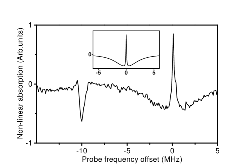
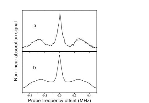
An example of a low resolution nonlinear absorption spectrum as a function of the frequency difference between the probe and the pump fields is presented in Fig. 1. In this spectrum, the pump frequency is kept fixed at the center of the transition of . The spectrum presents three distinct features. Two of these are centered around . There is a broad resonance (dip) whose width is given by the excited state width . At the bottom of this resonance there is a much narrower one with opposite sign. The broad resonance corresponds to an increase in the transmission due to the saturation of the sample by the pump field while the narrow feature represents an increase of the absorption. The latter corresponds to EIA. The sub-natural width of this resonance is an indication of its coherent nature. The inset in Fig. 1 shows the calculated signal around using the model described in [3, 6]. An interesting feature in the spectrum in Fig. 1 is the large dip appearing around . This resonance occurs when the probe frequency equals the frequency of the trapping beams (red-detuned with respect to the atomic transition). Since, as a consequence of the technique used for the fields preparation, the probe, pump and trapping beams are highly correlated, it is rather natural to observe a coherent resonance involving the probe and the trapping fields. However, keeping in mind the two-frequencies detection technique used, the pump field must also participate in the generation of any detected signal. Consequently the peak observed around corresponds to a nonlinear process involving at least one photon from each of the tree fields (pump, probe and trapping). The study of this multiphoton effect is beyond the scope of this paper. We focus our attention on the EIA structure observed around . Fig. 2(a) presents a detailed view of this structure observed in a MOT with a total trapping beams power attenuated to approximately . Under this condition the linear absorption by the cold atoms was maximum () at the trapping transition. A triplet structure is clearly visible which is reminiscent of the EIA spectrum obtained in a homogeneous sample for perpendicular pump and probe polarizations in the presence of a magnetic field orthogonal to both polarizations [6]. The observed value of the frequency separation between the center and the two sidebands would correspond in that case to a magnetic field of . This figure is of the order of the typical magnetic field magnitude within the cold atoms volume due to the quadrupolar distribution. Since in our experiment the pump and probe beams overlap a significant fraction () of the cold atomic sample, the EIA spectrum is due to the average contribution from atoms in different magnetic environments.
The hypothesis that the observed structure of the EIA spectrum is determined by the magnetic field present at the MOT can be tested by adding a small bias magnetic field at the sample. This was achieved by varying the current in the Helmholtz coils surrounding the trap that are normally used to compensate the earth field. The effect of the bias field is to change the magnetic field distribution at the position explored by the pump and probe waves. It also produces a translation of the cloud that results in a variation in the total number of atoms interacting with the pump and probe waves. Significant changes in the relative weight and positions of the resonances occur for different bias magnetic fields. Since in these measurements the pump, probe and trapping fields are kept unchanged, we conclude that the observed spectral structure is mainly determined by the magnetic field. When the pump-probe beam was carefully aligned through the atomic cloud for maximum linear absorption, the observed structure of the EIA spectrum was a triplet [Fig. 2(a)]. This structure represents the total response of the magnetically inhomogeneous sample. We have compared this observation with the numerical calculation of the response arising from atoms in different positions in an ideal MOT. We used a Monte Carlo procedure where the position of the atom is randomly chosen (uniform distribution) within an oblate ellipsoidal volume, representing the trapped cloud, centered at the zero of a quadrupolar magnetic field. The vertical dimension (along the symmetry axis) of the ellipsoid is shorter by a factor of two than the horizontal one. The absolute value of the magnetic field at the points where the principal axis intercept the ellipsoid surface is . The pump field polarization is vertical and the probe polarization horizontal. For each atomic position the local magnetic field is evaluated [8] and the corresponding nonlinear response is calculated using the model described in [6]. Then the total absorption is calculated as the sum of the individual atomic contributions. The result of the simulation is shown in Fig. 2(b) where a realistic value of has been chosen. The total absorption spectrum is a triplet in agreement with our observation. Unlike, the ideal ellipsoidal sample assumed for the calculation, the actual cloud of cold atoms is not symmetric around the zero of the magnetic field distribution. This is due to power imbalance and wavefront irregularities in the counterpropagating trapping beams. This produces a cold cloud displaced from the zero of the magnetic field with an asymmetric shape and an irregular atomic density distribution. This explains the fact that varying the position of the pump-probe beam across the atomic sample we were able to observe significant variations of the nonlinear absorption shape ranging from a single-peak spectrum, a doublet, a triplet or even a five-peaks structure.
Considering that the EIA spectra were recorded with the trapping field on, it is somehow surprising to observe spectral structures determined by the Zeeman shift of ground-state sublevels by the quadrupolar magnetic field. In the trapping region, the resonant interaction of the atoms with the trapping field results in significant displacement of the atomic levels due to (AC Stark) light shift. The magnitude of the light shift is different for different ground-state sublevels and depends on the local trapping field intensity and polarization experienced by the atom. The six trapping beams are responsible for a rather complicate interference pattern resulting in three dimensional modulation of the trapping field intensity and polarization with a spatial period of half wavelength [9]. The importance of the ground-state sublevels light shifts in a MOT is well illustrated by the peculiar shape of the absorption spectra of the trapped atoms in the MOT [10] analyzed by Grison and coworkers [11]. The influence of the ground-state Zeeman sublevels light shifts on the absorption spectra of strongly driven degenerate two-level systems was studied in [12]. Since the light shifts are uncorrelated with the Zeeman shift produced by the quadrupolar magnetic field, one should expect that the former will result in an inhomogeneous dispersion of the energies of the ground-state sublevels producing a smearing of the EIA resonance frequencies.
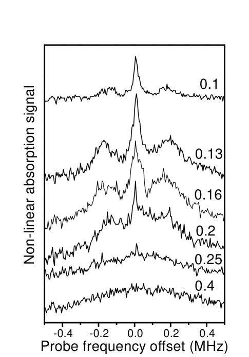
We have observed the variation of the EIA spectrum with the intensity of the trapping field (Fig. 3). For low trapping field intensities a well resolved triplet was obtained. The width of the central peak of the upper trace in Fig. 3 is . This is to our knowledge the narrowest coherent resonance observed to date in an operating MOT [10, 11, 13, 14, 15]. As the trapping field intensity is increased, the EIA resonances broaden. For large enough trapping field intensity, the triplet structure transforms into a unique broad peak. It is worth noticing that a regime can be found where the trapped cloud is rather dense (linear absorption around ) while at the same time the magnetic-field-dependent structure of the EIA spectrum remains clearly resolved. In this regime the modification of the ground-state sublevels by the light shift produced by the trapping field is small compared to the Zeeman shift.
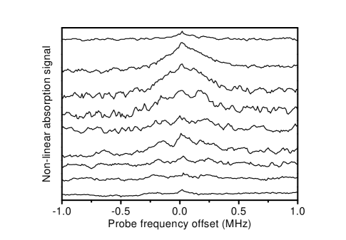
The fact that the EIA spectrum reflects the magnetic-field distribution within the trapped atoms cloud may be used for mapping the magnetic-field variations inside the cold atoms sample. This possibility is illustrated in figure 4 showing the modification of the EIA spectrum as the pump-probe beam is translated in the equatorial plane of the cold atom cloud. Between two consecutive traces of Fig. 4 the pump-probe beam was translated by . From bottom to top, the spectra in figure 4 evolve from a well resolved tripled into a broad unresolved peak. The series in Fig. 4 may be interpreted as a mapping of the magnetic field inside the cold atoms cloud averaged over the volume explored by the pump-probe beam. In these measurements the pump-probe beam diameter at the sample is a figure which is only a factor of smaller than the estimated cold atoms cloud diameter. This limits in our case the spatial resolution of the magnetic field mapping. A two orders of magnitude improvement in the resolution is in principle possible with a tight focussed pump-probe beam. The spectra presented in Fig. 4 clearly indicate the lack of symmetry of our MOT. As a matter of fact, the zero of the magnetic field, around which symmetric variations of the EIA spectrum should take place, is located outside the region of large atomic density.
The mapping of the magnetic field using EIA spectroscopy, appear as a rather simple and straightforward technique. It can be achieved in a cw regime without the need for turning off the MOT trapping beams and with very low-power pump and probe fields. As demonstrated here, these fields can be easily obtained from the trapping laser. As in the example shown, qualitative information about the atomic density distribution with respect to the quadrupolar magnetic field is directly inferred from the spectra. The fitting of the experimental EIA spectra to theoretical predictions (based on consistent assumptions for the trapped cloud density distribution) can in principle be used as a tomographic technique for the characterization of the atoms in the MOT [8]. This approach was not developed in the present case in view of the irregular atomic density distribution observed in our MOT.
IV Conclusions.
We have observed EIA resonances on a sample of cold atoms in a MOT in the presence of the trapping light beams. An operating regime of the MOT (with moderate trapping beams intensity) could be found where the atomic density is quite large while at the same time the atomic ground state is weakly perturbed by the trapping field. In this regime the energy shift of the ground state sublevels is essentially governed by the Zeeman effect. Coherent resonances as narrow as were observed for the first time in an operating MOT. EIA absorption spectroscopy, performed in a cw regime with very low optical intensities, appears as a suitable tool for the inspection of the Zeeman shift produced for the quadrupolar magnetic field at different positions of the cold atom sample.
V Acknowledgments.
The authors are thankful to A. Akulshin for his contribution to the early stages of this study and to H. Failache for helpful discussions. This work was supported by the Uruguayan agencies: CONICYT, CSIC and PEDECIBA and by the ICTP/CLAF.
REFERENCES
- [1] See M.O. Scully and M.S. Zubairy, Quantum Optics, Cambridge University Press, Cambridge (1997) and references therein.
- [2] A.M. Akulshin, S. Barreiro and A. Lezama, Phys. Rev. A 57, 2996 (1998).
- [3] A. Lezama, S. Barreiro and A.M. Akulshin, Phys. Rev. A. 59, 4732 (1999).
- [4] A. Akulshin, S. Barreiro and A. Lezama, Phys. Rev. Lett. 83, 4277 (1999).
- [5] see S. E. Harris, Physics Today, 50(7) 36 (1997) and references therein.
- [6] A. Lezama, S. Barreiro,. A. Lipsich and A.M. Akulshin, Phys. Rev. A 61, 013801 (2000).
- [7] M.O. Scully and M. Fleishhauer, Phys. Rev. Lett. 69, 1360 (1992). H. Lee, M. Fleishhauer and M.O. Scully, Phys. Rev. A 58, 2587 (1998).
- [8] T.A. Savard, S.R.. Granade, K.M. O’Hara, M.E. Gehm and J.E. Thomas, Phys. Rev. A 60, 4788 (1999).
- [9] S.A. Hopkins and A.V. Durrant, Phys. Rev. A 56, 4012 (1997).
- [10] J.W.R. Tabosa, G. Chen, Z. Hu, R.B. Lee and H.J. Kimble, Phys. Rev. Lett. 66, 3245 (1991).
- [11] D. Grison, B. Lounis, C. Salomon, J.Y. Courtois and G. Grynberg, Europhys. Lett. 15, 149 (1991).
- [12] A. Lipsich, S. Barreiro, A.M. Akulshin and A. Lezama, Phys. Rev. A 61, 053803 (2000).
- [13] T. van der Veldt, J.F. Roch, P. Grelu and P. Grangier, Opt. Commun. 137, 420 (1997).
- [14] S.A. Hopkins, E. Usadi, H.X. Chen and A.V. Durrant, Opt. Commun. 138, 185 (1997).
- [15] A.V. Durrant, H.X. Chen, S.A. Hopkins and J.A. Vaccaro, Opt. Commun. 151, 136 (1998).