The Physical Origin of Intrinsic Bends in Double Helical DNA
Abstract
The macroscopic curvature induced in the double helical B-DNA by regularly repeated adenine tracts (A-tracts) is a long known, but still unexplained phenomenon. This effect plays a key role in DNA studies because it is unique in the amount and the variety of the available experimental information and, therefore, is likely to serve as a gate to the unknown general mechanisms of recognition and regulation of genome sequences. We report the results of molecular dynamics simulations of a 25-mer B-DNA fragment with a sequence including three A-tract phased with the helical screw. It represents the first model system where properly directed static curvature emerges spontaneously in conditions excluding any initial bias except the base pair sequence. The effect has been reproduced in three independent MD trajectories of 10-20 ns with important qualitative details suggesting that the final bent state is a strong attractor of trajectories form a broad domain of the conformational space. The ensemble of curved conformations, however, reveals significant microscopic heterogeneity in contradiction to all existing theoretical models of bending. Analysis of these unexpected observations leads to a new, significantly different hypothesis of the possible mechanism of intrinsic bends in the double helical DNA.
Introduction
The intrinsic sequence dependent curvature of the DNA molecule is likely to be involved in fundamental mechanisms of genome regulation. The possibility of strong static bends in the B-DNA double helix has been proven for sequences containing regular repeats of , called A-tracts [1]. This effect plays an important role in DNA studies because it is unique in the amount and the variety of the available experimental information and, possibly, it can serve as a gate to the unknown general mechanisms of recognition and regulation of genome sequences. Every A-tract deviates the helical axis in a locally fixed direction by approximately 18∘, and, if the A-tracts are repeated in phase with the helical screw, a macroscopic curvature emerges. The effect was first noticed and identified in restriction fragments from the kinetoplast body of Leishmania tarentolae [2, 3], and confirmed by electric birefringence decay [4] and electron microscopy [5]. A large variety of interesting information has been obtained by biochemical methods. It appeared that the double helix bends towards the minor grooves of A-tracts [6, 7]. The curvature is reduced with the temperature above 40∘ and in high salt, but for some sequences it is increased in presence of divalent metal ions [8]. It depends upon the length and composition of A-tracts as well as on sequences between them [6]. Detailed analysis of these results can be found in comprehensive reviews published in different years [1, 9, 10, 11, 12, 13].
According to many independent experimental observations, the structure of A-tract sequences should differ significantly from the “random” B-DNA. It is well established that, in solution, the poly-dA double helix is overwound to a twist of around 36∘ from around 34∘ of a random sequence [14, 15, 16]. The models constructed from fiber diffraction data suggest consistently that the poly-dA double helix is characterized by a very narrow minor groove and a high propeller twist [17, 18, 19]. Yet another distinction is an apparently large negative inclination of base pairs [18]. Several A-tracts avaliable in single crystal structures of B-DNA oligomers have irregular conformations, but exhibit similar trends toward their centers [20, 21, 22, 23, 24]. Even though the curvature is apparently caused by A-tracts, in Xray structures, A-tracts look generally less prone to bending than other sequences. Some indirect observations also support this view, notably, poly-dA fragments move faster than random DNA in gel migration assays [6] and avoid wrapping around nucleosome particles [25, 26].
In spite of a large body of the experimental information accumulated during the last twenty years, the possible physical mechanism of this effect remains unclear. Since every base pair in a stack interacts only with the two neighbors, any sequence specificity in the DNA structure should mainly depend upon the stacking interactions in one base pair step. Non-local effects are also possible, however, due to base-backbone interactions and propagation of correlations along the backbone. The initial experimental data on A-tract bending were interpreted in terms of two alternative mechanisms, namely, the wedge model [27] and the junction model [28]. Both had to be modified significantly as and when new experimental data appeared and some other theories were discussed as well. The possible mechanisms of bending considered in the literature will be discussed below. Here we note only that none of them explains all experimental data and can be definitely preferred [1]. The overall pattern has been additionally complicated when it was found that certain non A-tract sequences also exhibit distinguishable curvature [29].
Here we report new results obtained in free unbiased molecular dynamics simulations of a DNA oligomer with phased A-tracts. We managed to find computational conditions in which stable sequence dependent static curvature emerges spontaneously in good agreement with experimental observations in terms of both the bending direction and magnitude. It is found that three independent long time trajectories converge to conformations with similar bends, but rather different local structural parameters, in evident contradiction with the common views of the origin of curvature. Analysis of these discrepancies leads to a new, significantly different hypothesis of the possible mechanism of intrinsic bends in the double helical DNA.
Theoretical Background
As a theoretical problem, the phenomenon of the sequence dependent DNA bending presents a challenge in many aspects similar to the protein folding problem. In order to understand the underlying physical mechanism, one has to analyze terribly noisy experimental data, with the noise being due to the biological diversity and, therefore, unremovable. If and when the physical mechanism is understood one will have to tackle this diversity again, because it will be necessary to analyze specific DNA sequences. The atom level molecular modeling is virtually the only theoretical approach that is potentially able to treat these difficulties. Ideally, we would like to have a model where the base pair sequence represents the only initial bias towards a specific conformation. If it could reproducibly yield curved DNA conformations in agreement with experimental data we should be able disclose the mechanism of bending in the model, and hope that a similar mechanism takes place in the nature.
The foregoing scheme, however, is too difficult and, until now, most of the modeling studies used other strategies. Much has been learned about the DNA bending mechanics by using energy minimization [30, 31, 32, 33] and Monte Carlo [34]. Unfortunately, the possibilities of such studies are limited by the multiple minima problem especially when it is necessary to take into consideration specific interaction with solvent molecules. The proposed alternative strategies commonly involved some bias towards specifically bent conformations introduced either explicitly, by imposing restraints, or implicitly, by choosing particular initial conditions, which made impossible unequivocal conclusions concerning the possible mechanism of bending. In the recent years, owing to the progress achieved in improving the full atom force fields, multi-nanosecond free MD simulations of DNA became feasible [35, 36]. A few such studies of phased A-tract sequences have been already reported [37, 38]. It has been demonstrated that, without any a priori bias, the DNA double helix bends anisotropically, and certain sequence specific features of A-tracts were at least qualitatively reproduced. At present, the free MD simulations represent the most promising line of research in this field, and we continue it here by using the recently proposed minimal model of B-DNA [39, 40].
The minimal B-DNA consists of a double helix with the minor groove filled with explicit water. Unlike the more widely used models, it does not involve explicit counterions and damps long range electrostatic interactions in a semi-empirical way by using distance scaling of the electrostatic constant and reduction of phosphate charges. We have earlier found that the minimal model gives B-DNA conformations which better compare with experimental structures than DNA structures obtained with other computational methods. Notably, it is free from a systematic negative bias of the average twist observed in simulations with full hydration and explicit counterions [39]. This factor is likely to be involved in DNA bending because, as noted above, A-tracts are overwound with respect to the average B-DNA. For the standard test case of the EcoRI dodecamer structure, the dynamics of the minimal model reproducibly converged to structures slightly bent towards the minor groove which was narrowed in excellent agreement with the single crystal conformation [39, 40]. All these preliminary observations suggested that the minimal model was a good choice for studying the DNA bending induced by A-tracts.
We report here simulations of the bending dynamics of a 25-mer B-DNA fragment. Its sequence, , has been constructed after many preliminary tests with shorter sequence motives, and it includes three A-tracts separated by one helical turn. Our general strategy came out from the following considerations. Although the A-tract sequences that induce the strongest bends are known from experiments, probably not all of them would work in simulations. There are natural limitations, such as the precision of the model, and, in addition, the limited duration of trajectories may be insufficient for some A-tracts to adopt their specific conformation. Also, there is little experimental evidence of static curvature in short DNA fragments, notably, one may well expect the specific A-tract structure to be unstable near the ends. That is why we did not simply take the strongest experimental “benders”, but looked for sequence motives that in calculations readily adopt the characteristic local structure, with a narrow minor groove profile and high propeller twist, both in the middle and near the ends of the duplex. The complementary duplex has been constructed by repeating and inverting one such motive.
Our goal was to find sequences that would appear statically bent in these conditions. It means that, in dynamics, the structure should fluctuate around a state with a distinguishable bend and a definite bending direction. Since any MD simulation is limited in time, there is no way to prove rigorously that some specific conformation is representative. Some degree of confidence can be achieved, however, if independent trajectories are able converge to the same state, and this is exactly what we tried to obtain. We found important to place A-tracts at both ends because of the two reasons. First, we could study only short DNA fragments, therefore, it was preferable to place A-tracts at both ends in order to maximize the possible bend. Second, in calculations with short sequences, it is important to be sure that the boundary conditions are correct. Since the A-tracts have a characteristic local structure, the boundary conditions could be at least qualitatively verified, which would not be the case for GC-rich sequences.
Results
Three long MD trajectories were computed for a complementary DNA duplex with the sequence . The model system employed was same in all three simulations, with only the starting states varied. The first trajectory referred to below as TJBa started from the fiber canonical B-DNA structure [41] and continued to 10 ns. When it was found that TJBa converged to a statically bent conformation, in good qualitative agreement with expectations based upon experimental data, another trajectory (TJBb) was computed in order to verify the reproducibility of the results. It started with random velocities from a re-minimized straight conformation taken from the initial phase of TJBa and was also continued to 10 ns. Simultaneously, in order to remove any initial bias implicitly involved in the choice of the starting state, the third trajectory (TJA) was obtained which started form the fiber canonical A-DNA conformation [41]. Initially, we computed 10 ns of TJA and found that it sampled conformations rather dissimilar from those observed in TJBa and TJBb. A careful analysis revealed, however, certain slow structural trends and prompted us to continue TJA to 20 ns. The first two trajectories (TJBa and TJBb) have been the subject of our initial report [42]. Therefore, below we describe in detail only TJA and use the corresponding data from TJBa and TJBb in comparisons. The structures referred to as the final MD states are the conformations averaged over the last nanosecond of the corresponding trajectory. The detailed computational protocols are described in Methods.
All Three Trajectories Converge to Similar Structures within B-DNA Family
Table I presents atom rmsd comparison between the final MD states of the tree trajectories and canonical A and B-forms of this 25-mer duplex. All computed structures are clearly different from the standard A-DNA, even though for TJA it was the starting point. Moreover, the TJA final state appears to be the less similar of the three, demonstrating that the trajectories were not trapped kinetically in the vicinities of their starting points. At the same time, the final MD states are evidently close to the canonical B-DNA. When only the central undecamer is considered, the rmsd values are in the same range as those observed in our earlier MD simulations of dodecamer duplexes [39, 40]. They are low, and the three computed conformations seem to form a single cluster around the canonical B-DNA. The rmsd naturally increases with the helix length, but it should be noted that the somewhat larger values obtained for the whole structures are much lower than ever observed in free MD simulations of long DNA helices [37]. It appears, however, that, if taken as a whole, the TJA state is as close to the canonical B-DNA as to the TJBa and TJBb states, while the latter two are yet closer to each other.
| Xdispa | Inclina | Risea | Twista | Bendb | Bendingc | |
|---|---|---|---|---|---|---|
| angle | direction | |||||
| A-DNAd | -5.4 | +19.1 | 2.6 | 32.7 | 0.0 | – |
| B-DNAd | -0.7 | -6.0 | 3.4 | 36.0 | 0.0 | – |
| TJAe | -0.7—-0.3 | +0.0—-1.2 | 3.5—3.5 | 34.3—34.8 | 35.8—30.4 | -136.8—+110.4 |
| TJBae | -1.0—-0.3 | -6.9—-4.5 | 3.6—3.5 | 34.1—34.8 | 13.2—29.5 | 22.3—42.9 |
| TJBbe | -1.0—-0.7 | -4.6—-1.8 | 3.5—3.5 | 34.3—34.2 | 18.4—28.4 | 35.6—53.7 |
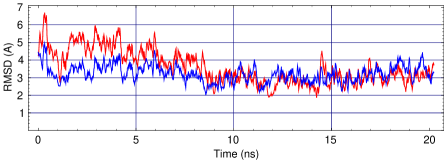
The kinetics of the structural convergence in terms of atom rmsd is illustrated in Fig. 1 for TJA. It is seen that the trajectory rapidly went from the initial A-DNA conformation towards the B-DNA form and, after the equilibration, the rmsd from B-DNA have already lost a half of the initial 8.6 Å. Starting from the second nanosecond it fluctuated between 2 and 4 Å. The corresponding kinetics for TJBa and TJBb were very similar except for the initial fall of the rmsd value [42]. The rmsd from the TJBa state also shown in Fig. 1 falls down to similar final values, but exhibits a somewhat different kinetics. Namely, an overall negative drift occurred during the first ten nanoseconds followed by random fluctuations during the second half of the trajectory. One may say, therefore, that TJA first quickly traveled from A-DNA towards the B-DNA family and next slowly refined its position within this family coming closer to other computed structures. This refinement was not complete, however, since, according to Table I, the final TJA-TJB difference is still larger than that between TJBa and TJBb. Figure 1 suggests that a more accurate convergence, if possible, would require much longer time.
Table II compares a few representative structural parameters of MD conformations with the corresponding standards. Already after the first nanosecond even TJA gave the helicoidals corresponding to the B-DNA family, and they exhibited no systematic change afterwards. All three final MD states have an overall bend of around 30∘. The bending direction is somewhat different between TJA and TJB, which is the main cause of the corresponding residual difference in terms of atom rmsd. Table II indicates that in all three trajectories both the magnitude and the direction of the bends changed significantly, and that very large variations in the bending direction apparently occurred in TJA. Thus, the slow rmsd kinetics considered above appear to be largely due to the bending of the double helices whereas the contribution from the variations of the helical parameters looks minor.
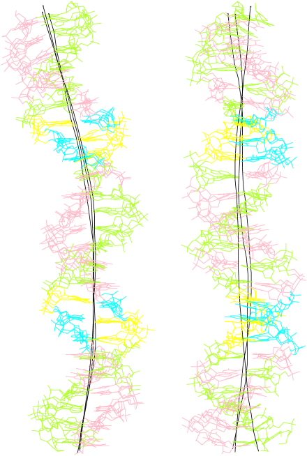
Figure 2 shows the three last nanosecond average structures superimposed. They all are evidently curved, with the bends being nearly planar in each structure. In agreement with Table II, the TJA bending plane slightly deviates from the other two. The bending planes intersect the minor groove in five points which alternate between the inside and the outside edges of the bend, and in each case the the three A-tracts appear at the inside edge. The tracts are approximately phased with the helical turn, but, since the lower one is inverted with respect to the other two, its 5’ end is phased with the 3’ ends of the other. The three inside intersection points are shifted within the A-tracts from their middle towards the 3’ end of the upper two and the 5’ end of the lower one. On the other hand, the minor grooves of the two AGGC tetraplets appear at the outside edge of the curved axis, and it is readily seen that the minor groove is widened here, especially at the upper tetraplet.
Quasi-Regular Rotation of The Bending Plane in TJA
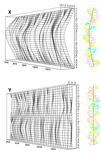
The two surface plots in Fig. 3 exhibit the time evolution of the shape of the helical axis for TJA. It is seen that the molecule was strongly bent after the initial equilibration, which was not observed in case of TJBa and TJBb [42]. One should note that considerable initial deformation of the double helix is common for trajectories starting from the A-DNA conformation. Apparently, the molecule is stressed because the transition to the B-form occurs in these conditions during unphysically short time with much energy released. During the next few nanoseconds the bend reduced and the axis acquired a more complex shape with wound profiles in both projections. After the fifth nanosecond the bending became more planar, with much smaller curvature in Y projection. A planar bent may just mean that the helical axis is kinked in a single point or, alternatively, a lager number of local bends are properly directed. Figure 3 indicates that there is probably a mixture of these two effects. During the last few nanoseconds the axis had one stable bending point shifted upwards from the middle while another bend in the lower half emerged from time to time. The two bends were slightly misaligned, therefore, the overall bending plane rotated a little when the second bend emerged, and, in the Y projection, one sees alternation of straight and S-shaped profiles.
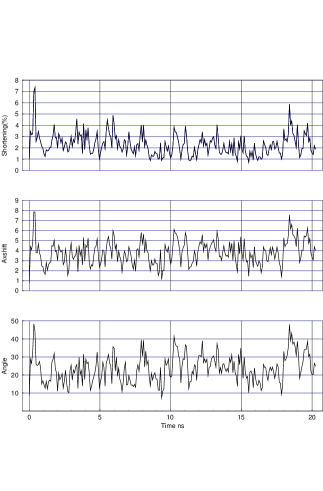
Figure 4 displays kinetics of several quantitative measures of the magnitude of bending. The three parameters used, namely, the total angle, the shortening, and the average shift of the curved axis, all exhibit a coherent pattern of fluctuations, which locally correlates also with the rmsd from the canonical B-DNA (see Fig. 1). This indicates that they all are produced by the same motion, namely, the axis bending. Comparison of the data in Figs. 3 and 4 with similar plots earlier reported for TJBa and TJBb [42] reveals little difference except the already mentioned initial deformation and the absence of a stable bend between the two lower A-tracts. Accordingly, TJBa and TJBb showed a somewhat stronger bending, with one-nanosecond average values usually beyond 35∘. In TJA, after the initial strong temporary bending, a comparable magnitude has been reached only during the last four nanoseconds.
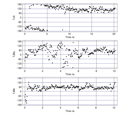
There is, however, a striking difference between TJA and the other two trajectories in the dynamics of the bend direction which is exhibited in Fig. 5. Both in TJBa and TJBb the final bending direction occurred early in the trajectories and remained quite stable although the molecule sometimes straightened producing broad scattering of points in Fig. 5. In contrast, during the first ten nanoseconds of TJA, the bending plane made almost a half turn with respect to the coordinate system bound to the molecule. It means that a transition occurred between the oppositely bent conformations, but, as seen in Fig. 3, the straight one was avoided. This rotation was very steady, almost regular. It gradually slowed down becoming indistinguishable in the last five nanoseconds. After this transition the directions of the bends in the three trajectories became much closer, and this quasi-regular motion is apparently responsible for the slow drift of the rmsd from the TJBa state in Fig. 1. The overall amplitude of this motion was around 150∘, that is the initial strong bend noticed in Fig. 3 was nearly opposite to that finally established.
The Rotation of the Bending Plane is Not Energy Driven
The overall character of motion revealed in Fig. 5, namely, the steady rotation of the bending plane, looks strange and counter-intuitive. A priori, we would rather expect to obtain random sampling of different bending directions, with the correct one statistically preferred due to its lower energy. The apparent quasi-regular dynamics exhibited in Fig. 4 might mean that our trajectory represents a downhill motion along a valley on a potential energy surface. Its steep borders would separate bent conformations from the straight one, with the bottom of this valley slightly inclined towards the preferred bending direction. In this case all bent conformations, including incorrect bends, should have been lower in energy than the straight one.
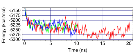
Figure 6 displays the time evolution of the potential energy in all three trajectories. It is seen that the energy dropped during the first nanosecond and later remained stable. No clear correlation is seen between the instantaneous magnitude of bending displayed in Fig. 4 and the potential energy, therefore, one cannot say that straight states have significantly different energies than the bent ones. Neither can we claim that the preferred bending direction is characterized by lower energy values than other bends. In Fig. 6, a slight decrease in energy is observed during the second half of TJA, but it occurred when the regular rotation of the bending plane has essentially finished. On the other hand, the lowest energy during the first half of the trajectory was observed at around 3.2 ns when the bending direction was completely different. Note also that, during the first ten nanoseconds, the traces of TJBa and TJBb go above the last one, although in these cases the correct bending direction has already established. We have to conclude, therefore, that the simple energetic al explanation of the observed effect does not work.
The Minor Groove Profiles of Converged Structures Are Similar But Not Identical
The surface plot in Fig. 7 exhibits the evolution of the profile of the minor groove during TJA. The initial A-DNA conformation is characterized by a uniformly wide minor groove of 13.6 Å. It is seen that after the equilibration period the groove was much narrower, but still wider than in the canonical B-DNA model. Moreover, a complex profile have emerged with three local widenings at A-tracts, which is exactly opposite to the expectations. The two terminal widenings reduced during the first ten nanoseconds whereas the maximum of the middle one gradually shifted from its 3’ end to 5’ end. This shift evidently accompanies the rotation of the bending plane described above.
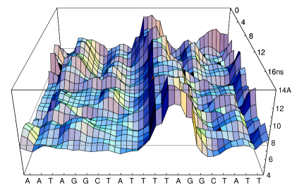
One can note that the maximal widening of the minor groove moved for only 2-3 base pair steps, which is less than a 150∘rotation seen in Fig. 5. It appears that, in fact, the maximal widenings and narrowings in the minor groove profile do not always correspond to the direction of local bends. The initial bend was directed towards the minor groove of the upper TAGG tetraplet where the minor groove was narrowed. The two neighboring widenings are shifted by three base pairs only and they appear at the opposite sides of the bending plane which is approximately collinear to the pseudodiad axis at the center of the middle ATT triplet. In contrast, in the last structure shown in Figs. 2 and 3 the bending plane passes exactly through the maximum widening of the minor groove. The overall rotation, therefore, corresponds to approximately four base pair steps which gives the observed turn by 150∘. It can be noted, finally, that although Fig. 5 indicates that the bending stabilized after ten nanoseconds, the profile of the minor groove in Fig. 7 continues to evolve slowly till the very end of the trajectory.

Figure 8 displays the minor groove profiles of the last average structures from the three trajectories. For TJBa and TJBb their kinetics was detailed in our first report [42], and we only note here that the corresponding profiles shown in Fig. 8 established during the first two nanoseconds and showed little variations afterwards [42]. The three traces evidently exhibit a certain similarity, but do not coincide. The TJBa and TJBb grooves have the same number of local narrowings and widenings which differ slightly between the two both in amplitude and in position. The TJA profile is similar in the right-hand half of the figure. One can notice that the change from TJBa to TJBb and next to TJA involves the growing widening at the TTAG tetraplet accompanied by a shift of the secondary maximum, and looks rather regular and concerted. At the opposite half of the structure, the TJA conformation shows a narrow minor groove without significant modulations of the width. This difference may be related with the smaller magnitude of the bending in the case of TJA where the second bending point appeared from time to time only and was less significant than in the other two trajectories.
Key Helicoidal Parameters Exhibit Consistent Regular Patterns Only after Window Averaging
Figure 9 shows variation of some helicoidal parameters along the duplex in the three structures. The two inter base pair parameters, namely, roll and tilt, are most often quoted in the literature in relation to the static DNA curvature. If one first takes an ideal straight column of stacked parallel base pairs and next introduce a non-zero roll value at a certain step, the structure will bend at this step towards the major groove, if the roll is positive, and to the minor groove if it is negative. A similar experiment with the tilt value would result in bending in the perpendicular direction. It seems obvious that, whatever the physical origin of the curvature, in a bent double helical DNA, the roll and tilt values must exhibit systematic variations phased with the helical turn. Moreover, it is often assumed that for some short DNA sequences certain non-zero roll and tilt values are strongly preferred energetically, which produces static bending when they are repeated appropriately.
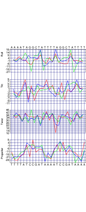
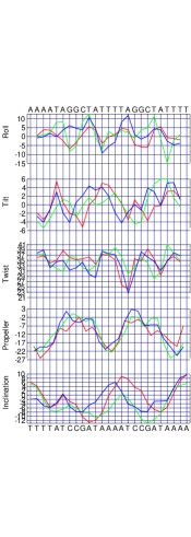
However surprising, although all three average structures are smoothly curved, only a few supporting signs for the foregoing paradigm are readily seen in Fig. 9. For the tilt, the three traces are very dissimilar and the only feature that repeats is the alteration of its values between consecutive steps. Namely, if the tilt is low at a given step it normally goes up at the next one, and vice versa. In the three average structures, however, these alterations are sometimes oppositely phased even in TJBa and TJBb where the overall structures look particularly similar.
The same is true for the roll and twist although, in these cases, some clear sequence preferences do exist. Note, for instance, that, in all four TpA steps, the roll is almost always positive and larger than in the neighboring steps. Paradoxically, two of these TpA steps occur almost exactly at the inside edge of the curved axis, that is a high positive roll accompanies the bending in an exactly opposite direction. This paradox is readily resolved when one looks at the roll values at the neighboring steps. A TpA step with a high positive roll is normally preceded or followed by a step with a low negative roll. The higher is the maximum, the lower is the neighboring minimum, so that the two nearly cancel each other. The other two TpA steps are found at the outside edge of the helical axis and their high roll probably contributes to bending. However, while a more or less repetitive pattern is observed around the third TpA step, the first one exhibits rather dissimilar pictures even for TJBa and TJBb structures which both have a widened minor groove here. Also, the roll values at the third TpA step differ considerably between the structures, but do not correlate with the bending magnitudes.
The twist, tilt, and roll values used for the plots in Fig. 9 are the so called “global” parameters from the outputs of the Curves program [43]. One may argue that they are not appropriate in the present context since they are computed by using local directions of already curved optimal helical axis. However, when “local” values are used instead, the amplitudes of the alternations in these profiles are only increased.
The last plot in Fig. 9 exhibits the variations of the propeller twist. Again one sees that its value alternates between consecutive base pairs, with little phase similarity between the three structures. At the same time, in this case, a consistent repetitive pattern is evident, with strong negative propeller values in all A-tracts. These regular patterns look even more similar than the structures themselves. For instance, there is no evident difference between the three traces that would correspond to that in the minor grove profiles in Fig. 8.
The apparent jumping alterations of the helicoidal parameters along the double helix naturally suggests that one should try to smooth them out by averaging the traces in Fig. 9 with a sliding window. Figure 10 shows the results of such treatment and also includes the corresponding data for the inclination which was, however, used without the smoothing. The difference between Figs. 9 and 10 is rather significant. Now all four helicoidal parameters considered in Fig. 9 exhibit regular, sometimes almost sinusoidal, oscillations. The phasing of these oscillations with the helical turn, however, is not always evident. The propeller and the inclination both exhibit approximately 2.5 periods, that is the dominating Fourier component has a wave length of approximately ten base pairs corresponding to one helical turn. For the roll, the dominating wave length apparently corresponds to 5-6 base pairs, that is a half of a helical turn. When different structures are compared, however, it is seen that only for propeller the maxima and minima coincide well. A more complex pattern is observed for the twist, and one can notice a correlation between its traces and the minor groove profiles shown in Fig. 8. Namely, the twist is lower in the widenings of the groove and higher in its narrowings.
These results suggest that the relationship between the helicoidal parameters and the bending is complex and cannot be reduced to simple models of roll-like or tilt-like bends outlined above. Accumulation of the regular variations revealed in Fig. 10 probably gives the correct overall bend angles and directions, but neither can be easily predicted just by looking at these traces.
The Distributions of Backbone Conformers in Bent Structures are Surprisingly Dissimilar
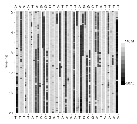
In all three trajectories, dynamics of backbone transitions was qualitatively similar in a few aspects. Consider Fig. 11a, for instance, where the results are shown for TJA. The overall pattern reveals rather slow dynamics, suggesting that MD trajectories in the 10 ns time scale are not long enough to sample all relevant conformations. A somewhat higher activity was observed during the first half of the trajectory, when the rotation of the bending plane occurred. It is seen that, in A-tracts, the BII conformers are preferably found in ApA steps and that they tend to alternate with BI within the same strand. There are many examples of concerted transitions, when a given step switches from simultaneously with an opposite transition in one of the neighboring steps. Sometimes three consecutive steps are involved and, less often, the opposite strand as well. Many transitions are reversed within hundreds of picoseconds, but there are also very long-living conformers and sites where either states are preferred. A strong preference of state is observed for all TpT steps, for example. However, it seems to be the only case when the effect repeats at a base pair step level. In a few steps where the conformation is preferred this is apparently determined by a broader sequence context.
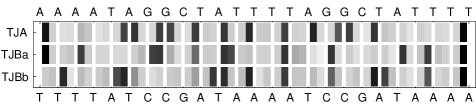
The corresponding data for TJBa and TJBb were included in our first report and they revealed the same qualitative features [42]. It was very surprising for us, however, that, in spite of the good convergence in terms of the overall bent shape of the molecule, the three trajectories gave rather dissimilar distributions of conformers along the sequence. Fig. 11b compares these distribution in the final backbone conformations in the three trajectories. There are 14 non TpT steps where the conformation is found in all three structures. However, since our trajectories started from states, this number hardly tells us something. On the other hand, the number of conformers found in each structure and in each strand is similar and roughly corresponds to 25% of phosphate groups. Assuming that the states are evenly distributed in the sequence one gets the expectation value of 0.75 for the number of cases when the conformer should be found in the same base pair step in all three structures. The observed number of such sites is three. Note, however, that they all are found in A-strands of A-tracts where, as noted above, the conformers tend to alternate. This, together with the strong preference of TpT steps for , increases the probability of matching.
These results suggest that the relationship between the bending of the DNA double helix and the backbone transitions, if any, is loose in the sense that a given bent shape does not impose a fixed distribution upon the backbone.
Discussion
This study gives the first example of a successful implementation of the general strategy outlined in Theoretical Background. Namely, we showed that the minimal model of B-DNA, which is biased only by the nucleotide sequence, in dynamics, reproducibly converges to a single state characterized by an ensemble of similar statically bent conformations. The effect has been demonstrated here for one sequence only. Moreover, this sequence was specifically constructed rather than taken from experimental studies. Nevertheless, the sequence motive AnTAG used in construction was found in the center of the first bent DNA fragment studied experimentally [3]. In addition, the character of bending in the computed conformations, notably, its direction with respect to the A-tracts, and modulations of the groove width, qualitatively agree with the rules derived from experiments. These observations validate an attempt to make the next step of the above strategy, namely, below we try to disclose the mechanism responsible for the bending within the framework of the minimal model. We believe that, in spite of the obvious limitations of this model, its main features responsible for the bending correspond to reality. At the same time, the real situation is certainly more complex.
Results Poorly Agree with Earlier Theories of Bending
All theories proposed during the last 20 years to explain intrinsic bends in DNA double helices agree with some experimental observations and disagree with the other and, probably, each of them continues to attract some proponents. Here we compare our results with the most popular models of bending regardless of their experimental validation. Comparisons with experimental data have been the subject of many reviews [1, 9, 10, 11, 12, 13].
The wedge model of DNA bending [27] resulted from merging of ideas developed in seventies to explain the ability of a double helix to wrap around nucleosome particles. The first idea was that this can occur due to kinks of the helical axis phased with the helical screw [46, 30], with kinks implying destacking of base pairs in fable points in order to maintain perfect stacking elsewhere. The second idea was that the double helix can be smoothly bent, without destacking, by small deformations in every base pair step [47]. The wedge model merges the two by postulating that, in every specific dinucleotide, the preferred stacking of bases is slightly non-parallel and this causes bending in the same way as kinks do. It can be further developed by increasing the number of wedge degrees of freedom, by considering triplets, tetraplets, and so forth instead of dinucleotides, and by assuming that the non-zero average wedges result from random sampling from asymmetrical energy valleys around local energy minima, rather than from minimum energy configurations [48]. Depending upon the specific wedge parameters, this model can place the curvature inside A-tracts or between them [48, 49] and also explain bending in non A-tract sequences [29]. For the present discussion, it is convenient to unite all such mechanisms in one group characterized by the tacit emphasis upon the specific base pair stacking preferences as the source of the DNA bending.
The results shown in Figs. 9 and 10 obviously disagree with these views. There is little similarity between matching dinucleotides in the same structure and, moreover, base pair steps put in the same sequence context in three closely similar bent conformations exhibit broadly different helical parameters. The last observation means that even a generalized wedge model with dinucleotide blocks replaced by triplets, tetraplets, and so forth, would disagree with our results.
The junction model [28] postulates that there is a distinct specific A-tract form of the double helix characterized by a stronger inclination of base pairs with respect to the helical axis than in the normal B-DNA. In this case, planar stacking at the junction between the two structures would result in a kink of the helical axis. Formally, such geometry can also be obtained with the generalized wedge model above, but the junction theory puts an emphasis upon the specific A-tract form of DNA as the principal physical cause of bending. Its structure can be due to cooperative interactions in long DNA stretches and its environment. Within the framework of the junction model, particular roles were sometimes attributed to bifurcated hydrogen bonds [17], the water spine in the minor groove [32], or the NH2 groups of adenines [31].
It is evident that the junction model also poorly agrees with our results. In dynamics, conformations, both smoothly bent and kinked at the two insertions between the A-tracts, are observed periodically. The kinks, however, are not centered at the boundaries between A-tracts and the flanking sequences, and they are not sharp. In such conformations, A-tracts are less bent than regions between them, that is the bend is localized but still smooth. In Fig. 10 the inclination shows smooth oscillations, even without window averaging, with no kinks. It decreases from 5’ to 3’ ends of A-tracts, and since the 3’ ends of A-tracts are dephased and positioned differently with respect to the bending plane, no evident relationship to bending can be readily seen. All helical parameters vary along the sequence so that there is no A-tract fragment where they repeat at two consecutive steps. Thus, although the structures are bent, the specific regular A-tract structure is not seen, as well as the “random B-DNA”, though, which are the two key components of the junction model.
The third model, which was first mentioned in the context of the junction theory [50], but became popular only in the recent years [51], attributes the cause of bending to solvent counterions. If they are specifically bound by minor grooves of A-tracts, in a phased sequence, phosphate groups would appear partially neutralized at one side of the double helix, and the repulsion between the opposite phosphates would bend DNA towards minor grooves of A-tracts. The very fact that the minimal model of B-DNA, without explicit counterions, produces static bends, in good agreement with experiments, strongly suggests that the counterions hardly play a key role in the A-tract induced bending.
At the same time, our results do not contradict less specific non-local theories of A-tract bending. The modified junction model [52] assumes that the deformations at the boundary between the two conformations can propagate for several base pair steps. The A-tracts in the sequence studied here may be too short for their ingenious structure to establish. The second such theory [53] proposed that the bending is caused by the modulations of the minor groove. Really, the double helix is usually bent towards the major groove at the minor groove widenings, and in the opposite direction at its narrowings. In TJA, for instance, this relationship is maintained during the rotation of the bending plane. Sometimes, however, the double helix straightens and remains unbent during nanoseconds, while the minor groove profile does not change [42].
It is understood, however, that the last two non-local models are incomplete. Actually, they cannot be verified or disproved because the issue of the physical origin of bending is tacitly dropped. Simple geometrical considerations dictate that the grooves must be narrower at the inside edge of the bend [54]. One may postulate, therefore, that groove modulations cause bending or, vice versa, that it is bending that causes groove modulations, but the physical origin of the phenomenon remains obscure. Similarly, the modified junction model essentially discards the essence of the original theory, which considers the specific poly-dA structure as the source of the bend. If the “boundary deformations” can exist without the structures and boundaries themselves one should look for another force that maintains these deformations.
Generally speaking, the results presented here are best interpreted if one assumes that there is an external force that imposes a bent shape upon the double helix as a series of mechanical constraints. The double helix is allowed to move, but so that these constraints would remain fulfilled. Thus, the bases can change their mutual orientation and the backbone can switch between conformations, but the overall proportion of the conformers remains constant, and fluctuations of helical parameters in the neighboring base pair steps tend to compensate.
Possible Physical Origin of Spontaneous Static Bends in Double Helices
The hypothesis outlined below is based upon our computational results as well as upon analysis of well-known experimental data. Although it does not answer all unclear questions concerning DNA bending we consider it most likely and describe it here for discussion and further investigation.
Let us first ask the following simple geometric question: “What is the shortest line that joins two points on a surface of a cylinder?” To answer it, one should first cut the cylinder parallel to its axis, unfold its surface onto a plane, join the two points by a straight line and then fold the surface back upon the cylinder. The resultant curve represents an interval of a spiral trace with a constant inclination to the cylinder axis. Now consider an ideal canonical B-DNA model of a double helix. The stacked base pairs form the core of a cylinder and the sugar-phosphate backbone forms an ideal spiral trace on its surface, that is the shortest line that joins the “surface” nitrogens of the bases. If we now assume that the backbone is a stiff elastic that can be characterized by a certain specific length, we are obliged to conclude that this model implicitly assumes that the backbone is stretched and tends to reduce its length on the surface of this cylinder. Our last question is: “What would happen if the preferred backbone length appeared to be longer that allowed by the canonical model?” A simple answer is: it would try to extend by pushing bases. They can accommodate this extention within the framework of a regular helical structure by increasing the helical twist and changing other helical parameters. This option, however, is opposed by the loss in the stacking energy and, when it becomes difficult to extend in this way, the backbone will try to deviate from the ideal spiral trace. In this case the the grooves on the surface of the Watson-Crick double helix can no longer maintain a constant width.
It seems possible to assume that, in physiological conditions, the equilibrium specific length of the DNA backbone is actually larger than that allowed by a regular B-DNA structure with the average helical twist of 34.5∘. Its further extention is opposed by the limit of the tolerance of the pase pair stacking and, as a result, the backbone appears “frustrated” and is forced to wander on the cylindrical surface formed by base pair stack. The ideal parallel stacking has to be perturbed and we believe that it is this effect that eventually bends the double helix. Modulations of the DNA grooves, which is a well-known ubiquitous feature of the single crystal B-DNA structures, is a natural indicator of this particular state of the backbone. It is observed for very different sequences as an apparently general attribute of the B-DNA form in physiological conditions. Thus, if we had to decide whether the DNA backbone in stretched, relaxed or compressed by looking only at the single crystal B-DNA structures, we would be obliged to conclude that the first option looks unlikely, the second is possible, while the third is the most probable. A compressed backbone is more likely to cause smooth groove modulations found in experimental structures than a relaxed one, which would rather be controlled by the local sequence effects.
Let us consider an ideal B-DNA model, with planar base pairs perpendicular to the helical axis, and try to imagine how wandering of the backbone traces can emerge. For simplicity, we first consider the helical twist as the only variable parameter. Obviously, by smoothly increasing and decreasing the twist we obtain, respectively, narrowings and widenings of the minor groove. The desired backbone waving emerges, and a larger its length can be accommodated on the same cylindrical surface. It is easy to see, however, that, if the parallel stacking is maintained, the backbone must be compressed when the twist is reduced and stretched in the opposite phase. In reality, however, the backbone is stiff and it cannot be compressed significantly, therefore, it is the stacking that suffers when the twist is reduced. Although this description is simplistic, and other base pair degrees of freedom in addition to the twist can contribute to the wandering, it captures the essence of the underlying mechanics. In the widenings of the minor groove, where the twist is reduced, the backbone pushes off the neighboring base pairs at C1’ atoms, causing various perturbations of the parallel stacking. On average, they are likely to deviate the helical axis towards the major groove because C1’ atoms are at the minor groove side. These perturbations are delocalized and involve rolling, tilting, as well as other relative motions of base pairs, and there is an ensemble of orientations that fulfill the constraints imposed by the backbone lengths, rather than a single preferred bent conformation. The modulations of the minor groove width and the bending of the double helix appear related, as was suggested earlier [53], because they represent two consequences of a single cause. They are related as brothers rather than as a parent and a child and, probably, are not bound to always appear together.
The major component of the backbone stiffness is the electrostatic repulsion between the charged phosphate groups. Even though this repulsion is shielded by water and counterions, the experiments where bending in B-DNA was induced by specific neutralization of phosphates [55] proved that they are not shielded even when separated by two helical turns. Complete neutralization, therefore, is hardly imaginable. The local electric field around a pair of neighboring phosphates in the same strand is created by all surrounding charges, including the phosphates of the opposite strand, and it favors maximal possible separation between . In B-DNA, this distance is close to the absolute maximum, which is achieved by putting all relevant backbone torsions except one in the trans configuration [56]. The maximal extention gives the ground energy state with the distance around 7.7 Å. The corresponding thermodynamic average for a free backbone in solution is Å, where is the temperature, and is the effective dielectric constant that depends upon the concentration of counterions. In the single crystal structures, the largest distances observed are in the range of 7.3 – 7.6 Å suggesting that there are no prohibitive steric obstacles for a completely extended backbone. At the same time, the distances most commonly found are around 6.7 Å while in the fiber canonical structure it is 6.5 Å. Apparently, with normal temperature in physiological conditions , and the backbone is forced to wander. should be a decreasing function of both arguments, therefore, the backbone stiffness and, accordingly, the curvature should decrease as the temperature grows and/or as the phosphate shielding is improved by increasing the ionic force. These two non sequence-specific effects have been found in experiments [8]. With the backbone relaxes and the phenomenon of DNA bending vanishes.
According to this hypothesis, the transition of A-tracts in a specific DNA form is not an indispensable prerequisite of the bending. Moreover, it suggests that the regular poly-dA structure may not exist in solution because, with the average twist increased to 36∘, the backbone is, possibly, still compressed and continues to wander, although with a longer characteristic wave lengths. The structures of short A-tracts computed here are rather variable and it is not clear how they can be extended to longer poly-dA double helices. We believe that the A-tracts rather label the regions where higher twist values are allowed by the base stacking interactions. The backbone prefers to compress the minor groove here, thus fixing the phase of its modulations along the double helix. In random and homopolymer sequences the minor groove probably also narrows and widens, but the corresponding maxima and minima can migrate along the double helix in a way similar to that observed here during the rotation of the bending plane in TJA.
Our model considers the bending of a DNA double helix as a deformation imposed upon the stem of the stacked base pairs by interactions external to it. The bases are forced to “forget” their preferred stacking orientations and look for a possibility to maintain the overall structure by sampling the orientations at the limit of destacking. At the same time, it is the broad “tolerance” of the base pair stacking that makes all this game possible. If true, this theory gives a slightly different overall view of the DNA molecule in physiological conditions and entails important biological consequences. Notably, it suggests that the local DNA structure is not simply determined by the stacking preferences of base pairs in dinucleotides, trinucleotide, and so forth. The two waving backbone profiles on the surface of the helix impose a medium range context upon the local base pair stacking because the phases of these modulations can well correlate over several DNA turns. This makes possible mutual dependence of local conformations in sites separated by considerable DNA stretches. Fine tuning of phases of these modulations may be the function of single short A-tracts as well as of some regulatory proteins. The degree of the backbone compression is connected with supercoiling and can be controlled in this way, which gives yet another possible instrument of structural regulation. One may note also that this theory offers a unified model which explains static bends in A-tract and non A-tract sequences as well as the bending induced by the negative supercoiling in circular DNA.
Conclusions
The static curvature spontaneously emerges in free MD simulations of an atom level molecular model of B-DNA double helix, with the nucleotide sequence as a single structural bias. Convergence to similar statically bent states have been demonstrated in three independent MD trajectories of 10-20 ns. The bending direction and its magnitude are in good agreement with experimental observations. Unexpectedly, the three computed MD structures exhibit a striking microscopic heterogeneity as regards variations of helical and conformational parameters along the molecule. Based upon the computational results as well as the literature experimental data a new possible mechanism of bending in a double helical DNA is proposed. It postulates that, in physiological conditions, the equilibrium specific length of the DNA backbone is larger than is admissible in the regular B-DNA form, which forces it to fold in a wavy trace on the cylindrical surface of the double helix. This results in modulations of the minor groove width, slight asymmetrical destacking of base pairs at the groove widenings and, eventually, in bending of the DNA molecule.
Methods
Molecular dynamics simulations have been performed with the internal coordinate method (ICMD) [57, 58] including special technique for flexible sugar rings [59]. The so-called minimal B-DNA model was used [39, 40] which consists of a double helix with the minor groove filled with explicit water. It does not involve explicit counterions and damps long range electrostatic interactions in a semi-empirical way by using linear distance scaling of the electrostatic constant and reduction of phosphate charges. The DNA model was same as in earlier reports, [39, 40] namely, all torsions were free as well as bond angles centered at sugar atoms, while other bonds and angles were fixed, and the bases held rigid. Molecular dynamics calculations were carried out with a time step of 10 fsec and the effective inertia of planar sugar angles increased by 140 amuÅ2 as explained elsewhere [39]. The coordinates were saved once in 2.5 ps. AMBER94 [35, 60] force field and atom parameters were used with TIP3P water [61] and no cut off schemes.
The initial conformation for TJBa was prepared by vacuum energy minimization starting from the fiber B-DNA model constructed from the published atom coordinates [41]. The subsequent hydration protocol to fill up the minor groove [39] normally adds around 16 water molecules per base pair. The starting state for TJBb was obtained by energy minimizing an equilibrated straight structure taken from the initial phase of TJBa. The initial conformation for TJA was prepared by hydrating the minor groove of the corresponding A-DNA model [41] without the preliminary energy minimization. In TJA, the necessary number of water molecules was added after equilibration to make it equal to that in TJBa and TJBb.
The heating and equilibration protocols were same as before [39, 40]. During the runs, after every 200 ps, water positions were checked in order to identify those penetrating into the major groove and those completely separated. These molecules, if found, were removed and next re-introduced in simulations by putting them with zero velocities at random positions around the hydrated duplex, so that they could readily re-join the core system. This procedure assures stable conditions, notably, a constant number of molecules in the minor groove hydration cloud and the absence of water in the major groove, which is necessary for fast sampling [40]. The interval of 200 ps between the checks is small enough to assure that on average less then one molecule is repositioned and, therefore, the perturbation introduced is considered negligible.
REFERENCES
- [1] D. M. Crothers and Z. Shakked, in Oxford Handbook of Nucleic Acid Structure, edited by S. Neidle (Oxford University Press, New York, 1999), pp. 455–470.
- [2] J. C. Marini, S. D. Levene, D. M. Crothers, and P. T. Englund, Bent helical structure in kinetoplast DNA, Proc. Natl. Acad. Sci. USA 79, 7664 (1982).
- [3] H.-M. Wu and D. M. Crothers, The locus of sequence-directed and protein-induced DNA bending, Nature 308, 509 (1984).
- [4] P. J. Hagerman, Evidence for the existence of stable curvature of DNA in solution, Proc. Natl. Acad. Sci. USA 81, 4632 (1984).
- [5] J. Griffith, M. Bleyman, C. A. Rauch, P. A. Kitchin, and P. T. Englund, Visulatization of the bent helix in kinetoplast DNA by electron microscopy, Cell 46, 717 (1986).
- [6] H.-S. Koo, H.-M. Wu, and D. M. Crothers, DNA bending at adenine-thymine tracts, Nature 320, 501 (1986).
- [7] S. S. Zinkel and D. M. Crothers, DNA bend direction by phase sensitive detection, Nature 328, 178 (1987).
- [8] S. Diekmann and J. C. Wang, On the sequence determinants and flexibility of the kinetoplast DNA fragment with abnormal gel electrophoretic mobilities, J. Mol. Biol. 186, 1 (1985).
- [9] S. Diekmann, in Nucleic Acids and Molecular Biology, Vol. 1, edited by F. Eckstein and D. M. J. Lilley (Springer-Veralg, Berlin Heidelberg, 1987), pp. 138–156.
- [10] P. J. Hagerman, Sequence-directed curvature of DNA, Annu. Rev. Biochem. 59, 755 (1990).
- [11] D. M. Crothers, T. E. Haran, and J. G. Nadeau, Intrinsically bent DNA, J. Biol. Chem. 265, 7093 (1990).
- [12] D. M. Crothers and J. Drak, Global features of DNA structure by comparative gel electrophoresis, Meth. Ensymol. 212, 46 (1992).
- [13] W. K. Olson and V. B. Zhurkin, in Structure and Dynamics. Vol. 2: Proceedings of the Ninth Conversation, State University of New York, Albany, NY 1995, edited by R. H. Sarma and M. H. Sarma (Adenine Press, New York, 1996), pp. 341–370.
- [14] L. J. Peck and J. C. Wang, Sequence dependence of the helical repeat of DNA in solution, Nature 292, 375 (1981).
- [15] D. Rhodes and A. Klug, Sequence-dependent helical periodicity of DNA, Nature 292, 378 (1981).
- [16] F. Strauss, C. Gaillard, and A. Prunell, Helical periodicity of DNA, poly(dA).poly(dT) and poly(dA-dT).poly(dA-dT) in solution, Eur. J. Biochem 118, 215 (1981).
- [17] J. Aymami, M. Coll, C. A. Frederick, A. H.-J. Wang, and A. Rich, The propeller DNA conformation of poly(dA):poly(dT), Nucl. Acids Res. 8, 3229 (1989).
- [18] A. A. Lipanov, V. P. Chuprina, D. G. Alexeev, and I. Y. Skuratovskii, Bh-DNA: Variations of the poly[d(A)]:poly[d(T)] structure within the framework of the fiber diffraction studies, J. Biomol. Struct. Dyn. 7, 811 (1990).
- [19] R. Chandrasekaran and A. Radha, Structure of poly d(A):poly d(T), J. Biomol. Struct. Dyn. 10, 153 (1992).
- [20] R. E. Dickerson and H. R. Drew, Structure of a B-DNA dodecamer. II. Influence of base sequence on helix structure, J. Mol. Biol. 149, 761 (1981).
- [21] H. C. M. Nelson, J. T. Finch, B. F. Luisi, and A. Klug, The structure of an oligo(dA):oligo(dT) tract and its biological implications, Nature 330, 221 (1987).
- [22] A. D. DiGabriele, M. R. Sanderson, and T. A. Steitz, Crystal lattice packing is important in determining the bend of a DNA dodecamer containing an adenine tract, Proc. Natl. Acad. Sci. USA 86, 1816 (1989).
- [23] K. J. Edwards, D. G. Brown, N. Spink, J. V. Skelly, and S. Neidle, Molecular structure of the B-DNA dodecamer d(CGCAAATTTGCG)2. An examination of propeller twist and minor-groove water structure at 2.2A resolution, J. Mol. Biol. 226, 1161 (1992).
- [24] A. D. DiGabriele and T. A. Steitz, A DNA dodecamer containing an adenine tract crystallizes in a unique lattice and exhibits a new bend, J. Mol. Biol. 231, 1024 (1993).
- [25] R. T. Simpson and P. Künzler, Chromatin and core particles formed from the inner histones and synthetic polydeoxyribonucleosides of defined sequence, Nucl. Acids Res. 6, 1387 (1979).
- [26] D. Rhodes, Nucleosome cores reconstituted from poly (dA-dT) and the octamer of histones, Nucl. Acids Res. 6, 1805 (1979).
- [27] E. N. Trifonov and J. L. Sussman, The pitch of chromatin DNA is reflected in its nucleotide sequence, Proc. Natl. Acad. Sci. USA 77, 3816 (1980).
- [28] S. D. Levene and D. M. Crothers, A computer graphics study of sequence-directed bending of DNA, J. Biomol. Struct. Dyn. 1, 429 (1983).
- [29] A. Bolshoy, P. McNamara, R. E. Harrington, and E. N. Trifonov, Curved DNA without A-A. Experimental estimation of all 16 DNA wedge angles, Proc. Natl. Acad. Sci. USA 88, 2312 (1991).
- [30] V. B. Zhurkin, Y. P. Lysov, and V. I. Ivanov, Anisotropic flexibility of DNA and the nucleosomal structure, Nucl. Acids Res. 6, 1081 (1979).
- [31] E. von Kitzing and S. Diekmann, Molecular mechanics calculations of and of the curved molecule , Eur. Biophys. J. 14, 13 (1987).
- [32] V. P. Chuprina and R. A. Abagyan, Structural basis of stable bending in DNA containing tracts. Different types of bending, J. Biomol. Struct. Dyn. 1, 121 (1988).
- [33] S. R. Sanghani, K. Zakrzewska, S. C. Harvey, and R. Lavery, Molecular modelling of : Sequence elements responsible for curvature, Nucl. Acids Res. 24, 1632 (1996).
- [34] V. B. Zhurkin, N. B. Ulyanov, A. A. Gorin, and R. L. Jernigan, Static and statistical bending of DNA evaluated by Monte Carlo simulations, Proc. Natl. Acad. Sci. USA 88, 7046 (1991).
- [35] W. D. Cornell, P. Cieplak, C. I. Bayly, I. R. Gould, K. M. Merz, D. M. Ferguson, D. C. Spellmeyer, T. Fox, J. W. Caldwell, and P. A. Kollman, A second generation force field for the simulation of proteins, nucleic acids and organic molecules, J. Am. Chem. Soc. 117, 5179 (1995).
- [36] A. D. MacKerell, Jr, J. Wiórkiewicz-Kuczera, and M. Karplus, An all-atom empirical energy function for the simulation of nucleic acids, J. Am. Chem. Soc. 117, 11946 (1995).
- [37] M. A. Young and D. L. Beveridge, Molecular dynamics simulations of an oligonucleotide duplex with adenine tracts phased by a full helix turn, J. Mol. Biol. 281, 675 (1998).
- [38] D. Sprous, M. A. Young, and D. L. Beveridge, Molecular dynamics studies of axis bending in : Effects of sequence polarity on DNA curvature, J. Mol. Biol. 285, 1623 (1999).
- [39] A. K. Mazur, Accurate DNA dynamics without accurate long range electrostatics, J. Am. Chem. Soc. 120, 10928 (1998).
- [40] A. K. Mazur, A minimal model of B-DNA, Preprint http: // xxx.lanl.gov/abs/ physics/9907028, (1999).
- [41] S. Arnott and D. W. L. Hukins, Optimised parameters for A-DNA and B-DNA, Biochem. Biophys. Res. Communs. 47, 1504 (1972).
- [42] A. K. Mazur, A-tract induced DNA bending is a local non-electrostatic effect, Preprint http: // xxx.lanl.gov/abs/ physics/0002010, (2000).
- [43] R. Lavery and H. Sklenar, The definition of generalized helicoidal parameters and of axis curvature for irregular nucleic acids, J. Biomol. Struct. Dyn. 6, 63 (1988).
- [44] R. E. Dickerson, M. Bansal, C. R. Calladine, S. Diekmann, W. N. Hunter, O. Kennard, R. Lavery, H. C. M. Nelson, W. K. Olson, W. Saenger, Z. Shakked, H. Sklenar, D. M. Soumpasis, C.-S. Tung, E. von Kitzing, A. H.-J. Wang, and V. B. Zhurkin, Definitions and nomenclature of nucleic acid structure parameters, J. Mol. Biol. 205, 787 (1989).
- [45] A. K. Mazur, Internal correlations in minor groove profiles of experimental and computed B-DNA conformations, J. Mol. Biol. 290, 373 (1999).
- [46] F. H. C. Crick and A. Klug, Kinky helix, Nature 255, 530 (1975).
- [47] N. Z. Namoradze, A. N. Goryunov, and T. M. Birshtein, On conformations of the superhelix structure, Biophys. Chem 7, 59 (1977).
- [48] R. C. Maroun and W. K. Olson, Base sequence effects in double-helical DNA. III. average properties of curved DNA, Biopolymers 27, 585 (1988).
- [49] D. S. Goodsell and R. E. Dickerson, Bending and curvature calculations in B-DNA, Nucl. Acids Res. 22, 5947 (1994).
- [50] S. D. Levene, H.-M. Wu, and D. M. Crothers, Bending and flexibility of kinetoplast DNA, Biochemistry 25, 3988 (1986).
- [51] N. V. Hud, V. Sklenár̆, and J. Feigon, Localization of ammonium ions in the minor groove of DNA duplexes in solution and the origin of DNA A-tract bending, J. Mol. Biol. 286, 651 (1999).
- [52] J. G. Nadeau and D. M. Crothers, Structural basis for DNA bending, Proc. Natl. Acad. Sci. USA 86, 2622 (1989).
- [53] A. M. Burkhoff and T. D. Tullius, The unusual conformation adopted by the adenine tracts in kinetoplast DNA, Cell 48, 935 (1987).
- [54] H. R. Drew and A. A. Travers, DNA bending and its relation to nucleosome positioning, J. Mol. Biol. 186, 773 (1985).
- [55] J. K. Strauss and L. J. Maher, III, DNA bending by asymmetric phosphate neutralization, Science 266, 1829 (1994).
- [56] W. Saenger, Principles of Nucleic Acid Structure (Springer Verlag, New York, 1984).
- [57] A. K. Mazur and R. A. Abagyan, New methodology for computer-aided modelling of biomolecular structure and dynamics: 1. Non-cyclic structures, J. Biomol. Struct. Dyn. 6, 815 (1989).
- [58] A. K. Mazur, Quasi-Hamiltoian equations of motion for internal coordinate molecular dynamics of polymers, J. Comput. Chem. 18, 1354 (1997).
- [59] A. K. Mazur, Symplectic integration of closed chain rigid body dynamics with internal coordinate equations of motion, J. Chem. Phys. 111, 1407 (1999).
- [60] T. E. Cheatham, III, P. Cieplak, and P. A. Kollman, A modified version of the Cornell et al. force field with improved sugar pucker phases and helical repeat, J. Biomol. Struct. Dyn. 16, 845 (1999).
- [61] W. L. Jorgensen, Transferable intermolecular potential functions for water, alcohols and ethers. application to liquid water., J. Am. Chem. Soc. 103, 335 (1981).
Appendix
This section contains comments from anonymous referees of a peer-review journal where this and a closely related paper entitled “Molecular Dynamics Studies of Sequence-directed Curvature in Bending Locus of Trypanosome Kinetoplast DNA” has been considered for publication, but rejected.
A Journal of Molecular Biology
1 First referee
These companion manuscripts describe a series of molecular dynamics trajectories obtained for DNA sequences containing arrangements of oligo dA - oligo dT motifs implicated in intrinsic DNA bending. Unlike previous MD studies of intrinsically bent DNA sequences, these calculations omit explicit consideration of the role of counterions. Because recent crystallographic studies of A-tract-like DNA sequences have attributed intrinsic bending to the localization of counterions in the minor groove, a detailed understanding of the underlying basis of A-tract-dependent bending and its relationship to DNA-counterion interactions would be an important contribution.
Although the MD calculations seem to have been carried out with close attention to detail, both manuscripts suffer from some troubling problems, specifically:
The DNA sequence in question is a 25-bp deoxyoligonucleotide that contains 3 A/T tracts. Two of these are arranged in phase with the helix screw with the third tract inverted with respect to the other two. Extrapolating from available experimental data, this sequence is expected to confer some degree of intrinsic bending. The main focus of this manuscript is the comparison of data obtained for an MD trajectory computed from an A-form starting conformation (TJA) with two other trajectories that begin with B-form structures (TJBa and TJBb).
Significant differences in behavior and in time-averaged helical parameters are observed for the TJA trajectory compared with both TJBa and TJBb, suggesting that the structures are not fully equilibrated. This is particularly evident in the computed bending direction, which varies dramatically during early times in the TJA trajectory. Even after 15 ns, when the orientations of bending planes appear to have approached asymptotic values, the TJA plane is displaced by between 30 and 60 degrees from those of TJBa and TJBb, which are quite similar to one another. This fact strongly suggests that the MD-simulation results depend nontrivially on initial conditions, even after 15-20 ns, which calls into question most of the results obtained from the computed trajectories.
2 Second referee
Dr. Mazur reports the results of MD simulations of DNA 25-mers sequences that contain three phased A-tracts. He believes that he has obtained the first model system in which properly directed static curvature emerges spontaneously in conditions excluding any initial bias except for base pair sequence. He observes that the ensemble of curved conformations reveals significant microscopic heterogeneity, which he believes is in contradiction to existing theoretical models of DNA bending. In CAM110/00 he performs a series of simulations on a DNA fragment that has not been shown experimentally to bend in solution. In this case the DNA sequence was chosen based on its propensity to adopt a characteristic structure during simulations. In CAM167/00 he performs a similar investigation on B-DNA fragment composed of a sequence that has been shown experimentally to bend. My view is these two papers should be combined as one, and the review will treat them as one.
I found this paper to be interesting and possibly worthy of publication in JMB, even as I took issue with a substantial portion of it. The basic premise of the paper is that a model lacking realistic electrostatics can provide meaningful information about long range DNA conformation. In Mazur’s model, long range electrostatic interactions are dampened and phosphate charges are attenuated.
I had some concerns about the basic rationale for the non-electrostatic model. I initially assumed that it’s greater simplicity would allow longer trajectories, etc. But the trajectories of Dr. Mazur are not substantially longer than those described by Beveridge, Pettitte, etc. And in fact that seems not to be the rationale. Dr. Mazur believes that full atom force fields, with explicit ions, give less realistic results than his electrostatic-attenuated model. In particular he says that full atom force fields give slightly overwound DNA, which camouflages DNA bending. What is the cause of this? A problem in the force field? Why not fix that instead of going the non-electrostatic route? I am not comfortable enough with the world of MD pass judgment on this issue, but think someone who is should evaluate that prior to publication.
I just went back and re-read Diekmann’s classic 1985 JMB paper [Diekmann, S., & Wang, J. C. ”On the sequence determinants and flexibility of the kinetoplast DNA fragment with abnormal gel electrophoretic mobilities” (1985) J. Mol. Biol. 186, 1-11.] Diekmann shows clearly that the electrophoretic anomaly of kinetoplast DNA decreases with increasing Na, and increases very dramatically with increasing Mg. His experiments seem well-conceived , well-conducted and well-analyzed. For example he implants a temperature sensor within his gels to insure constant, fixed temperature. One has to believe Diekmann’s results. My fundamental problem with Mazur’s model is that it cannot account for experimental data. How can bending be cation-dependent, but the mechanism not be electrostatic in nature?
Mazur does concede that experimentally ”curvature is reduced in high salt, but for some sequences it is increased in the presence of divalent metal ions” (cites Diekmann). [page 2 MS CODE CAM110/00]. But the implication here is that the observation of Diekmann is not general to all A-tracts. The next sentence of the manuscript may be read as confirming that the cation effect is not general, but is length and composition dependent (the text is a little confusing here). However the Woo and Crothers citation, used as support for that, does not discuss the cation effect. If there are data somewhere suggesting that the cation effects are not general, they should be cited and discussed. That would really increase the strength of Mazur’s argument. If not, the text should be clarified.
An additional issue that is not illuminated much here is the comparison of Mazur’s model with the results of x-ray crystallography. In crystals of oligonucleotides, A-tracts are straight (”less prone to bending than other sequences” is rather understating it). To accommodate this observation, Dickerson (JMB 1994) proposed a model in which A-tract DNA curvature results from roll-bending of non-A-tracts, and linear A-tracts. Crothers (JMB 1994) is contemptuous of that model, and believes that the linear conformations of A-tracts observed thus far in crystals are not those associated with the curvature ’observed’ in gel mobility experiments. In fact such a discrepancy between dilute solution (where intramolecular forces would dominate) and condensed states (such as crystals, where intermolecular forces dominate) is expected if long-range electrostatics play a key role in curvature. Those long range forces are turned off in Mazur’s model. (He does seem to allude to the crystallography/solution discrepancy on page 19). So again his model does not account for experimental data.
As an aside: Mazur believes that groove narrowing and bending are coupled. How does one then explain the observation that A-tracts in crystals have narrow minor grooves, yet are not bent?
Finally, some aspects of Mazur’s (combined) model seems to be inconsistent and self-contradictory. In his model (as I understand it), (1) electrostatic repulsion between adjacent phosphate groups drives helical twisting, (2) A-tracts are regions where higher helical twist is facilitated by lower stacking energies in comparison to those of G-C base pairs, (3) higher helical twist narrows the minor groove, and (4) groove narrowing is somehow related to axial curvature (this is a little unclear; the description ”as brothers rather than a parent and a child” did not enlighten me). This model has certain attractive features, [the idea that electrostatic repulsion between adjacent phosphate groups drives helical twisting while stacking opposes it was previously presented by Alex Rich in 1992 in a chapter of Structure & Function, Volume I: Nucleic Acids pp. 107-125 (from a Sarma meeting)] but some deficiencies also. If electrostatic repulsion between adjacent phosphate groups drives helical twisting, then how can correct values of helical twist be obtained with attenuated phosphate charges? Or restated: Does this model not ascribe electrostatic forces as the ultimate cause of static bends, contradicting the non-electrostatic assumption? And I just checked in one crystal structure and found a place where OP to OP (phosphate oxygens, where the negative charge resides) across the minor groove are less than those between adjacent phosphates. How can electrostatic repulsion between adjacent phosphate groups drive other phosphate groups together like that, especially if stacking forces are working in opposition? How can one understand such phenomena without explicitly considering electrostatic interactions?
Although the bulk of this review might appear rather critical, a model can be useful even if it does not account for all data. And that may be the case here. If Mazur has indeed obtained the first model system where properly directed static curvature emerges spontaneously, then his model clearly has utility. If a reviewer who specializes in MD simulations (not this reviewer) would confirm that, and support the utility of the approach, then publication may be in order. However I would like the paper more if it were reformulated as an exploration of possible models rather than the last word on the physical origin of intrinsic bends.
Re: measurement of the groove width: Is the some reason that an old version of Curves was used? The newer versions fit a surface to the groove, rather than just measure phosphate-phosphate distances, and provide a much finer view of groove width.