AS-TEXONO/05-10
Measurement of Trace 129I Concentrations in
CsI Powder and Organic Liquid Scintillator
with Accelerator Mass Spectrometry
K.J. Donga,b,
M. Hea,
S. Jianga,
H.T. Wongb,***Corresponding author
Mailing Address:
Institute of Physics, Academia Sinica, Taipei 11529, Taiwan;
Email: htwong@phys.sinica.edu.tw;
Tel:+886-2-2789-6789;
FAX:+886-2-2788-9828.,
J.Z. Qiua,c,
Y.J. Guana,
S.H. Lia,
S.Y. Wua,
M. Lina,
Q.B. Youa,
Y.W. Baoa,
Y.M. Hua,
D. Zhoua,
X.Y. Yina,
J. Yuana,
aDepartment of Nuclear Physics, Institute of Atomic Energy, Beijing 102413.
bInstitute of Physics, Academia Sinica, Taipei 11529.
cArmed Police Force Academy, Langfang 065000.
Abstract
Levels of trace radiopurity in active detector materials is a subject of major concern in low-background experiments. Procedures were devised to measure trace concentrations of in the inorganic salt CsI as well as in organic liquid scintillator with Accelerator Mass Spectrometry (AMS) which leads to improvement in sensitivities by several orders of magnitude over other methods. No evidence of their existence in these materials were observed. Limits of g/g and g/g on the contaminations of in CsI and liquid scintillator, respectively, were derived. These are the first results in a research program whose goals are to develop techniques to measure trace radioactivity in detector materials by AMS.
PACS Codes:
07.75.+h,
92.20.Td,
41.75.-i
Keywords:
Mass spectrometers,
Radioactivity,
Charged-particle beams
1 Introduction
Measurement of trace concentrations of naturally-occurring and cosmic-ray induced radioactive isotopes is an important technique with major impact to low background experiments, such as those for Dark Matter searches, as well as the studies of double beta decays, reactor and solar neutrinos [1]. The TEXONO Collaboration is pursuing a research program in low energy neutrino and astroparticle physics [2]. One of the efforts is to perform such trace radiopurity measurements using the techniques of Accelerator Mass Spectrometry (AMS) [3, 4], which can potentially lead to significant improvements in sensitivities and flexibilities over existing methods. In this article, we reported on the measurement of trace in inorganic crystal and organic liquid scintillators with the AMS facility at the China Institute of Atomic Energy (CIAE) [5], shown schematically in Figure 1.
The isotope is a long-lived (half-life years) fission product with yields of 0.74% and 1.5% for thermal neutron-induced fissions of 235U and 239Pu, respectively. It is commonly found in the environment, iodine being readily soluble in water. Measurements of trace concentration are adopted world-wide for nuclear safeguards, in the detection and prevention of accidental or deliberate discharge of nuclear waste debris into the environment [6]. Another application is on radioactive dating of, for instance, oil field materials [7].
The isotope decays via
Such processes can contribute to the background in Dark Matter [1, 8] and low energy neutrino experiments, such as the searches of neutrino magnetic moments [1, 9]. Concentrations of can also indicate the contamination levels of other problematic fission fragments such as 137Cs inherently present in the materials.
The techniques of measuring with AMS are by now matured, following early works in the 80’s [10]. They improve over the various other measurement methods with Radio-chemical Neutron Activation Analysis [11], Inductively Coupled Plasma Mass Spectrometry [12] and Liquid Scintillation Counting [13]. The AMS technique has strong rejection capabilities for isobaric, molecular and isotopic interferences, providing powerful background suppression. Consequently, AMS is usually taken as the best among the various measurement methods, exceeding the others by three-to-four orders of magnitudes. It is commonly adopted for the tasks of environment monitoring. For instance, anomalous concentrations of in rainwater samples collected shortly after the Chernobyl accident were measured by AMS [14].
Measurement of trace radiopurity in detector materials is a subject of great importance in low-background experiments. It is usually performed by high-purity germanium detectors [15] or, in the most elaborate case, with dedicated big-volume liquid scintillator [16]. Both of these techniques are not applicable to . We extended the list of measurable isotopes to include by the AMS methods.
An organic liquid and an inorganic salt were selected for studies since they require different experimental procedures and systematic effect considerations. The organic liquid studied is the standard mesitylene(1,3,5-trimethylbenzene)+PPO liquid scintillator (LS) mixture†††Supplier: Gaonengkedi Science & Technology Co. Ltd., China. The processing and measurement procedures with other organic solvent and dyes are expected to be very similar. The inorganic salt selected was CsI powder‡‡‡Supplier: Chemtall GMBH, Germany, since CsI(Tl) crystal scintillators are being used in reactor neutrino [17] and dark matter [18] experiments. Being iodine based, the contaminations are expected to more likely compared to the other materials.
2 Experimental Set-Up Procedures
In a typical AMS facility, the samples to be measured are ionized by a Cs sputtering negative ion source. The and 127I ions are selected and accelerated alternatively. The ions are eventually detected by a detector, while the 127I current is measured by a Faraday cup.
The overall transmission efficiency common to both isotopes from the ion source to the detector is about 1%. This was determined with a silver iodide (AgI) sample by comparing the currents between the “Low Energy Cup” and “AMS Cup” at the initial and final stages, respectively, as depicted in Figure 1.
We report on the experimental details in the following sub-sections.
2.1 Pre-Processing
No chemical procedures are necessary for the CsI powder which was directly used in the AMS measurement. However, CsI is a hygroscopic material which can easily lead to injector magnet excursion. Accordingly, the CsI samples were deposited quickly on to a cathode of electrolytic copper in a dry box. The cathode was then baked in an oven at 100oC for two days prior to the measurement.
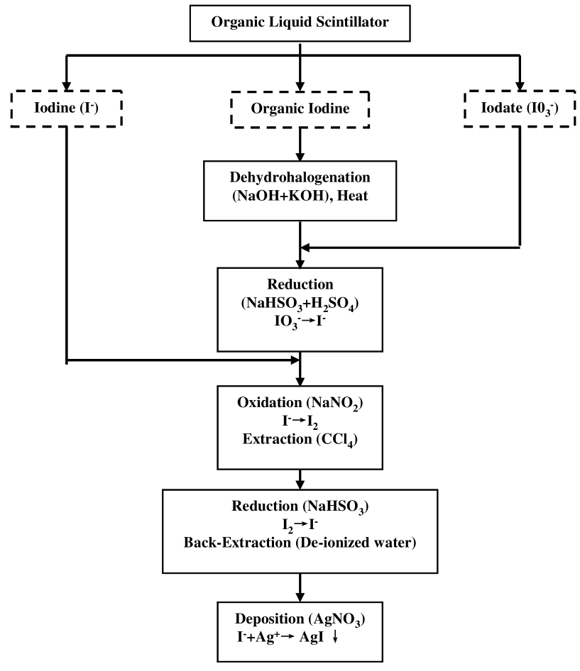
For the organic LS, an extraction procedure for the possible iodine contaminations has to be devised. The adopted sample preparation procedure is shown schematically in Figure 2. Similar schemes have been devised and studied in previous work [19]. A volume of 100 ml of LS was evaporated under vacuum. The residuals left behind consisted mostly of the solid PPO powder, as well as trace concentrations of the other impurities.
A KI carrier solution of mass 10 mg and a solution mixed with 2 mol/l NaOH and 2 mol/l KOH in a 3:2 ratio were added to the residual solid. After stirring to ensure a homogeneous solution, the mixture was transferred into a crucible and put onto a sand bath to dry. The dried sample was ashed into a muffle furnace at 600oC, and then leached with de-ionized water. After being processed by a centrifuge, the iodate in the leached solutions was reduced to iodide with sulfuric acid and sodium hydrogen sulfite (NaHSO3). The iodide was oxidized to I2 by the addition of sodium nitrite and then extracted with carbon tetrachloride (CCl4) and back-extracted into de-ionized water by reduction of the I2 with 5% NaHSO3.
These extraction and back-extraction steps were repeated until the purple color of carbon tetrachloride disappeared. The aqueous phase was boiled for a short time to remove the residual CCl4. After cooling, a silver nitrate (AgNO3) solution was added immediately and processed with a centrifuge. The end-product AgI was rinsed by de-ionized water, dried and collected. Finally, the AgI was mixed with Nb with a ratio of 1:2 and kept for subsequent AMS measurement.
As illustrated in Figure 2, the three possible forms where iodine may exist in LS can all be extracted into the AgI samples for measurement. The extraction efficiency of this procedure was 80%, determined by comparing the ratio, after proper normalizations, of the mass of the extracted AgI compared to that of KI initially introduced.
2.2 Injection and Accelerator
The concentration in the CsI powder and the LS extracted as AgI were measured with CIAE-AMS facility depicted schematically in Figure 1. The tandem accelerator was operated at a terminal voltage of 8.0 MV. A “Multi-Cathode Source of Negative Ions by Cesium Sputtering” was used as the negative ion source. Forty samples were positioned on the target wheel at one time. The wheel could be rotated without affecting the vacuum conditions such that stable operating configurations were maintained during measurements of a group of samples.
The I- negative ions extracted from cesium sputter source were focused by a trim einzel lens and a double focusing 90o deflecting magnet where momentum analysis selected the negative ion beams of interest. The ions were guided to an aperture of 2 mm diameter located at the entrance of the pre-acceleration tube, and then accelerated up to about 120 keV kinetic energy with a terminal voltage of about 8 MV. A carbon foil was attached at the head of accelerator. The molecular background was eliminated due to break-up of molecular ions.
After passing through the accelerator tank, ions with charged state 11+ were selected by a 90o double focusing analyzing magnet with a mass energy product (ME/Z2) at 200 to suppress the isotopic background. A high-resolution electrostatic deflector was placed at a branch beam line to further reduce the isotopic background and other undesired beams.
2.3 Detector
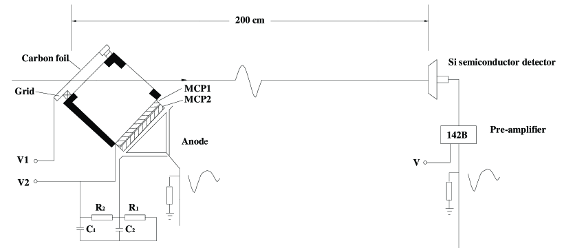
Particle detection and identification of was performed via Time-of-Flight (TOF) detector. A detailed layout of the TOF system is shown in Figure 3. A Micro-Channel Plate (MCP) detector provided the “” signal while a gold-silicon surface barrier detector located 200 cm downstream was used to to give a “”signal. The resolution of the TOF system is 600 ps. The difference between the flight time of and 127I was 2 ns under the conditions of equal momentum at a kinetic energy of about 96 MeV.
2.4 Calibration and Cross-Checks
Standard AgI samples with were prepared and verified by the procedures described in Ref. [20]. According to the contrast results, an uncertainty better than 1% for the source strength in the samples could be measured. The samples were subsequently measured at the AMS facility under the conditions discussed. The derived /127I ratios in several measurements were consistent to 10% with the reference values varying from to .
The energy of the 127I ions are 1.5% higher than that of ions at the same momentum. Accordingly, the 127I ions would deflect more than those of by the electrostatic analyzer, as depicted in Figure 4, such that an energy resolution (E/E) better than 0.5% was achieved. The TOF selection suppressed the 127I by another two orders of magnitude, as shown in Figure 5a for the standard sample with known concentration of /127I at . An “-signal-box” region can be defined from this measurement to locate the candidate events.
2.5 Results
The suppression factors for 127I due to the various AMS components are summarized in Table 1. An efficient transmission of at 60% was achieved, as demonstrated by the measurements with the AgI calibration samples with known absolute strengths and /127I ratios.
| Components | 127I Rejection | 129I Transmission |
|---|---|---|
| Deflecting Magnet | 1 | |
| Analyzing Magnet | 1 | |
| Electrostatic Deflector | 0.8 | |
| TOF Detector | 0.8 | |
| Total | 0.6 |
The TOF scattered plots for the CsI and LS samples are presented in Figures 5b and c, respectively. In both of these cases, as well as in other control measurements with commercially available KI and AgI powder, the measured -signal-box/127I ratios are all . In comparison, a “blank measurement” of only the copper cathode without samples gave zero counts in the -signal-box, indicating that the events are iodine-related. These events can be due to actual contaminations in the samples as well as background from spurious effects or tail distributions of the dominant 127I. Therefore, conservative limits of
can be derived in all four cases (CsI, LS with KI as carrier, as well as the KI and AgI control samples).
This limit is directly applicable to characterize the contaminations in CsI. It can be alternatively expressed as g/g. Accordingly, the -decay background due to in CsI is less than 83 kg-1day-1. In comparison, a recent measurement of 137Cs contaminations in CsI(Tl) crystals [21] was g/g. Both 137Cs and are fission fragments found in the environment, such that their contamination levels in CsI are expected to be similar. Measurement sensitivity for 137Cs is much enhanced due to its much shorter half-life and the emissions of mono-energetic -rays which are easily identified.
For the LS measurement, the volume of the LS and the mass of the KI carrier were known, from which the limit of concentration in LS of g/g can be derived, implying a background -decay rate in LS of less than 15 ton-1day-1.
Further improvement on the sensitivities in the LS measurements is possible, through the use of larger initial LS samples as well as reduced KI mass in the carrier solution. In addition, if the residual events in the -signal-box can be identified to be background from 127I through more detailed studies of the TOF system response, the limits can also be improved.
3 Conclusion
Measurements on the concentrations in an inorganic salt and organic liquid scintillator were performed with the AMS techniques. No evidence were observed for contaminations and sensitive limits were derived. The limits are relevant to the design and interpretation of various low background experiments.
The measurements of are the first “demonstration-of-principle” efforts of devising techniques and procedures in the trace radiopurity measurements of naturally-occurring isotopes using AMS. Research program on the applications of AMS techniques to 40K and 87Rb are being pursued, while those for heavier isotopes like 238U and 232Th series are being planned.
The authors gratefully acknowledge Y.D Chen, B.F. Ni, H.Q. Tang, W.Z. Tian and Z.Y. Zhou for helpful discussions, as well as to the technical staff at CIAE for smooth accelerator operation. This work is supported by contract 10375099 from the National Natural Science Foundation, China.
References
- [1] See the respective sections in Review of Particle Physics, Phys. Lett. B 592, 1 (2004), for details and references.
- [2] H.T. Wong, Mod. Phys. Lett. A 19, 1207 (2004).
- [3] “Accelerator Mass Spectrometry”, C. Tuniz et al., CRC Press (1998).
- [4] D. Elmore and F.M. Phillips, Science 346, 543 (1987).
-
[5]
S. Jiang et al.,
Nucl. Instrum. Methods B 52, 285 (1990);
S. Jiang et al., Nucl. Instrum. Methods B 92, 61 (1994). - [6] R. Molina, in Proc. Int. Symposium on the monitoring of radioactive airborne and liquid releases from nuclear facilities, IAEA-SM-217-15, 181 (1978).
- [7] U. Fehn et al, Nucl. Instrum. Methods B 52, 446 (1990).
- [8] See, for example, J. Gascon, Nucl. Phys. B (Proc. Suppl.) 143, 423 (2005).
- [9] See, for example, H.T. Wong, Nucl. Phys. B (Proc. Suppl.) 143, 205 (2005).
- [10] D. Elmore et al., Nature 286, 138 (1980).
-
[11]
L. C. Bate and J. R. Stokely,
J. Radioanal Chem. 72, 557 (1982);
Y. Muramatsu, Y. Ohmomo and D. Christoffers, Radioanal. Nucl. Chem. 82 2, 353 (1984);
R. S. Strebin et al., J. Radioanal. Nucl. Chem. Lett. 127, 59 (1988);
C. L. Tseng and J. H. Chao, Appl. Radiat. Isot. 47 8, 723 (1996). -
[12]
J. J. Stoffels, Radiochem. Radioanal. Letters,
55 2, 99 (1982);
R. J. Cox and C. J. Pickford, J. Anal. Atom. Spec. 7, 635 (1992). - [13] J.J. Gabay et al., Health Phys. Soc. (C) 26(l), 89 (1974).
- [14] M. Paul et al., Nucl. Instrum. Methods B 29, 341 (1987).
-
[15]
G. Heusser, Nucl. Instrum. Methods B 58, 79 (1991);
P. Jagam and J.J. Simpson, Nucl. Instrum. Methods A 324, 389 (1993). - [16] G. Alimonti et al., Nucl. Instrum. Methods A 406, 411 (1998).
-
[17]
H.B. Li et al.,
Nucl. Instrum. Methods A 459, 93 (2001);
Y. Liu et al., Nucl. Instrum. Methods A 482, 125 (2002). -
[18]
H. Park et al., Nucl. Instrum. Methods A 491, 460 (2002);
T.Y. Kim et al., Nucl. Instrum. Methods A 500, 337 (2003). - [19] K.A. Schwehr and P.H. Santschi, Anal. Chimica Acta 482, 59 (2003).
- [20] S.S. Jiang et al., J. Radioanal. Nucl. Chem. 264 3, 549 (2005).
- [21] Y.F. Zhu et al., nucl-ex/0511001, Nucl. Instrum. Methods A, in press (2006).