Conflict of interest footnote placeholder
This paper was submitted directly to the PNAS office.
Abbreviations: FvK, Föppl-von Kármán number
Submitted to Proceedings of the National Academy of Sciences of the United States of America
Mechanical limits of viral capsids
Abstract
We study the elastic properties and mechanical stability of viral capsids under external force-loading with computer simulations. Our approach allows the implementation of specific geometries corresponding to specific phages such as and CCMV. We demonstrate how in a combined numerical and experimental approach the elastic parameters can be determined with high precision. The experimentally observed bimodality of elastic spring constants is shown to be of geometrical origin, namely the presence of pentavalent units in the viral shell. A criterion for capsid breakage is defined, which explains well the experimentally observed rupture. From our numerics we find for the dependence of the rupture force on the Föppl-von Kármán (FvK) number a crossover from to . For filled capsids high internal pressures lead to a stronger destabilization of viruses with a buckled ground state than unbuckled ones. Finally, we show how our numerically calculated energy maps can be used to extract information about the strength of protein-protein interactions from rupture experiments.
keywords:
thin shells—membranes—biomaterialsBacteriophage capsids have astonishing elastic properties. They withstand extreme internal pressures exerted by their densely packed DNA. DNA packaging experiments on phage [1] and theoretical arguments [2, 3, 4, 5, 6] imply that phage capsids store their DNA under 50atm. This pressure is necessary to inject the DNA into the prokaryotic host cell [7]. It has been shown experimentally that the ejection pressure from -phages is 20atm [8]. The remarkable robustness under high pressures motivated recent nanoindentation studies [9], which showed that capsids also resist external point forces up to 1nN.
In contrast, viruses that infect eukaryotic cells, e.g. CCMV and polyoma, penetrate their host cell. Their DNA is released by subsequent disassembly of the shell. Correspondingly, these viruses do not store their DNA under high pressure. Nevertheless, their resistance to external forces is remarkably strong [10].
Viral capsids are composed of a small number of different proteins, which cluster to morphological units (“capsomers”). These units are put together in a highly regular fashion described by the “quasi-equivalence” principle by Caspar and Klug [11]. Due to the presence of capsomers the surface of capsids has a discrete structure. Mathematically, these capsomers correspond to the vertices of a regular triangulation of a sphere. For such a triangulation the number of vertices , connecting edges , and associated faces have to fulfill Euler’s theorem . This theorem implies that capsids of icosahedral symmetry have 12 pentavalent morphological units (“pentamers”) embedded in an environment of hexavalent units (“hexamers”).
Viral capsids can undergo elastic and bending deformations. Generally, the elastic properties of such thin shells (of typical length scale ) depend only on a single dimensionless parameter, the FvK number set by the ratio between elastic modulus and bending rigidity . It has been shown in Ref. [12], that at a critical spherical shells undergo a buckling transition, in which the shell acquires a more faceted shape. This transition is caused by large strains associated with the pentamers which are reduced upon buckling into a conical shape.
Typical values of viral shells lie in the range from below the buckling threshold (e.g. alfalfa mosaic virus with [13]) up to several thousand (e.g. phage with [14]). An extreme example is the giant mimivirus (diameter of nm) with a Fvk number [15]. Even higher -values are possible for artificial capsules such as vesicles with crystallized lipid membrane [16] () or polyelectrolyte capsules [17] ().
The elastic properties of capsids can be probed in scanning-force microscopy (SFM) experiments [9, 10]. Depending on the strength of loading two regimes are explored. Small forces lead to a completely reversible deformation of capsids. Here, the linear and nonlinear regime of thin spherical shell elasticity can be studied. In this regime the deformations explore the global elastic properties. Larger forces (1nN) cause irreversible changes in the shell structure commonly attributed to bond rupture. Rupturing studies therefore give information about the molecular interactions between capsomers thus elucidating local features of shell mechanics.
Viral shells have also been the subject of numerical investigations. The dependence of virus shape on the FvK number was analyzed in Ref. [12]. Only recently, the elasticity of capsids has come into the focus of numerical studies [18] which are based on a discretization scheme introduced for crystalline membranes in Ref. [19]. In other approaches, the shells are directly built up of proteins [20] or capsomers [21] with specified (spatially varying) interactions. On this basis the stability of capsids against internal pressure has been studied [22].
Here, we generalize a numerical approach developed for the investigation of vesicles [23] to viral capsids. The big advantage of our method is its applicability to arbitrary geometries. In particular, our discretization of the bending energy does not depend on the underlying triangulation of the viral surface. Therefore, our method produces highly stable and reliable results even under high local strain which allows us to investigate the rupture of mechanical shells. In our simulations, we can systematically vary the elastic moduli and geometry of the capsids and probe their mechanical response to external disturbances. We can even implement specific geometries, corresponding to specific phages and viruses, and determine (by a direct comparison with experimental indentation experiments) with high precision their elastic parameters, such as linear and nonlinear spring constants, and the FvK number. In our simulations, we are also able to access features which are not observable in experiments. For example, by measuring the local strain we are able to determine numerically the spatial distribution of rupture probabilities across the capsid surface. We will show that experimental deviations from this distribution may be used to draw conclusions about the spatial variation of protein binding strength.
This paper is organized as follows: after a short summary of the numerical methods, we first study the global response of capsids to externally applied point forces. The numerical results for (empty) 29 and CCMV are compared with experimental data. Next, we analyze the local, irreversible response to indentation forces and we discuss the dependence of local rupture-probability on the geometry of the capsid. Finally, our analysis is extended to filled capsids whose elastic parameters and mechanical stability is studied in the last 2 sections.
1 Methods
We have performed numerical minimization simulations of triangulated surfaces representing the surface of capsids with elastic (stretching) and bending energy. In our (small mesh-size) triangulation every capsomer is represented by several vertices. Therefore, these units also have some flexibility and elasticity. Such a discretization is suitable to determine the shape of viral shells [12] and yields results which do not depend on the number of vertices (in contrast to coarse triangulations in which each capsomer is represented by a single vertex [18]). The triangulation represents the underlying icosahedral (quasi-)symmetry of the capsid. In particular, the pentavalent vertices correspond to the centers of the pentamers.
In our model, the vertices are connected by harmonic springs. Any deviation from the preferred inter-vertex distance gives rise to an elastic energy [19],
| (1) |
where vertex is at position and the sum extends over nearest neighbor pairs only. is related to the 2D Young modulus via .
The bending energy is given by
| (2) |
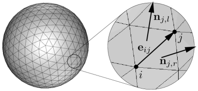
where the sum extends over all vertices . Here, is the area assigned to the -th vertex given by 1/3 of the area of all adjacent triangles. is the mean curvature of vertex (with coordination number ) given by [23]
| (3) |
with , and and are the non-normalized surface vectors of the facets left and right of the edge connecting vertex and , see Fig. 1.
Starting from a triangulated sphere the shape of minimal energy is found by minimizing the total energy using a conjugate gradient algorithm [24]. In order to simulate the SFM-experiments mentioned in the introduction vertices at the point of loading were moved away from their equilibrium position . We thus work in an ensemble of prescribed indentation rather than in an ensemble of applied force. All vertices are constrained to lie above a (virtual) plane at . The shape of the triangulated surface under these constraints is again found by minimizing the total energy.
The shapes of bacteriophage 29 and the CCMV plant virus have been quantitatively characterized by Cryo-EM and X-ray studies [25, 26]. Phage 29 has an average equatorial radius nm and a shell thickness of nm. Its FvK number is implying that the shape of 29 is noticeably buckled. For CCMV the corresponding values are nm and nm. Thus, and CCMV is slightly buckled.
2 Results and Discussion
Spring Constants. As mentioned in the introduction loading with small forces probes the global elastic behavior of viral shells. Here, we analyze the influence of internal structure on the elastic response of capsids by comparing the numerical simulations with analytical results for small deformations. Furthermore, we extract the values of the elastic moduli of and CCMV by a direct comparison of numerical and experimental force-distance relations.
In a first step we have numerically calculated force-distance curves for viruses indented by a distance for (varying with step-size ). By minimizing the total energy the (dimensionless) force is obtained by , where is the change in total energy caused by the increase in . Both hexamers and pentamers were displaced. The indented hexamers discussed here all lie in the center of the facet spanned by their three neighboring pentamers.
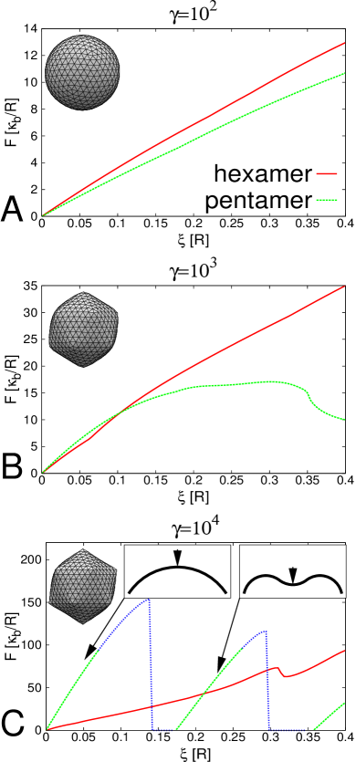
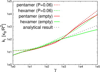
As can bee seen from Fig. 2 the elastic response of the capsid to the deformation strongly depends on the ratio of bending and elastic energy characterized by the FvK number . For small capsids have a nearly spherical shape [12] and behave like homogeneous continuous shells. For the (dimensionless) linear spring constants of both hexameric and pentameric regions follow a square root law for sufficiently small
| (4) |
see Fig. 3. The transition to the nonlinear regime (at ) takes place at smaller the larger , see Fig. 2A and B. For small FvK number corrections to thin shell theory of order become relevant.
For the capsid has a strongly faceted shape since the 12 disclinations buckle. Due to this structural inhomogeneity the elastic response of pentamers and hexamers is different, i.e. pentamers generally become stiffer than hexamers with increasing . Furthermore, the abrupt shape transformation shown in Fig. 2C only occurs for loading on pentamers.
As Fig. 3 shows, the difference between the spring constants for pentamers and hexamers increases with increasing . At the spring constants are already well separated explaining the experimentally measured bimodality of spring constants of [9]. From our numerical data we find for hexamers and for pentamers. When loading on pentamers is parallel to ridges then in agreement with the findings of Refs. [27, 28].
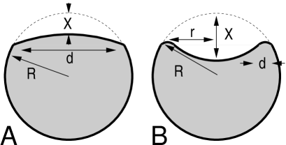
The elastic behavior of spherical capsids under axial point force can be understood by simple scaling arguments [29] of an elastic sphere with energy . For small indentations the top of the sphere is flattened in a circular region of diameter , see Fig. 4A. In the flattened region the radius of curvature and meridians are compressed by an amount . In equilibrium one then finds yielding a total energy of the deformed sphere. Thus, the force-distance relation is linear with .
In fact, a more rigorous analysis of the linear regime yields [30] Eq. (4) for , see Fig. 1 in the supplementary material. For the scaling picture breaks down since the dominant bending energy is no longer concentrated in a small region around the pole.
At a critical indentation the shell undergoes a larger shape transformation in which a circular region in the upper part inverts its shape, see Fig. 4B. Here, the deformation energy is concentrated in a ring of radius and width . The radius of curvature of the ring is and meridians are shortened by a factor . By using again to equilibrate bending and elastic energy (i.e. ) one finds leading to a nonlinear force-distance relation .
For sufficiently large the inversion transition of pentamers (shown in Fig. 4B) is a first order transition (in ) as can be seen from the -curve in Fig. 2C. There, only the green lines correspond to conformations of minimal energy. The blue lines represent intermediate (metastable or unstable) conformations connecting the weakly and strongly deformed capsid shapes. For smaller the transition is continuous, see Fig. 2B.
With our numerical simulations it is also possible to extract precise material parameters of experimentally investigated viruses. To do so we have simulated the conditions corresponding to the SFM experiments on [9]. By using the FvK number as fit parameter for the measured bimodality ratio one finds , in good agreement with the value estimated above from the geometrical parameters of Ref. [26].
Then, mN/m can be extracted directly from the dimensionless spring constant shown in Fig. 3 by using, e.g., the softest experimentally measured value of N/m (with corresponding N/m) [9]. The force scale is then pN. For empty CCMV the bimodality cannot be resolved experimentally (N/m [10]), while numerically we find N/m and N/m. In this case, we extract mN/m and pN from the numerical data.
With these material parameters a direct comparison with SFM experiments can be achieved. Fig. 5 shows as an example the force-distance curves for hexamers and pentamers of . Within our numerical analysis it is even possible to take into account the elastic spring constant of the cantilever (measured in units of ). Then, a measured force corresponds to a displacement of the shell. Thus, as Fig. 5 shows for sufficiently stiff cantilevers the shape inversion of the viral shell leads to a discontinuous force-displacement curve. The numerical results (shown in the inset) are in excellent agreement with the experimentally measured force-distance curves [9]. In particular, for both numerical and experimental deformations the shape instability occurs at

Rupture. We have also analyzed the local response of elastic shells to loading by investigating the conditions under which rupture occurs. This process takes place in regions of high in-plane stress where bonds break due to over-stretching or compression. Thus, this analysis provides information about the interaction between capsomers.
As experimentally observed, phage 29 breaks at a critical indentation [9]. To determine the associated change in bond length we have calculated numerically the shape of a shell with at this indentation yielding a maximal compression of . The corresponding numerical rupture force is nN in agreement with experimental findings [9]. Using this criterion, the numerics for empty CCMV predicts a rupture force nN while the experimentally determined value is nN [10].
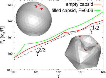
In the following, we analyze rupture of viral shells as a function of . Fig. 6 shows the -dependence of . For rupture occurs in the linear regime, see Fig. 4A. Here, rupture is caused by compression of meridians, see left inset in Fig. 6. In the linear regime, we observe that the rupture force scales approximately . However, most viruses have higher FvK numbers and will therefore rupture in the nonlinear regime after shape inversion, see Fig. 4B. Here, rupture is caused by circumferential compression in the highly bent rim. In this regime, the rupture force scales (roughly) .
We have also investigated the influence of the capsid geometry on rupture by comparing an icosahedral and a sphero-cylindrical capsid. This is motivated by the fact that even-numbered -phages and have additional capsomers in the equatorial region elongating the icosahedral shape. For example, the head of is an icosahedron with triangulation number 13 and one additional ring of hexamers [31]. The geometry of the sphero-cylinder used in our simulations is that of 29, i.e. an icosadeltahedron with triangulation number , elongated by two additional equatorial rings of radius [26]. The force was applied perpendicularly to the axis of symmetry. In order to avoid tilting of the structure, the lowest 10% of vertices of the shell were kept fixed in the simulations.
Fig. 7 shows the rupture force mapped across the surface for and . In the corresponding numerical simulations every vertex was indented in steps of followed by the calculation of the new equilibrium conformation. When stretching of some bond exceeded 4.5% the applied force was determined. For below the buckling threshold the shells have a uniform elastic behavior across the surface. Since the force exerted on the caps has a tangential component, part of the displacement directly leads to bond stretching. Therefore, the caps are the regions of highest instability. Above the buckling threshold, the pentamers are rigid cones but the space between them becomes more flexible. When pushing a pentameric region the displacement is mainly transformed into compression of the ridges. Pentamers are thus the most unstable region. On average a larger (scale-free) force is required to break the capsid for larger FvK number .
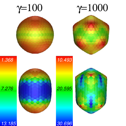
The spatial distribution of deformation energy can serve as a measure for the tendency to rupture. To mimic an ensemble of indentation experiments, we carried out simulations in which each vertex of the shell was indented by a constant (). For all vertices the deformation energy was recorded for all deformations. In order to find the spatial distribution of deformation energy in the ensemble, the elastic energy per vertex was averaged over the ensemble measurement. Fig. 8 shows a map of the average spatial distribution of elastic energy, normalized by the total elastic energy put into the system during the measurement on the ensemble.
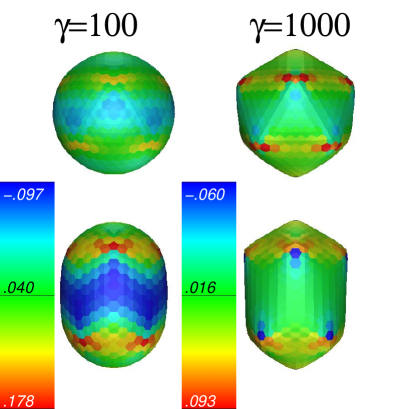
In our simulations we assume a uniform distribution of binding energies between capsomers. In reality, binding energies will show a spatial variation and an ensemble of indentation experiments on capsids will yield different rupture probabilities than the numerical data presented in Fig. 8. However, with a direct comparison of the two approaches conclusions about the distribution of binding potentials can be drawn.
Filled Capsids. Phages infect their host cells by penetrating the cell membrane and rapidly injecting the DNA into the cell plasma. Therefore, their DNA is stored under high pressure. The pressure of DNA inside is of the order of 6MPa [1, 5] or in units of elastic parameters .
Other viruses, like CCMV, self-assemble inside the host cell and enclose the DNA. Correspondingly, their internal pressure is much lower than that of phages. Typically, 1atm is a good estimate for the internal pressure of a filled CCMV capsid.
To investigate the influence of internal pressure on the elastic properties we have numerically simulated filled capsids with the methods described above. However, here the additional constraint has to be taken into account that due to the presence of DNA inside the capsids the enclosed volume is fixed. In order to mimic a packed phage, first the equilibrium conformation under pressure was determined by minimizing the total energy with an additional pressure contribution with .
Experiments on full CCMV were performed in Ref. [10]. A direct comparison of our numerical simulations with these experiments yields (for and volume const.) the spring constants N/m, N/m, and a rupture force nN. These values are again in good agreement with the experimental findings (N/m and nN). There are no SFM-experiments on filled 29 phages to which we could compare our numerical simulations.
Generally, the volume constraint leads to increased linear spring constants, see Fig. 3. This can be understood in the scaling picture: In order to preserve constant cross-sectional area, the local compression at the point of loading must be compensated by an expansion of the equatorial area. Therefore the influence of the volume constraint becomes more apparent for larger , i.e. for shells dominated by elastic energy.
At high pressures the circumferential stress at the fivefold disclinations is balanced by the volume contribution . Thus, pentamers do not form rigid buckles and hexamers remain flat. For example, the DNA pressure of 29-phages reduces its aspherity from for an empty capsid to . This similarity between hexamers and pentamers is reflected by the fact, that the elastic response of hexamers and pentamers becomes similar for high internal pressure, see Fig. 3.
The rupturing behavior of filled capsids is shown in Fig. 6. The rupture force is larger for filled capsids than for empty ones. Due to the internal pressure the non-indented shell is already stretched. This compensates the force-induced compression in meridional direction at the poles thus reducing their tendency to rupture. Rupture of filled capsids is caused by circumferential expansion at the equator.
Osmotic Shock. One way to extract DNA from viral capsids is to put them under osmotic shock. Under these conditions some viral capsids (e.g. -even phages) rupture [32] while others (e.g. -odd phages) stay intact. Thus, these experiments also elucidate details about capsomer-capsomer interactions and we have analyzed whether rupture induced by internal pressure and by external force are related.
To do so we have numerically determined the shape of the capsid under internal pressure and measured the bond length between neighboring vertices. It was assumed that at rupture pressure the critical bond stretching is 4.5% as for rupturing due to bond compression. Fig. 9 shows the corresponding rupture pressure (in units of ) as function of for an icosahedral shell.
As can be seen from Fig. 9, capsids with a buckled ground state () rupture at lower than unbuckled ones. One should note, that for rupture induced by an external force, rupture forces are generally higher for than for , see Fig. 6. However, as can be seen from the inset in Fig. 6, in the buckled configuration the force-induced deformation propagates over a larger area than in the unbuckled state. Therefore, it is possible that the external rupture pressure for force-induced rupture is also lower for buckled capsids. But this questions needs to be addressed in a more detailed analysis.
For small the rupture pressure reaches a constant value, see Fig. 9. In this limit deviations from spherical shape are small and the internal pressure simply leads to a form-invariant up-scaling of the viral shell. Thus, changes in bending energy can be neglected. By equilibrating the expanding pressure force and the restoring force arising from bond stretching for a triangulated sphere the relative change of bond length is found to be . The numerically found value is somewhat lower caused by the small deviations from spherical shape. The same calculation shows that a triangulated sphere with a harmonic stretching energy is only stable for pressures , independently of , see supporting information.

Summary and Conclusions. Recent SFM-measurements have revealed the elastic properties and mechanical limits of viral capsids. In this paper, we have shown that numerical simulations are a powerful tool in complementing these experimental efforts. We have examined the reversible and irreversible mechanical behavior on local and global scales. In particular, we have shown that the elastic parameters characterizing the mechanical properties of phages and viruses can be determined very precisely by a direct comparison of numerical and experimental data. With the numerical methods presented here, one is able to resolve the mechanical response of viral shells to an external force in high detail. In particular, since we are able to distinguish the response of hexamers and pentamers we can identify the origin of the experimentally observed bimodality of the elastic spring constants. We also make predictions for the elastic response and rupturing behavior of both empty and filled capsids as function of the FvK number . Furthermore, in our simulations the mechanical limits of viral shells can be probed in an ensemble where every capsomer is indented. The comparison of the corresponding rupture map of the shells with experimental data on SFM-induced rupturing offers new methods in experimentally probing the local protein-protein interactions. We can also use the rupture criterion to predict the maximal sustainable internal pressure of capsids.
So far, we have focused on shells with the shape of an icosadeltahedron. However, many viruses such as phage HK97 [33] or the animal virus polyoma [34] are chiral. The influence of chirality on the elastic properties and mechanical stability will be addressed in a forthcoming publication [30].
Acknowledgment. PL gratefully acknowledges support through the Fonds der Chemischen Industrie and the Center for Theoretical Biological Physics (National Science Foundation Grants Nos. PHY0216576 and PHY0225630).
References
- [1] Smith, D. E, Tans, S. J, Smith, S. B, Grimes, S, Anderson, D. L, & Bustamante, C. (2001) Nature 413, 748–752.
- [2] Riemer, S. C & Bloomfield, V. A. (1978) Biopolymers 17, 785–794.
- [3] Kindt, J, Tzlil, S, Ben-Shaul, A, & Gelbart, W. M. (2001) Proc. Natl. Acad. Sci. 98, 13671–13674.
- [4] Tzlil, S, Kindt, J. T, Gelbart, W. M, & Ben-Shaul, A. (2003) Biophys. J. 84, 1616–1627.
- [5] Purohit, P. K, Kondev, J, & Phillips, R. (2003) Proc. Natl. Acad. Sci. 100, 3173–3178.
- [6] Purohit, P. K, Inamdar, M. M, Grayson, P. D, Squires, T. M, Kondev, J, & Phillips, R. (2005) Biophys. J. 88, 851–866.
- [7] Atlas, R. M. (1998) Principles of Microbiology. (Wm.C. Brown Publishers).
- [8] Evilevitch, A, Lavelle, L, Knobler, C. M, Raspaud, E, & Gelbart, W. M. (2003) Proc. Natl. Acad. Sci. 100, 9292–9295.
- [9] Ivanovska, I, de Pablo, P, Ibarra, B, Sgalari, G, MacKintosh, F, Carrascosa, J, Schmidt, C, & Wuite, G. L. (2004) Proc. Natl. Acad. Sci. 101, 7600–7605.
- [10] Michel, J, Ivanovska, I, Gibbons, M, Klug, W, Knobler, C, Wuite, G. L, & Schmidt, C. (2006) Proc. Natl. Acad. Sci. 103, 6184–6189.
- [11] Caspar, D & Klug, A. (1962) Cold Spring Harb. Symp. Quant. Biol. 27.
- [12] Lidmar, J, Mirny, L, & Nelson, D. R. (2003) Phys. Rev. E 68, 051910.
- [13] Kumar, A, Reddy, V. S, Yusibov, V, Chipman, P. R, Hata, Y, Fita, I, Fukuyama, K, Rossmann, M. G, Loesch-Fries, L. S, Baker, T. S, & Johnson, J. E. (1997) J. Virol. 71, 7911–7916.
- [14] Olson, N. H, Gingery, M, Eiserling, F. A, & Bakera, T. S. (2001) Virology 279, 385–391.
- [15] Xiao, C, Chipman, P. R, Battisti, A. J, Bowman, V. D, Renesto, P, Raoult, D, & Rossmann, M. G. (2005) J. Mol. Biol. 353, 493–496.
- [16] Dubois, M, Deme, B, Gulik-Krzywicki, T, Dedieu, J.-C, Vautrin, C, Desert, S, Perez, E, & Zemb, T. (2001) Nature 411, 672–675.
- [17] Elsner, N, Dubreuil, F, Weinkamer, R, Wasicek, M, Fischer, F, & Fery, A. (2006) Prog. Colloid Polym. Sci. 132, 117–123.
- [18] Vliegenthart, G. A & Gompper, G. (2006) Biophys. J. 91, 834–841.
- [19] Seung, H & Nelson, D. R. (1988) Phys. Rev. A 38, 1005–1018.
- [20] Reddy, V, Giesing, H, Morton, R, Kumar, A, Post, C, Brooks III, C, & Johnson, J. (1998) Biophys. J. 74, 546–558.
- [21] Zandi, R, Reguera, D, Bruinsma, R. F, Gelbart, W. M, & Rudnick, J. (2004) Proc. Natl. Acad. Sci. 101, 15556–15560.
- [22] Zandi, R & Reguera, D. (2005) Phys. Rev. E 72, 021917.
- [23] Wintz, W. (1997) Ph.D. thesis (Universität Potsdam).
- [24] Press, W, Teukolsky, S. A, Vetterling, W. T, & Flannery, B. P. (1992) Numerical Recipes in C. (Cambridge University Press).
- [25] Speir, J. A, Munshi, S, Wang, G, Baker, T. S, & Johnson, J. E. (1995) Structure 3, 63–78.
- [26] Tao, Y, Olson, N. H, Xu, W, Anderson, D. L, Rossmann, M. G, & Baker, T. S. (1998) Cell 95, 431–437.
- [27] Lobkovsky, A. E & Witten, T. (1997) Phys. Rev. E 55, 1577–1589.
- [28] DiDonna, B & Witten, T. (2001) Phys. Rev. Lett. 87, 206105.
- [29] Landau, L & Lifshitz, E. (1975) Theory of Elasticity. (Pergamon, New York).
- [30] Bünemann, M & Lenz, P. (2006) to be published.
- [31] Fokine, A, Chipman, P. R, Leiman, P. G, Mesyanzhinov, V. V, Rao, V. B, & Rossmann, M. G. (2004) Proc. Natl. Acad. Sci. 101, 6003–6008.
- [32] Anderson, T. F. (1950) J. Appl. Phys. 21, 70.
- [33] Helgstrand, C, Wikoff, W, Duda, R, Hendrix, R, Johnson, J, & Liljas, L. (2003) J. Mol. Biol. 334, 885–899.
- [34] Stehle, T & Harrison, S. (1996) Structure 4, 183–194.