Incident-energy and polarization-dependent resonant inelastic x-ray scattering study of La2CuO4
Abstract
We present a detailed Cu -edge resonant inelastic X-ray scattering (RIXS) study of the Mott insulator La2CuO4 in the 1-7 eV energy loss range. As initially found for the high-temperature superconductor HgBa2CuO4+δ, the spectra exhibit a multiplet of weakly-dispersive electron-hole excitations, which are revealed by utilizing the subtle dependence of the cross section on the incident photon energy. The close similarity between the fine structures for in-plane and out-of-plane polarizations is indicative of the central role played by the core hole in inducing charge excitations within the CuO2 planes. On the other hand, we observe a polarization dependence of the spectral weight, and careful analysis reveals two separate features near 2 eV that may be related to different processes. The polarization dependence indicates that the 4 electrons contribute significantly to the RIXS cross section. Third-order perturbation arguments and a shake-up of valence excitations are then applied to account for the final-energy resonance in the spectra. As an alternative scenario, we discuss fluorescence-like emission processes due to transitions into a narrow continuum band.
pacs:
74.25.Jb, 74.72.-h, 78.70.Ck, 71.35.-yI Introduction
With the advent of third generation synchrotron sources, inelastic X-ray scattering has emerged as a powerful probe of momentum- and energy-dependent charge and lattice dynamics. This development has led to new insight into low-density metallic electrodynamics Burns et al. (1999), valence fluctuating compounds Dallera et al. (2002), H2O molecular correlations P. Wernet, D. Nordlund, U. Bergmann, M. Cavalleri, M. Odelius, H. Ogasawara, L. A. Naslund, T. K. Hirsch, L. Ojamae, P. Glatzel, L. G. M. Pettersson, and A. Nilsson (2004); Sette et al. (1995), phonon dynamics d’Astuto et al. (2002), and the Mott physics of correlated electron systems such as the lamellar copper oxides Hill et al. (1998); Abbamonte et al. (1999); Hämäläinen et al. (2000); Hasan et al. (2000); Kim et al. (2002, 2004); L. Lu, X. Zhao, G. Chabot-Couture, J. N. Hancock, N. Kaneko, O. P. Vajk, G. Yu, S. Grenier, Y. J. Kim, D. Casa, T. Gog, and M. Greven (2005); K. Ishii, K. Tsutsui, Y. Endoh, T. Tohyama, S. Maekawa, M. Hoesch, K. Kuzushita, M. Tsubota, T. Inami, J. Mizuki, Y. Murakami, and K. Yamada (2005); K. Ishii, K. Tsutsui, Y. Endoh, T. Tohyama, K. Kuzushita, T. Inami, K. Ohwada, S. Maekawa, T. Masui, S. Tajima, Y. Murakami, and J. Mizuki (2005) and the manganites K. Ishii, T. Inami, K. Ohwada, K. Kuzushita, J. Mizuki, Y. Murakami, S. Ishihara, Y. Endoh, S. Maekawa, K. Hirota, and Y. Moritomo (2004); S. Grenier, J. P. Hill, V. Kiryukhin, W. Ku, Y. J. Kim, K. J. Thomas, S. W. Cheong, Y. Tokura, Y. Tomioka, D. Casa, and T. Gog (2005). Resonant inelastic X-ray scattering (RIXS) provides a considerable advantage over ordinary inelastic scattering since, at resonance, the inelastic signal is significantly enhanced Kao et al. (1996). In the lamellar copper oxides, this resonance condition can be readily met by tuning the incoming photon energy to the vicinity of the Cu edge. The RIXS cross section sensitively depends on the incident photon energy and on the nature of the intermediate states L. Lu, X. Zhao, G. Chabot-Couture, J. N. Hancock, N. Kaneko, O. P. Vajk, G. Yu, S. Grenier, Y. J. Kim, D. Casa, T. Gog, and M. Greven (2005); Krisch et al. (1995); Döring et al. (2004); Glatzel and Bergmann (2005a).
If viewed as a two-stage process, the intermediate state in RIXS is the same as the final state of x-ray absorption: for example, in Cu K-edge RIXS, a localized core hole is created through photoexcitation. The core hole interacts strongly with the valence electron system, generating a strong response that corresponds to the many-electron bound states of the local, nascent core-hole potential.Tranquada et al. (1991) In RIXS, the relaxation of these highly excited states leads to the emission of photons and leaves the valence system in an excited state. One usually identifies energy-loss features with the excitations of the valence electrons. When viewed as a second-order optical process, there exists a close connection between the initial absorption and final emission stages in RIXS. Accordingly, the spectra simultaneously depend on both the incident and final photon energies.Kotani and Shin (2001)
In the present work, we investigate these energy dependences as well as the polarization dependence of the cross section in La2CuO4, the best-characterized lamellar copper oxide. This Mott insulator is the parent compound of the original high-temperature superconductor (La,Ba)2CuO4, and it has been the subject of a number of prior RIXS studies.Kim et al. (2002); L. Lu, X. Zhao, G. Chabot-Couture, J. N. Hancock, N. Kaneko, O. P. Vajk, G. Yu, S. Grenier, Y. J. Kim, D. Casa, T. Gog, and M. Greven (2005); Collart et al. (2006) Exploiting the incident-energy sensitivity, we are able to identify additional charge excitation features. We demonstrate that the fine structure is present for photon polarization both parallel and perpendicular to the CuO2 planes, and suggest that the subtle differences between the two polarization conditions can be explained in terms of models in which the electrons play a significant role.
This paper is organized as follows. After the discussion of the experimental details in the next section, we present our results for out-of-plane and in-plane polarization in Secs. III and IV, respectively. Section V contains a discussion of our data, and we summarize our work in Sec. VI.
h
II Experimental Details
The focus of this work is on the collective electronic excitations of La2CuO4 in the 1 7 eV range using incident photon energies in the vicinity of the Cu -edge absorption threshold. We have measured the RIXS response at several high-symmetry positions in the Brillouin zone, covering a fine mesh of incident and scattered photon energies around the Cu edge.
Two sets of measurements were taken, one collected at the Advanced Photon Source with incident photon polarization vector perpendicular to the CuO2 planes (E), and the other at SPring-8 (Japan) with incident photon polarization parallel to the CuO2 planes (E). The measurements with in-plane polarization [Fig. 1 (a)] were taken in horizontal scattering geometry at beamline BL11XU at SPring-8, with the tetragonal reciprocal lattice point G=(3,0,0) as Brillouin zone center, and a scattering angle of . A Si(111) main monochromator and a Si(400) secondary monochromator were used to obtain an incident energy resolution of 220 meV. A bent Ge(733) analyzer crystal situated at the end of a 2 m four-circle diffractometer arm selected the energy of the photons scattered from the sample, which were then collected by a solid state detector. In this geometry, the polarization vector of the incident photon was always parallel to the CuO2 planes, with a typical angle of with respect to the tetragonal axis, i.e., the planar Cu-O bond direction. The overall energy resolution was about 400 meV [full width at half maximum (FWHM)], as determined from the energy width of the elastic line.
Out-of-plane polarization measurements [Fig. 1 (b)] were performed at beamline 9-ID-B at the Advanced Photon Source in a vertical scattering geometry. The reciprocal lattice vectors (3,0,0) and (1,0,0) were chosen as reference zone centers to reduce the contribution from the elastic tail, because Bragg scattering at these reflections is forbidden. The setup employed a Si(111) primary monochromator, a Si(333) secondary monochromator, and a spherical diced Ge(733) analyzer crystal with a 1 m radius, and yielded an overall energy resolution of about 300 meV (FWHM). For data collected that were measured at the reduced wave vector (,0), or absolute momentum of (1.5,0,0), we used a primary Si(111) and a second Si(444) channel-cut monochromator in conjunction with a diced analyzer on a 2 m diameter Rowland circle. This configuration can provide at best an energy resolution of 110 meV, but we chose wide aperture slits in front of the detector to obtain a significant signal boost and a comparable resolution of 300 meV for better comparison with measurements at other wave vectors.
Data were taken at ambient temperature on the same single-crystalline sample in both polarization geometries. La2CuO4 undergoes a tetragonal-to-orthorhombic structural phase transition at K associated with the staggered tilting of the CuO6 octahedra.Birgeneau et al. (1999) We note that our crystal is twinned, and hence we do not distinguish between the two inequivalent planar orthorhombic directions. The crystal was grown in an image furnace at Stanford University. As-grown crystals are known to contain excess oxygen, and hence hole carriers. In order to assure that the sample was free of any carriers it was annealed for 24 h in Ar flow at 950∘C. This reduction treatment resulted in a Néel temperature of K, as determined from a measurement of the magnetic susceptibility.Vajk et al. (2002)

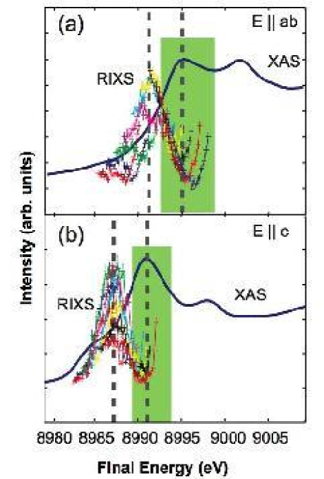
Figure 2 shows the x-ray absorption spectra (XAS) for each polarization condition as measured by total fluorescence yield. For each polarization, there are two peaks, at 8991 and 8998 eV for E, and at 8995 and 9002 eV for E. The lower of these resonances is usually identified with a transition into a well-screened state,”Hill et al. (1998); Hämäläinen et al. (2000); Kim et al. (2002) a many-body excitation which effectively screens the 1s core hole and has significant character. The higher resonance is identified with a transition into a poorly screened state, Hill et al. (1998); Hämäläinen et al. (2000); Kim et al. (2002) another bound state of the many-electron system in the core hole potential which has predominantly character. For each polarization, the resonance peaks are separated by approximately 7 eV. The resonance energies differ by about 4 eV between the two geometries, which may be primarily due to the larger Cu-O distance for the negatively charged apical oxygens, rendering the 4 electronic orbitals lower in energy than their 4 counterparts.Heald et al. (1988); Lee and Pickett (2006) The spectra presented in this paper were taken in the vicinity of the well-screened condition for both polarization geometries.
III Incident Energy Dependence, Out-of-plane polarization
RIXS spectra obtained in early work in the soft x-ray regime exhibited a clear incident and final photon energy dependence.Kotani and Shin (2001) The incident-photon-energy dependence was recently employed in the hard x-ray regime (at the Cu edge) in a study of both La2CuO4 and the single-layer hightemperature superconductor HgBa2CuO4+δ (Hg1201).L. Lu, X. Zhao, G. Chabot-Couture, J. N. Hancock, N. Kaneko, O. P. Vajk, G. Yu, S. Grenier, Y. J. Kim, D. Casa, T. Gog, and M. Greven (2005) This study revealed additional features in the 2-5 eV range, an observation that necessitates a new interpretation of the charge dynamics in these materials. For example, a eV feature was identified in Hg1201. It was argued in Ref. L. Lu, X. Zhao, G. Chabot-Couture, J. N. Hancock, N. Kaneko, O. P. Vajk, G. Yu, S. Grenier, Y. J. Kim, D. Casa, T. Gog, and M. Greven, 2005 that this feature is not likely a excitation, but rather indicates the presence of a remnant charge-transfer gap even at optimal doping in this model superconductor. The presence of an additional feature at 3 eV, which was only identified through inspection of multiple spectra obtained with different photon energies, constrains the dispersion of the 2 eV feature to be less than 500 meV. The same approach was applied to La2CuO4, and preliminary data revealed charge-transfer features that are remarkably similar to those in Hg1201,L. Lu, X. Zhao, G. Chabot-Couture, J. N. Hancock, N. Kaneko, O. P. Vajk, G. Yu, S. Grenier, Y. J. Kim, D. Casa, T. Gog, and M. Greven (2005) a result that is qualitatively different from prior work on the Mott insulators La2CuO4 Kim et al. (2002) and Ca2CuO2Cl2 Hasan et al. (2000) The small dispersion of the 2 eV feature was further confirmed by subsequent measurements.Collart et al. (2006); Kim (2006) Our primary focus here is to investigate in greater detail the incident-energy and polarization dependence of the inelastic cross section near the absorption threshold in La2CuO4.
The molecular orbital excitation at eV was studied in detail in Ref. Kim et al., 2004 and is most prominently observed near = 8998 eV, an incident photon energy for which the lower-lying charge-transfer excitations in the 2-6 eV range do not resonate. We therefore limit our attention to the incident energy range = 8989-8994 eV [shaded area in Fig. 2 (a)] and to energy transfers below 7 eV for out-of-plane polarization. A fine step size of meV was chosen, allowing us to demonstrate the high sensitivity of the RIXS cross section to the incident photon energy.
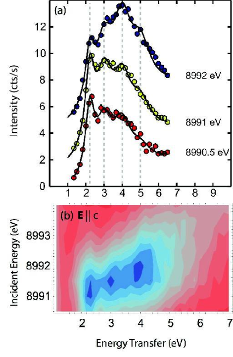
Figure 3 (a) shows representative line scans at the zone center, taken at three different incident energies with out-of-plane polarization. These data resemble previous work Kim et al. (2002), yet closer inspection reveals additional features. At low incident energy, the most distinct feature is that at 2.25 eV. As the incident energy is increased, this sharp feature gradually weakens relative to those at higher energy. Another new feature at 3 eV, which was not observed in prior work Kim et al. (2002), can be discerned at all three incident photon energies. The feature at eV, also seen in previous zone-center data Kim et al. (2002), becomes dominant at eV. Finally, a comprehensive analysis of all data L. Lu, X. Zhao, G. Chabot-Couture, J. N. Hancock, N. Kaneko, O. P. Vajk, G. Yu, S. Grenier, Y. J. Kim, D. Casa, T. Gog, and M. Greven (2005) reveals a second new feature at eV. We discuss below how the systematic center-of-mass shift with incident energy suggests a modulation of the inelastic cross section through final photon-energy-dependent denominators.
Figure 3 (b) shows a contour plot constructed from all line scans at the zone center. This mode of representation is similar to the “RIXS plane” of incident photon energy versus energy transfer in Ref. Glatzel and Bergmann, 2005b. By extending the energy-transfer spectra into the incident-energy dimension, features at , 3 and 4 eV are readily apparent.
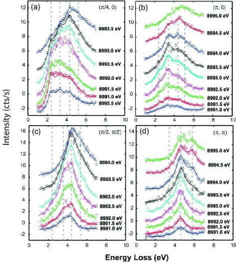
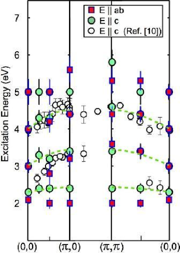
Figure 4 shows additional results at high-symmetry points of the two-dimensional Brillouin zone. Using the fit procedure defined in Ref. L. Lu, X. Zhao, G. Chabot-Couture, J. N. Hancock, N. Kaneko, O. P. Vajk, G. Yu, S. Grenier, Y. J. Kim, D. Casa, T. Gog, and M. Greven, 2005, the spectra at each momentum transfer are fitted simultaneously to obtain the peak positions that determine the energy transfer of the corresponding charge excitations. Specifically, we assume that the peak positions do not vary with incident energy and that each energy transfer feature is represented by a Lorentzian line shape. The number, energy-transfer positions, and energy widths of the features are considered to be shared parameters for all spectra at the same momentum transfer. At different incident energies, on the other hand, the spectral weight of each component is allowed to vary. We also use spectra on the energy gain side (not shown in Fig. 4) for background subtraction, and allow for linear slopes to approximate the continuum due to transitions to continuous unoccupied states. A simultaneous least-squares fit of all spectra at each momentum transfer results in the lines in Figs. 3 (a) and 4 and allowed us to extract the peak positions plotted in Fig. 5.
Although the different components are not as easily distinguishable as at the zone center, the relative strengths of the four features identified below 6 eV exhibit distinct dependences on momentum transfer and incident energy, which may be indicative of different excitation symmetry. The 5 eV component is most pronounced at and at relatively high incident energies. Away from the zone center, the relative weight of the 2 eV feature quickly decreases. While the 2 and 3 eV features are still separable and comparable in strength at , the 2 eV component is only barely visible (at low incident energies) at , the 3 and 4 eV features maintain comparable weight along . In contrast, along , the overall response away from the zone center appears to be dominated by the (approximately Lorentzian-shaped) response just above 4 eV. As for the fine structure, the momentum dependence away from the zone center is again consistent with what is observed in Hg1201, although that study was carried out with in-plane polarization.L. Lu, X. Zhao, G. Chabot-Couture, J. N. Hancock, N. Kaneko, O. P. Vajk, G. Yu, S. Grenier, Y. J. Kim, D. Casa, T. Gog, and M. Greven (2005)
IV Incident Energy Dependence, in-plane polarization
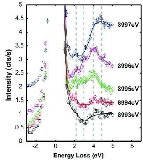
Figure 6 shows line scans at the zone center for the complementary in-plane-polarized (SPring-8) experiment. As indicated in Fig. 2 (b), the incident photon energy was chosen to lie in the range 8993 8999 eV. The fine structure revealed through the incident energy dependence of the spectra is very similar to that found for out-of-plane polarization. Differences in the configuration between the two experiments make a direct comparison of signal levels difficult. Overall, there are still four major features present at , 3, 4 and 5 eV. The close similarity of the spectra supports the intuitive notion that the spherically-symmetric core hole potential dominates the generation of the valence excitations in both cases.Mahan (2000); deGroot (2001); Nomura and Igarashi (2005); Hasan et al. (2000); Tsutsui et al. (2003)
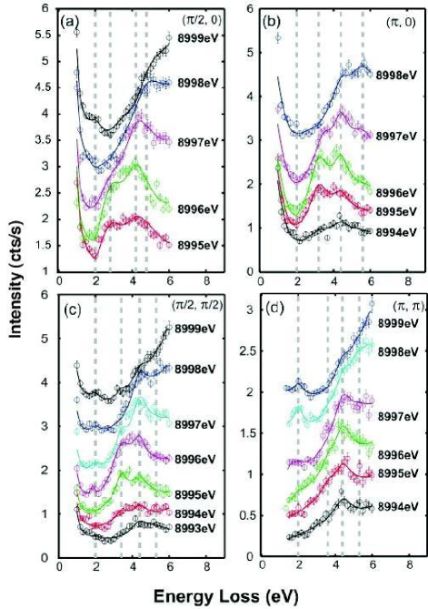
There also exist similarities between the two polarization geometries in the momentum dependence of the multiplet structure. Figure 7 shows line scans with fits at four reduced momentum transfer values away from the zone center. As in the out-of-plane polarized experiment, the strength of the 2 eV feature decreases toward the zone boundary. In the present case, it is no longer visually observable at . Also, the 3 eV and 4 eV features have comparable spectral weight along , and remain comparable up to along . Between (/2,) and (,), however, the 3 eV feature is quickly suppressed between and, as in the case for out-of-plane polarization, the 4 eV spectral weight becomes dominant.
Differences between the spectra obtained in the two geometries are also noticeable. First, with in-plane polarization, the intensity of the 2 eV feature relative to that of spectral features with higher energy transfer is much smaller than for the out-of-plane polarized experiment. However, the 2 eV feature is still observable even up to , reaching a maximum at an incident energy of 8997 eV, slightly above the absorption threshold (8995 eV) for in-plane polarization. Second, for in-plane polarization, as we increase in incident energy, the center of mass of the spectra continuously shifts to higher energy transfers. For out-of-plane polarization, on the other hand, it peaks between 4 and 5 eV.
As for the E data, all spectra acquired at the same momentum transfer were simultaneously fit assuming the same set of peak positions. The results of the fits are shown by the lines in Figs. 6 and 7 and the peak positions are compared to those for out-of-plane polarization in Fig. 5. While there is an overall good agreement, the 2 eV features have significantly different excitation energies for the two polarization conditions. We note that the dispersions of all four features summarized in Fig. 5 are weak. These observations will be discussed in more detail in the next section.
V Discussion
The presence of a 2 eV resonance feature has been discussed in connection with excitations across the charge transfer gap.Kotani and Shin (2001); Hasan et al. (2000); Abbamonte et al. (1999); Kim et al. (2002); L. Lu, X. Zhao, G. Chabot-Couture, J. N. Hancock, N. Kaneko, O. P. Vajk, G. Yu, S. Grenier, Y. J. Kim, D. Casa, T. Gog, and M. Greven (2005) In La2CuO4, this excitation is observed only for transitions into well-screened states, in which the core hole is screened by a valence electron from a neighboring CuO4 plaquette, leaving a doubly occupied Cu+ ion () and a hole on the neighboring plaquette. It has been suggested that the nonlocal hole can form a Zhang-Rice singlet,Zhang and Rice (1988) which can propagate efficiently through the antiferromagnetic background, and that this singlet could form a strong bond with the Cu+ quasiparticle and become even more dispersive as a bound exciton.Zhang and Ng (1998) However, high-resolution electron-energy-loss spectroscopy (EELS) on the related Mott insulator Sr2CuO2Cl2 suggests the existence of another charge-transfer excitation, with slightly lower energy, that involves only the local CuO4 plaquette. These latter findings were argued to be consistent with embedded molecular cluster calculations.Moskvin et al. (2002) Indeed, our data reveal that the actual excitation energies of the 2 meV feature differ by as much as 300 meV for in-plane and out-of-plane polarization conditions. While the former excitation has no discernible dispersion, the latter appears to disperse by 100 150 meV toward the zone boundary. In addition to these differences, we also find that the spectral weights of these two low-energy features exhibit rather different momentum dependences, especially along . We note that it is not likely that the 2 eV features are excitations, since the latter lie below 2 eV and are expected to be much weaker at the edge than at the and edges.Kotani and Shin (2001)
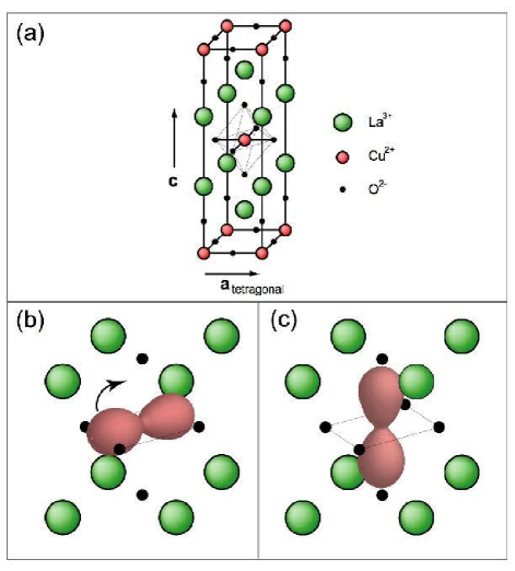
In an attempt to understand the differences between the two 2 eV features, we consider a possible photon polarization effect. When the polarization vector of the incident photon lies within the CuO2 plane, the electron in the intermediate state is in the orbital [see Fig. 8 (b)] and overlaps with the electrons. The repulsive Coulomb interaction between O and Cu therefore tends to suppress the OCu charge transfer. On the other hand, for out-ofplane polarization, the orbital is oriented orthogonally to the electrons [see Fig. 8 (c)] and the Coulomb interaction has a limited effect on the charge-transfer process. That difference in intermediate states may explain why we observe a second (local) “2eV” component for in-plane polarization, since the component that involves a nonlocal charge-transfer process from a neighboring CuO4 plaquette to the central core hole site could be spectroscopically suppressed for in-plane polarization. The lower of the two 2 eV features may be intrinsically weaker, which could explain why we are unable to discern it for out-of-plane polarization. As an alternative scenario, consistent with the identification of two distinct features, a detailed analysis of the scattering configurations reveals that the optical-limit Raman efficiencies set by geometry are quite different in each case. It is possible that the suppression of the higher energy feature for the plane-polarized experiment reflects details of the symmetry of this excitation. Further experimentation is required to resolve this possibility. The above is consistent with earlier suggestions based on Cu K-edge XAS.Tolentino et al. (1992) Especially for in-plane polarization, the 2 eV excitation still maintains its strength along [], whereas it is weakened at the same location for out-of-plane polarization. Therefore, it indeed appears that these two low-energy excitations have a distinct physical origin, consistent with the high-resolution EELS work.Moskvin et al. (2002)
The magnitude of the dispersion will be an important factor in the eventual determination of the origin of the charge excitations. Our La2CuO4 data reveal charge-transfer features that are remarkably similar to those for Hg1201,L. Lu, X. Zhao, G. Chabot-Couture, J. N. Hancock, N. Kaneko, O. P. Vajk, G. Yu, S. Grenier, Y. J. Kim, D. Casa, T. Gog, and M. Greven (2005) a result that is qualitatively different from prior work on La2CuO4.Kim et al. (2002) As summarized in Fig. 5, below 4 eV we identify one additional branch at 3 eV for both polarization conditions. For example, for out-of-plane polarization, simple fits to a sinusoidal form (shown in Fig. 5) yield dispersions of 120(30), 410(110), and 490(70) meV for the 2, 3, and 4 eV features along both high-symmetry directions. The 5 eV feature is dispersion-less within the experimental uncertainty, and we note a possible anomaly at (, ). Below, we discuss the possibility that the 5 eV feature may actually be the result of a shake-up excitation at 7.2 eV. The 100 meV dispersion of the nonlocal 2 eV excitation is less than the Zhang-Rice singlet bandwidth of 250 meV identified by angle resolved photoemission spectroscopy.Wells et al. (1995); C. Dürr, S. Legner, R. Hayn, S. V. Borisenko, Z. Hu, A. Theresiak, M. Knupfer, M. S. Golden, J. Fink, F. Ronning, Z.-X. Shen, H. Eisaki, S. Uchida, C. Janowitz, R. Müller, R. L. Johnson, K. Rossnagel, L. Kipp, and G. Reichardt (20); F. Ronning, C. Kim, K. M. Shen, N. P. Armitage, A. Damascelli, D. H. Lu, D. L. Feng, Z.-X. Shen, L. L. Miller, Y. J. Kim, F. Chou, and I. Terasaki (2003) This observation challenges the notion of an excitonic picture to explain the dispersion of the charge-transfer gap excitation. In principle, if the electronic states of the electron and hole are asymmetric,Ahn et al. (2004) the observed dispersion may either represent the bandwidth of the upper Hubbard band, if the electron is more mobile, or of the Zhang-Rice singlet band, if the hole is freer to move. However, it is difficult to reconcile the small electron-hole-pair dispersion of 100 meV with the relatively large Zhang-Rice singlet bandwidth, unless the observed behavior represents a significant core hole effect.
In order to understand the appearance of multiple charge excitations in the higher-energy (transfer) region, a more complex approach appears to be necessary. An initial suggestion concerning the 4 eV excitation invoked an excitonic state of unspecified origin,Kim et al. (2002) yet more recent workNomura and Igarashi (2005); Markiewicz and Bansil (2006); L. Lu, X. Zhao, G. Chabot-Couture, J. N. Hancock, N. Kaneko, O. P. Vajk, G. Yu, S. Grenier, Y. J. Kim, D. Casa, T. Gog, and M. Greven (2005) suggests that the higher-energy spectral features ought to be described in terms of a multiband picture. Considering the involvement of bonding and nonbonding oxygen and orbitals, as well as charge-transfer processes through local and nonlocal screening channels, there exist many candidate modes. Eskes and SawatzkyEskes and Sawatzky (1991) also find that triple-band physics, including the Zhang-Rice triplet states, as well as -orbitals are relevant up to about 7 eV in binding energy. Further experimentation, including symmetry analysis, is required to resolve the physical origin of the high-energy spectral features.
We will now discuss the photon energy and polarization dependence of the observed higher-energy features. Figure 9 (a) shows zone-center contour plots of incident energy versus energy transfer. The out-of-plane data are from Ref. Kim et al., 2002 and were taken with coarser incident energy step size (1 eV vs. 0.5 eV), but span a wider incident energy range than our data in Fig. 3 (b). The in-plane data (inset) are from our present study and were taken with incident-energy increments of 1 eV. As mentioned above, the center of mass of the RIXS spectra shifts to higher energy transfer as the incident photon energy increases. This variation is identified in Fig. 9 (a) as a “streak” of intensity from 2 to 7 eV which, instead of extending horizontally as one would expect for resonances associated with fixed incident photon energy, tilts toward the upper right corner. We will first discuss that this streak of intensity can be interpreted in a shakeup picture, which uses third-order perturbation theory. As an alternative scenario for the slope-1 component of this streak of intensity between 5 and 7 eV, we will then discuss fluorescence-like emission processes due to transitions into a narrow continuum band.
Pioneering work on La2CuO4 (Ref. Abbamonte et al., 1999) utilized in-plane polarization and revealed one single excitation between 3 and 6 eV, and it was found that the peak position varied nonlinearly with incident energy. These results were interpreted in terms of a shake-up of the electron system, and explained within third-order perturbation theory. Following this treatment, which was formulated in detail in Ref. Abbamonte et al., 1999; Döring et al., 2004; Platzman and Isaacs, 1998, the scattering amplitude in third-order perturbation theory is given by
| (1) | |||||
where and denote different intermediate states containing a virtual exciton, and are the incident and final photon energies, and the initial energy and final energies of the electron system, and and are absorption and emission operators Döring et al. (2004). A simplification is made by defining the constants , and , which can be considered incident and final resonant energies, respectively, and the respective inverse lifetimes and . Abbamonte et al. (1999); Döring et al. (2004). With this simplification, we ignore the details of the intermediate states. We note that this formula contains separate denominators involving incident and final photon energies. The scattering intensity is given by
| (2) | |||||
where represents a Lorentzian function with half width .
To model the shake-up process, we replace with a sum of Gaussian functions, each representing a distinct symmetry-allowed shake-up excitation with energy and heuristic inverse life-time :
| (3) |
The single excitation observed in prior work on La2CuO4 (Ref. Abbamonte et al., 1999) was described with eV, eV, eV and eV. Since we have been able to resolve a multiplet of excitations rather than a single excitation, we apply third-order perturbation theory to the entire multiplet. In our calculation, we find that four valence excitations with energies , 3, 4 and 7.2 eV are adequate to represent the spectra for both polarization conditions. The result of this calculation is shown in Fig. 9 (b). For this particular calculation, to be 0.4, 1.0, and 1.1 eV for the lower three excitations, respectively. We find that our data can be adequately represented if the 7.2 eV molecular orbital excitation resonates for transitions into both well- and poorly screened states, with variable characteristics. This excitation is represented by two Gaussians (with set to 3.9 eV (well-screened) and 1.9 eV (poorly screened)), and we therefore consider a total of five Gaussians for four excitations.
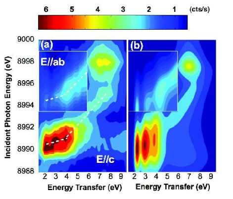
We note that the 5 eV feature discussed in Secs. III and IV was not considered in the above calculations since it is most prominent at high incident energies and shifts with . It therefore seems likely that this feature may actually be associated with a resonance of the 7.2 eV molecular orbital excitation at the well-screened state. Due to the doubleresonance denominator, each component may, in principle, show two separate resonances for and . For the final-energy resonance, as the incident energy changes, the peak position of the excitation shifts to so as to maintain the same final energy. In the twodimensional contour plot of Fig. 9, this manifests itself as a slope-1 streak of intensity.
The inverse lifetimes determine the shapes of the resonant spectral features. When , , and approach the value of excitation energy , the two resonances merge and become indistinguishable. If or is small compared to and , one of the two resonances nearly disappears, and the response either is either a circular region or a slope-one streak. By carefully choosing and () for each component, we we were able to emulate the main characteristics of the experimental data. As seen from Fig. 9 (a), the lowest three excitations are only strongly resonant at the well-screened state, while the 7.2 eV excitation is associated with the poorly-screened state. Consequently, should be different for the latter. For the slope-1 streak component, the incident resonance energy is the same as for the lower three excitations, but we find it necessary to choose a slightly smaller .
For out-of-plane polarization, we set = = 2 eV and = = 8990 eV for the lower three excitations. For the 7.2 eV component that results in the diagonal streak, we set = 1.7 eV, = 1.5 eV, = 8990 eV, and = 8989 eV, and for the other 7.2 eV component we chose = 1.0 eV, = 1.5 eV and = = 8998.5 eV. For in-plane polarization, we simply shifted all values by 4 eV, and adjusted the relative intensity between the three low-lying excitations and the two 7.2 eV components.
The above analysis has two important implications. One is that the “5 eV” feature is to be viewed as a shake-up excitation at 7.2 eV. The second important implication is that this local molecular-orbital excitation not only resonates at the poorly screened state at which copper has an open-shell configuration (), but also at the well-screened state (). This is different from findings for CuO and from the argument that the observed shake-up excitation requires the open-shell configuration and should be absent at the well-screened state Döring et al. (2004). However, we note that the molecular-orbital excitation was observed to be resonant at both well-screened and poorly-screened states in superconducting HgBa2CuO4+δ L. Lu, X. Zhao, G. Chabot-Couture, J. N. Hancock, N. Kaneko, O. P. Vajk, G. Yu, S. Grenier, Y. J. Kim, D. Casa, T. Gog, and M. Greven (2005).
We will now discuss a different interpretation for the slope-1 component of the streak of intensity that relates it to a resonant excitation into the continuum of unoccupied states.Gel’Mukhanov and gren (1998, 1999) Figure 2 shows the zone-center RIXS spectra together with the respective absorption spectra for both polarization condtions. The RIXS intensity is plotted versus final photon energy () instead of energy transfer (). We find that the spectra collapse to a peak, with an envelope of approximately Lorentzian shape, centered at about eV for in-plane polarization and eV for out-of-plane polarization. The peak position lies 4 eV below the photoabsorption threshold for both polarization conditions. This observation is consistent with the presence of slope-1 streaks in the contour plots for both polarization conditions: for in-plane polarization this streak starts at 8995 eV and for out-of-plane polarization it starts at 8991 eV.
The photon-absorption process near the Cu edge is comprised of transitions to either narrow molecular orbitals or continuum unoccupied states. For discrete levels, such as the well-screened and poorly screened states, induced valence excitations resonate and follow the Raman-Stokes law as crosses the discrete levels, i.e., the excitation energies do not vary with incident photon energy. On the other hand, a resonance due to transitions to continuum states behaves differently. Here, the resonance condition is fulfilled for every incident energy tuned to the continuum, and the subsequent emission is independent of once the incident energy increases above the lower edge of the continuum. When is below the edge, the resonant emission should also follow the Raman-Stokes behavior, but the spectral weight may be suppressed due to the small density of states below the edge. Our observation is consistent with the existence of a continuous unoccupied band of symmetry, with an edge at 8991 eV and 8995 eV for the respective polarizations, and a width of about 3 4 eV. This would support the view that a combination of an extended picture of itinerant electrons and of localized molecular orbitals is necessary to interpret the -edge absorption spectraTranquada et al. (1991) and the nature of the intermediate states in RIXS.
We note that the envelope of the in-plane data collapse shown in Fig. 2(a) is very similar in shape and position to the XAS corresponding to the well-screened state for out-of-plane polarization [Fig. 2(b)]. This leads us to a third interpretation of the slope-one contribution to the RIXS cross section shown in Fig. 9(a). Specifically, it suggests that part of the in-plane-polarized RIXS signal can be interpreted through the following complex dynamical process illustrated in Fig. 8: La2CuO4absorbs a photon of energy 8995 eV with polarization E, creating on a Cu site a well-screened core hole and an electron in a orbital [Fig. 8 (b)]. The electron then evolves into a state [Fig. 8 (c)] via a subsequent relaxation process. Finally, the 4pz electron recombines with the core hole, emitting a photon with energy 8991 eV. However, for out-of-plane polarization, electrons are already excited into a state, and this relaxation will not occur. Nevertheless, the envelope for out-of-plane polarization lies also 4 eV below that of the main edge. This interpretation therefore requires the existence of a discrete lower-energy state with energy 8987 eV for 4pz electron to relax into. Interestingly, in Ref. Abbamonte et al., 1999, a resonance of approximately this energy was observed with in-plane polarization. Further experimentation combined with theoretical modeling can be expected to resolve the origin of the slope-1 streak of intensity.
VI Summary
In summary, we have presented a detailed Cu -edge RIXS study of La2CuO4 in which we resolve a multiplet of charge-transfer excitations in the 1-7 eV range. We suggest several interpretations to explain the polarization-dependent spectra. A calculation applying third-order perturbation theory introduces a final-energy resonance and successfully simulates the main characteristics of the spectra for both polarization conditions. On the other hand, transitions to continuum bands that begin at the main absorption edge as well as relaxation are offered as alternative explanations to the fluorescence-like component in the contours. These proposals all emphasize the important role of the electrons in the RIXS cross section.
VII Acknowledgements
The authors gratefully acknowledge valuable discussions with P. Abbamonte, U. Bergmann, J. van den Brink, T. P. Devereaux, M. V. Klein, Y. J. Kim, K.-W. Lee, R. S. Markiewicz, W. E. Pickett, K. M. Shen, M. van Veenendaal, and F. C. Zhang. The work at Stanford University was supported by the DOE under Contract No. DE-AC02-76SF00515 and by the NSF under Grant No. 0405655. Work at the CMC-XOR Beamlines is supported in part by the Office of Basic Energy Sciences of the U.S. Dept. of Energy and by the National Science Foundation Division of Materials Research. Use of the Advanced Photon Source is supported by the Office of Basic Energy Sciences of the U.S. Department of Energy under Contract No. W-31-109-Eng-38
References
- Burns et al. (1999) C. A. Burns, P. Abbamonte, E. D. Isaacs, and P. M. Platzman, Phys. Rev. Lett. 83, 2390 (1999).
- Dallera et al. (2002) C. Dallera, M. Grioni, A. Shukla, G. Vanko, J. L. Sarrao, J. P. Rueff, and D. L. Cox, Phys. Rev. Lett. 88, 196403 (2002).
- P. Wernet, D. Nordlund, U. Bergmann, M. Cavalleri, M. Odelius, H. Ogasawara, L. A. Naslund, T. K. Hirsch, L. Ojamae, P. Glatzel, L. G. M. Pettersson, and A. Nilsson (2004) P. Wernet, D. Nordlund, U. Bergmann, M. Cavalleri, M. Odelius, H. Ogasawara, L. A. Naslund, T. K. Hirsch, L. Ojamae, P. Glatzel, L. G. M. Pettersson, and A. Nilsson, Science 304, 995 (2004).
- Sette et al. (1995) F. Sette, G. Ruocco, M. Krisch, U. Bergmann, C. Masciovecchio, V. Mazzacurati, G. Signorelli, and R. Verbeni, Phys. Rev. Lett. 75, 850 (1995).
- d’Astuto et al. (2002) M. d’Astuto, P. K. Mang, P. Giura, A. Shukla, P. Ghigna, A. Mirone, M. Braden, M. Greven, M. Krisch, and F. Sette, Phys. Rev. Lett. 88, 167002 (2002).
- Hill et al. (1998) J. P. Hill, C. C. Kao, W. A. L. Caliebe, M. Matsubara, A. Kotani, J. L. Peng, and R. L. Greene, Phys. Rev. Lett. 80, 4967 (1998).
- Abbamonte et al. (1999) P. Abbamonte, C. A. Burns, E. D. Isaacs, P. M. Platzman, L. L. Miller, S. W. Cheong, and M. V. Klein, Phys. Rev. Lett. 83, 860 (1999).
- Hämäläinen et al. (2000) K. Hämäläinen, J. P. Hill, S. Huotari, C. C. Kao, L. E. Berman, A. Kotani, T. Ide, J. L. Peng, and R. L. Greene, Phys. Rev. B 61, 1836 (2000).
- Hasan et al. (2000) M. Z. Hasan, E. D. Isaacs, Z. X. Shen, L. L. Miller, K. Tsutsui, T. Tohyama, and S. Maekawa, Science 288, 1811 (2000).
- Kim et al. (2002) Y. J. Kim, J. P. Hill, C. A. Burns, S. Wakimoto, R. J. Birgeneau, D. Casa, T. Gog, and C. T. Venkataraman, Phys. Rev. Lett. 89, 177003 (2002).
- Kim et al. (2004) Y. J. Kim, J. P. Hill, S. Komiya, Y. Ando, D. Casa, T. Gog, and C. T. Venkataraman, Phys. Rev. B 70, 094524 (2004).
- L. Lu, X. Zhao, G. Chabot-Couture, J. N. Hancock, N. Kaneko, O. P. Vajk, G. Yu, S. Grenier, Y. J. Kim, D. Casa, T. Gog, and M. Greven (2005) L. Lu, X. Zhao, G. Chabot-Couture, J. N. Hancock, N. Kaneko, O. P. Vajk, G. Yu, S. Grenier, Y. J. Kim, D. Casa, T. Gog, and M. Greven, Phys. Rev. Lett. 95, 217003 (2005).
- K. Ishii, K. Tsutsui, Y. Endoh, T. Tohyama, S. Maekawa, M. Hoesch, K. Kuzushita, M. Tsubota, T. Inami, J. Mizuki, Y. Murakami, and K. Yamada (2005) K. Ishii, K. Tsutsui, Y. Endoh, T. Tohyama, S. Maekawa, M. Hoesch, K. Kuzushita, M. Tsubota, T. Inami, J. Mizuki, Y. Murakami, and K. Yamada, Phys. Rev. Lett. 94, 207003 (2005).
- K. Ishii, K. Tsutsui, Y. Endoh, T. Tohyama, K. Kuzushita, T. Inami, K. Ohwada, S. Maekawa, T. Masui, S. Tajima, Y. Murakami, and J. Mizuki (2005) K. Ishii, K. Tsutsui, Y. Endoh, T. Tohyama, K. Kuzushita, T. Inami, K. Ohwada, S. Maekawa, T. Masui, S. Tajima, Y. Murakami, and J. Mizuki, Phys. Rev. Lett. 94, 919 (2005).
- K. Ishii, T. Inami, K. Ohwada, K. Kuzushita, J. Mizuki, Y. Murakami, S. Ishihara, Y. Endoh, S. Maekawa, K. Hirota, and Y. Moritomo (2004) K. Ishii, T. Inami, K. Ohwada, K. Kuzushita, J. Mizuki, Y. Murakami, S. Ishihara, Y. Endoh, S. Maekawa, K. Hirota, and Y. Moritomo, Phys. Rev. B 8, 224437 (2004).
- S. Grenier, J. P. Hill, V. Kiryukhin, W. Ku, Y. J. Kim, K. J. Thomas, S. W. Cheong, Y. Tokura, Y. Tomioka, D. Casa, and T. Gog (2005) S. Grenier, J. P. Hill, V. Kiryukhin, W. Ku, Y. J. Kim, K. J. Thomas, S. W. Cheong, Y. Tokura, Y. Tomioka, D. Casa, and T. Gog, Phys. Rev. Lett. 94, 047203 (2005).
- Kao et al. (1996) C. C. Kao, W. A. L. Caliebe, H. J. B, and J. M. Gillet, Phys. Rev. B 54, 16361 (1996).
- Krisch et al. (1995) M. H. Krisch, C. C. Kao, F. Sette, W. A. Caliebe, K. Hämäläinen, and J. B. Hastings, Phys. Rev. Lett. 74, 4931 (1995).
- Döring et al. (2004) G. Döring, C. Sternemann, A. Kaprolat, A. Mattila, K. Hämäläinen, and W. Schulke, Phys. Rev. B 70, 085115 (2004).
- Glatzel and Bergmann (2005a) P. Glatzel and U. Bergmann, Coord. Chem. Rev. 249, 65 (2005a).
- Tranquada et al. (1991) J. M. Tranquada, S. M. Heald, W. Kunnmann, A. R. Moodenbaugh, S. L. Qiu, Y. W. Xu, and P. K. Davies, Phys. Rev. B 44, 5176 (1991).
- Kotani and Shin (2001) A. Kotani and S. Shin, Rev. Mod. Phys. 73, 203 (2001).
- Collart et al. (2006) E. Collart, A. Shukla, J. P. Rueff, P. Leininger, H. Ishii, I. Jarrige, Y. Q. Cai, S. W. Cheong, and G. Dhalenne, Phys. Rev. Lett. 96, 157004 (2006).
- Birgeneau et al. (1999) R. J. Birgeneau, M. Greven, M. A. Kastner, Y. S. Lee, B. O. Wells, Y. Endoh, K. Yamada, and G. Shirane, Phys. Rev. B 59, 13788 (1999).
- Vajk et al. (2002) O. P. Vajk, P. K. Mang, M. Greven, P. M. Gehring, and J. W. Lynn, Science 295, 1691 (2002).
- Heald et al. (1988) S. M. Heald, J. M. Tranquada, A. R. Moodenbaugh, and Y. W. Xu, Phys. Rev. B 38, 761 (1988).
- Lee and Pickett (2006) K.-W. Lee and W. E. Pickett, Private Communication (2006).
- Kim (2006) Y. J. Kim, private communication (2006).
- Glatzel and Bergmann (2005b) P. Glatzel and U. Bergmann, Coord. Chem. Rev. 249, 65 (2005b).
- Mahan (2000) G. D. Mahan, Many-Particle Physics (Springer Verlag, New York, 2000).
- deGroot (2001) F. deGroot, Chem. Rev. 101, 1779 (2001).
- Nomura and Igarashi (2005) T. Nomura and J. Igarashi, Phys. Rev. B 71, 035110 (2005).
- Tsutsui et al. (2003) K. Tsutsui, T. Tohyama, and S. Maekawa, Phys. Rev. Lett. 91, 117001 (2003).
- Zhang and Rice (1988) F. C. Zhang and T. M. Rice, Phys. Rev. B 37, 3759 (1988).
- Zhang and Ng (1998) F. C. Zhang and K. K. Ng, Phys. Rev. B 58, 13520 (1998).
- Moskvin et al. (2002) A. S. Moskvin, R. Neudert, M. Knupfer, J. Fink, and R. Hayn, Phy. Rev. B 65, 180512 (2002).
- Tolentino et al. (1992) H. Tolentino, M. Medarde, A. Fontaine, F. Baudelet, E. Dartyge, D. Guay, and G. Tourillon, Phys. Rev. B 45, 008091 (1992).
- Wells et al. (1995) B. O. Wells, Z. X. Shen, A. Matsuura, D. M. King, M. A. Kastner, M. Greven, and R. J. Birgeneau, Phys. Rev. Lett. 74, 964 (1995).
- C. Dürr, S. Legner, R. Hayn, S. V. Borisenko, Z. Hu, A. Theresiak, M. Knupfer, M. S. Golden, J. Fink, F. Ronning, Z.-X. Shen, H. Eisaki, S. Uchida, C. Janowitz, R. Müller, R. L. Johnson, K. Rossnagel, L. Kipp, and G. Reichardt (20) C. Dürr, S. Legner, R. Hayn, S. V. Borisenko, Z. Hu, A. Theresiak, M. Knupfer, M. S. Golden, J. Fink, F. Ronning, Z.-X. Shen, H. Eisaki, S. Uchida, C. Janowitz, R. Müller, R. L. Johnson, K. Rossnagel, L. Kipp, and G. Reichardt, Phys. Rev. B 63, 014505 (2000).
- F. Ronning, C. Kim, K. M. Shen, N. P. Armitage, A. Damascelli, D. H. Lu, D. L. Feng, Z.-X. Shen, L. L. Miller, Y. J. Kim, F. Chou, and I. Terasaki (2003) F. Ronning, C. Kim, K. M. Shen, N. P. Armitage, A. Damascelli, D. H. Lu, D. L. Feng, Z.-X. Shen, L. L. Miller, Y. J. Kim, F. Chou, and I. Terasaki, Phys. Rev. B 67, 035113 (2003).
- Ahn et al. (2004) K. H. Ahn, A. J. Fedro, and M. van Veenendaal, cond-mat/0412635 (2004).
- Markiewicz and Bansil (2006) R. S. Markiewicz and A. Bansil, Phys. Rev. Lett. 96, 107005 (2006).
- Eskes and Sawatzky (1991) H. Eskes and G. Sawatzky, Phys. Rev. B 44, 9656 (1991).
- Platzman and Isaacs (1998) P. M. Platzman and E. D. Isaacs, Phys. Rev. B 57, 11107 (1998).
- Gel’Mukhanov and gren (1998) F. Gel’Mukhanov and H. gren, Phys. Rev. B 57, 2780 (1998).
- Gel’Mukhanov and gren (1999) F. Gel’Mukhanov and H. gren, Phys. Rep. 312, 87 (1999).