Chapter 0 19
Modeling molecular conduction in DNA wires: Charge transfer theories and dissipative quantum transport
Abstract
Measurements of electron transfer rates as well as of charge transport characteristics in DNA produced a number of seemingly contradictory results, ranging from insulating behaviour to the suggestion that DNA is an efficient medium for charge transport. Among other factors, environmental effects appear to play a crucial role in determining the effectivity of charge propagation along the double helix. This chapter gives an overview over charge transfer theories and their implication for addressing the interaction of a molecular conductor with a dissipative environment. Further, we focus on possible applications of these approaches for charge transport through DNA-based molecular wires.
1 Introduction
The discovery of long-range electron transfer processes in double stranded DNA [1] considerably attracted the attention of biologists, chemists and physicists. The motivation is threefold: i) the possible use of DNA-molecules in nanotechnology applications [2], ii) the biological role of electron transfer in, for example, radiation damage and repair, [3] and iii) the potentials of biochemical sensors based on electron transfer in DNA [8].
Despite the intensive experimental efforts, the results for electron transport still appear to be contradictory, ranging from metallic conduction [4, 5] to insulating behaviour with very large bandgaps [6, 7]. We refer the reader to Ref. [8] for a recent review. The measurements of electron transfer, on the other hand, appear to be much better controlled and earlier discrepancies on the distance dependence of the electron transfer rate are now attributed to the different experimental setups [3].
Theoretically, several classes of factors have been meanwhile identified, which considerably determine the effectivity of charge propagation along the double helix. They can be roughly classified as being related to (i) static disorder associated with the random or quasi-random sequence of bases in DNA oligomers [9, 10, 11], (ii) dynamical disorder arising from strong structural fluctuations of the molecular frame [12, 13, 14], and (iii) environmental effects related to the presence of an aqueous environment and counterions [15, 16, 17, 18, 19, 20, 21]. While the first two factors can still be addressed in a first approximation by considering only the atomic structure of isolated DNA oligomers, environmental effects require the consideration of the solvation shells and counterions and their interaction with the DNA molecules. Though the performance of ab initio approaches has considerably improved in the last years, the description of the dynamical interaction of DNA with an environment is still a formidable computational task involving at least several thousands of atoms. As a consequence, only relatively few first principle studies addressing this issue have been carried out in the past years [15, 16, 17, 18, 19, 20, 21]. Thus, model Hamiltonian approaches describing charge propagation in presence of a dissipative environment are very valuable and help to gain some insight into the subtleties of the physical behavior of a quantum mechanical system interacting with a macroscopic number of degrees of freedom.
This chapter will give an overview of different approaches to address charge propagation in a dissipative environment. In the next section, we discuss some results from ab initio calculations of DNA oligomers in presence of an aqueous environment. In section 3 some basic facts on how to model the interaction between an arbitrary quantum mechanical system in interaction with a dissipative environment are introduced. Finally, in subsection 3.1, a special application to a DNA model is discussed.

2 Environmental effects within ab-initio approaches
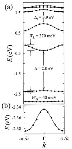
For the purpose of illustrating some basic facts concerning the electronic structure of a dried DNA oligomer, let us look at a recent band-structure calculation of poly(GC) carried out with the density-functional-based code SIESTA [23]. First-principle results for poly(AT) oligomers have also been recently presented [24, 25, 26]. The natural advantage of Poly(GC) or Poly(AT) is its periodic structure which considerably minimizes the computational efforts. In Fig. 1, the resulting band-structure is shown. From the practical point of view, it is also expected that this kind of periodic structures will have a higher potential applicability in molecular electronics than their disordered counterparts like -DNA.
The top-most valence band (HOMO) and the lowest conduction band (LUMO), both having -character, are separated by a bandgap of eV. The HOMO and LUMO bands are basically derived from the overlap of guanine and cytosine orbitals, respectively. As a consequence, the charge density of HOMO and LUMO bands is confined along the G- and C-strands, respectively. The Fermi level lies between these two bands so that the system appears to be a insulator.
Most striking are the very small bandwidths of the bands close to the Fermi level. The top-most valence band has a width of meV while the lowest conduction band has a somewhat broader bandwidth of meV. Part (b) of Fig. 2 of Ref. [23] (see Fig. 1, right panel, in this chapter) shows a tight-binding modeling of the top-most valence band using a single orbital per base pair. The resulting hopping matrix elements are meV (for nearest neighbor hopping) and meV (for next-nearest neighbor hopping).
In principle, such a scenario allows for electronic transport mediated by carriers which are introduced by doping either in the top of the valence band or the bottom of the conduction band. The extremely small bandwidths, however, suggest that Bloch states mediated transport cannot be stable under the various perturbations present in DNA [23].
One of the possible perturbations which have been studied in some detail is the role of the environment [15, 16, 17, 18, 19]. As shown in Ref. [15], the existence of rather different time scales of the environment may have a strong impact on a charge propagating along the DNA molecule. First-principle simulations were performed, including four base pairs of B-DNA in the sequence GAGG, together with Na+ counterions and the hydration shell. It turns out that holes can be gated by the temperature dependent dynamics of the environment, i. e. there may exist configurations that localize the hole. Dynamical fluctuations of the counterions can lead, however, to configurations which support hole motion. A hole thus experiences transitions between quantum-mechanical states that are correlated with different environmental configurations [27]. These results have been partly confirmed by recent ab initio simulations in Ref. [19]. The authors have additionally pointed out at a different, proton-mediated mechanism for hole localization, which may be quite effective in Poly(GC) DNA.
The ab initio-based studies in Refs. [16, 17, 18] have yielded further insight into the role played by water and counterions in modifying the low-energy electronic structure of DNA oligomers. Despite the differences in the DNA-conformations (Z- [16] vs. B-DNA [17, 18]) as well as in computational approaches (different basis sets and approximations for the exchange-correlation potentials), they nevertheless indicate that the environment can introduce midgap states. Though these electronic states do not form truly extended electronic bands, they may support activated charge hopping at high-temperatures and thus lead to an enhancement of the conductivity. In this respect, they resemble to some degree the defect levels induced by impurities in bulk semiconductors.
We can conclude from this that (i) the appearance of a band-gap is not at all a generic feature for the band-structure of DNA and (ii) the extremely small values of the bandwidths do appear to be generic. The general question which arises from that is the relation of the bandwidths and to other typical energy scales due to disorder effects, electron-phonon coupling, and Coulomb-correlations. Furthermore, the environment can have a dramatic influence on the electronic structure of the oligomer by inducing defect-like states within the gap. However, as previously stated, the complexity of the problem makes a full ab initio treatment rather difficult. This leads us to the issue of how the system-environment interaction can be modelled within a Hamiltonian model approach. Which are the essential ingredients that have to be taken into account?
3 Modeling the system-environment interaction
The importance of the system-environment interaction has long been recognized in biomolecules (such as proteins), in which electron transfer reactions take place. Within Marcus theory [28], the coupling of the electronic degrees of freedom to a reaction coordinate is the first step of a successful description of electron transfer processes. The quantum mechanical analog of the reaction coordinate is a phononic degree of freedom originating from vibrations of the protein matrix. In general, there might not be one dominating phononic mode; such a breakdown of the standard single reaction coordinate description has been suggested in the context of charge transfer between DNA base pairs [29]. More importantly, even a dominating reaction coordinate is coupled to the fluctuations of the environment, such as surrounding water molecules, so that the resulting spectral function of all relevant phononic modes can be regarded as continuous over a very broad energy range (in theoretical calculations, the low-energy cutoff is typically set to ).
The coupling of the electronic subsystem (the electron transfered between donor and acceptor site) to the environment leads to a very important effect: when an electron initially localized at the donor site tunnels to the acceptor site (which typically has a lower energy), the energy difference is dissipated to the environment so that the electron transfer process is irreversible [30]. If this friction would be too small, or if the electron would couple to a single phonon mode only, the electron would oscillate between donor and acceptor sites making the electron transfer process highly inefficient.
In the work of Garg et al. [30], the friction term has been modeled quantum mechanically via a coupling to a bath of harmonic oscillators. A minimal model for electron transfer processes, similar to the one proposed in [30], then takes the form:
| (1) | |||||
The operators denote annihilation (creation) operators for electrons on the donor () and acceptor () sites; is defined as . The first two terms of the Hamiltonian eq. (1) correspond to a two-site tight-binding Hamiltonian with the on-site energies and the hopping matrix element.
The last two terms in eq. (1) describe the free bosonic bath (with bosonic creation and annihilation operators and ) and the coupling between electrons and bosons, respectively. Assuming symmetric phonon displacements due to the electronic occupancy at donor and acceptor sites, one can set and [31].
The coupling of the electrons to the bath degrees of freedom is completely specified by the bath spectral function . The form of can, in principle, be calculated with molecular dynamics simulations (see, for example, [32]). To study the qualitative influence of the environment, an ohmic bath spectral function (with a suitable high-energy cut-off) is sufficient for most cases. In this description, dominant reaction coordinates lead to additional resonances in the bath spectral function.
Note that such a continuous bath spectral function enforces a quantum mechanical treatment of the phononic degrees of freedom since the temperature range always lies within the continuum of phononic modes.
The Hamiltonian eq. (1) can be viewed as a paradigm for modeling the system-environment interaction in biomolecules in which the electronic degrees of freedom couple to a dissipative environment. We should add here that the model eq. (1) can be exactly mapped onto the well-studied spin-boson model [33, 34] for the case of one electron in the system. In the spin-boson model description, the state () corresponds to the electron localized at the donor (acceptor) site. More complicated situations arise when the spin degree of freedom of the electron — not to be confused with the artificial spin in the spin-boson model — is taken into account, see the discussion in Ref. [[35]].
Calculations for these types of models in the context of electron transfer problems have been presented in [30, 35, 36]. It is natural to assume that the electron environment interaction plays an equally important role for electron transport through the DNA double helix. The main difference here is that electron transfer/transport occurs over very many sites so that the two-site model eq. (1) has to be suitably generalized. One such example is discussed in the following section.
1 Modeling the system-environment interaction: a DNA-wire in a dissipative bath
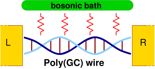
The first-principle calculations reviewed in Section 1 have shown that the environment in which DNA oligomers are placed may have a non-negligible influence on their electronic structure. In this section, we will illustrate within an effective model Hamiltonian approach, how the presence of a dissipative environment does affect the low-energy transport properties of a DNA molecular wire [37, 38]. Our reference system will be poly(GC) because of its periodic structure, which should make optimal the interbase electronic coupling along the strands. Moreover, recent experiments [39] on single poly(GC) molecules have shown non-zero current at low bias, which is at variance with the fact that the molecule should have a (rather large) HOMO-LUMO gap [40]. Since these experiments were performed in an aqueous environment and the authors excluded ionic current contributions, one may consider the possibility that the environment is modifying the molecule electronic structure.
In our model, we will exclusively focus on the low-energy transport, i. e. the charge injection energies are small compared with the molecular band gap of the isolated molecule (). Consequently, only equilibrium transport will be considered and a transmission-like function can still be defined [37, 38, imry04]. At low energies, only the frontier orbitals (HOMO and LUMO) of the molecule are expected to contribute to transport. As mentioned in sec. 1 these orbitals have -character and their charge densities extend along the G- and C-strands for the HOMO and the LUMO, respectively. Motivated by this, we have formulated a minimal tight-binding model [37, 38, 41], where a single electronic -orbital channel is connected to left and right electrodes. The sugar-phosphate backbones are assumed to locally perturb the electronic states and lead to the opening of a semiconducting gap for the infinite chain. As a result, the size of the band gap can be controlled by the strength of this perturbation, given by the parameter in Eq. (2) below. The environment is described by a collection of harmonic oscillators which linearly couple to the charge density on the backbone sites. Assuming zero onsite energies (the Fermi level thus lies at ), the Hamiltonian reads:
| (2) | |||||
In the above equation, is the Hamiltonian of the HOMO (or LUMO) channel () and the backbone sites (), contains both the Hamiltonian of the bath and the mutual interaction of the bath with the electronic degrees of freedom at the backbone sites (second row). Finally, contains the electrode Hamiltonians as well as the tunneling Hamiltonian describing the propagation of a charge from the leads onto the HOMO (or LUMO) channel and viceversa. In absence of coupling to the bath, the eigenstates of yield two manifolds containing states each and separated by a band gap, whose magnitude basically depends on the size of the tranversal coupling . The bath is completely described by introducing its spectral density as given by [34]: where is a high-frequency cut-off and we assume ohmic dissipation, . By performing a unitary transformation, the linear coupling to the bath can be eliminated. However, the transversal coupling terms will be renormalized by exponential bosonic operators [37, 38]. Using equation of motion techniques, one can show, to lowest order in , that the Green function of the wire satisfies the following Dyson-equation:
| (3) |
In this expression, the influence of the electrodes is captured by the complex self-energy functions . The function is an entangled electron-boson Green function: and . Note that acts as an additional self-energy and that the influence of the backbones is contained only in this function.
Several coupling regimes to the bath can be analyzed [38]. We focus here only on the strong-coupling limit (SCL), defined by the condition , which basically means that the time scales of the charge-bath interaction are much shorter compared with typical electronic time scales. We refer the reader to Refs. [37, 38] for technical details.
|
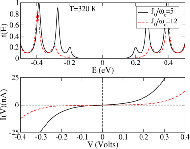
The impact of the bath on the electronic structure is twofold [37, 38]. On one side, the strong coupling to the bath leads to the emergence of new bath-induced electronic states inside the wire band gap. On the other side, however, these states are strongly damped by the dissipative action of the bath. In other words, the bath completely destroys the coherence of transport through the wire. This effect has also been discussed for transport through molecular chains under the influence of external time-dependent fields, see [42, 43]
As a result, the bath-induced states will not manifest as resonances in the transmission spectrum, see Fig. 3, left panel. Nevertheless, they induce a temperature dependent background which leads to a (small) finite density of states inside the gap. Charges injected at low energies will now find states supporting transport at high temperatures and thus, a finite current at low bias may flow. Hence we call the new gap a pseudogap, in contrast to the intrinsic band gap found in the isolated wire. Note that increasing the interaction with the bath (increasing ) does not necessarily lead to a global increase of the current, since the frontier orbitals of the wire are strongly damped with increasing coupling.
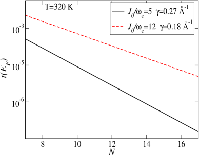
Signatures of this situation are seen in the length dependence of the transmission at the Fermi energy, see Fig. 3, right panel. Tunneling through an intrinsic gap would lead to a very strong exponential length dependence with with typical inverse decay lengths [40]. We find, however, much smaller values . With increasing bath coupling the exponential dependence weakens, reflecting the increase of the density of states in the pseudogap and the strong contribution of incoherent processes [44]
4 Conclusions and Outlook
In conclusion, we have shown that generic model approaches can deal with experimentally relevant situations. One of the major problems for the theoretical modeling of charge transport in DNA oligomers is the lack of a clear experimental picture of their transport signatures. Thus, focusing on individual factors affecting charge propagation helps to shed light onto the relevant mechanisms controlling the charge dynamics in DNA. We have addressed in this chapter environmental effects in the charge transport through DNA oligomers within a minimal Hamiltonian model approach. Obviously, other factors not treated here, like electronic correlations, static disorder or internal vibrational excitations can also have a non-negligible influence on charge propagation.
5 Acknowledgments
The authors thank A. Nitzan for useful comments and suggestions. This research was supported by the DFG through SFB 484, by the Volkswagen Foundation Grant Nr. I/78-340 and by the EU under contract IST-2001-38951.
References
- [1] C. J. Murphy, M. R. Arkin, Y. Jenkins, N. D. Ghatlia, S. H. Bossmann, N. J. Turro, and J. K. Barton, Science 262, 1025 (1993).
- [2] C. Dekker and M. Ratner, Physics World (August 2001).
- [3] E. M. Boon and J. K. Barton, Curr. Op. in Struct. Biol. 12, 320 (2002).
- [4] H.-W. Fink and C. Schönenberger, Nature 398, 407 (1999).
- [5] A. Y. Kasumov, M. Kociak, S. Gu ron, B. Reulet, V. T. Volkov, D. V. Klinov, and H. Bouchiat, Science 291, 280 (2001).
- [6] D. Porath, A. Bezryadin, S. D. Vries, and C. Dekker, Nature 403, 635 (2000).
- [7] A. J. Storm, J. V. Noort, S. D. Vries, and C. Dekker, Appl. Phys. Lett. 79, 3881 (2001).
- [8] D. Porath, G. Cuniberti, and R. Di Felice, Topics in Current Chemistry 237, 183 (2004).
- [9] S. Roche, D. Bicout, E. Macia, and E. Kats, Phys. Rev. Lett. 91, 228101 (2003).
- [10] S. Roche, Phys. Rev. Lett. 91, 108101 (2003).
- [11] D. Klotsa, R. A. Röemer, and M. S. Turner, q-bio.GN/0504004 (2005).
- [12] F. Palmero, J. F. R. Archilla, D. Hennig, and F. R. Romero, New J. Phys. 6, 13 (2004).
- [13] S. Komineas, G. Kalosakas, and A. R. Bishop, Phys. Rev. E 65, 061905 (2002).
- [14] F. C. Grozema, L. D. A. Siebbeles, Y. A. Berlin, and M. A. Ratner, ChemPhysChem 6, 536 (2002).
- [15] R. N. Barnett, C. L. Cleveland, A. Joy, U. Landman, and G. B. Schuster, Science 294, 567 (2001).
- [16] F. L. Gervasio, P. Carolini, and M. Parrinello, Phys. Rev. Lett. 89, 108102 (2002).
- [17] R. G. Endres, D. L. Cox, and R. R. P. Singh, Rev. Mod. Phys. 76, 195 (2004).
- [18] A. Hübsch, R. G. Endres, D. L. Cox, and R. R. P. Singh, Phys. Rev. Lett. 94, 178102 (2005).
- [19] F. L. Gervasio, A. Laio, M. Parrinello, and M. Boero, Phys. Rev. Lett. 94, 158103 (2005).
- [20] Ch. Adessi, S. Walch, and M. P. Anantram, Phys. Rev. B 67, 081405(R) (2003).
- [21] H. Mehrez and M. P. Anantram, Phys. Rev. B 71, 115405 (2005).
-
[22]
http://www.accessexcellence.org/RC/VL/GG/
http://www.genome.gov/ - [23] E. Artacho, M. Machado, D. Sanchez-Portal, P. Ordejon, and J. M. Soler, Mol. Phys. 101, 1587 (2003).
- [24] E.B. Starikov, PhysChemChemPhys, 4, 4523 (2002).
- [25] J. P. Lewis, J. Picus, Th. E. Cheatham III, E. B. Starikov, H. Wang, J. Tomfohr, and O.F. Sankey, Phys. Stat. Solidi B 223, 90 (2002).
- [26] J. P. Lewis, Th. E. Cheatham III, E. B. Starikov, O. F. Sankey, J. Phys. Chem. B107, 2581 (2003).
- [27] We note that a similar situation is known in electron transfer theories, where solvent reorganization brings into resonance the electronic states of donor and acceptor centers.
- [28] R. A. Marcus, J. Chem. Phys. 24, 966 (1956); Rev. Mod. Phys. 65, 599 (1999).
- [29] R. Bruinsma, G. Gruener, M. R. D’Orsogna, and J. Rudnick, Phys. Rev. Lett. 85, 4393 (2000).
- [30] A. Garg, J. N. Onuchic, and V. Ambegaokar, J. Chem. Phys. 83, 4491 (1985).
- [31] V. May and O. Kühn, Charge and Energy Transfer Dynamics in Molecular Systems (WILEY-VCH, Weinheim, 2004).
- [32] D. Xu and K. Schulten, Chem. Phys. 182, 91 (1994).
- [33] A. J. Leggett, S. Chakravarty, A. T. Dorsey, M. P. A. Fisher, A. Garg, and W. Zwerger, Rev. Mod. Phys. 59, 1 (1987).
- [34] U. Weiss, Quantum Dissipative Systems, Vol. 10 of Series in Modern Condensed Matter Physics (World Scientific, 1999).
- [35] S. Tornow, N.-H. Tong, and R. Bulla, Europhys. Lett. 73, 913 (2006).
- [36] L. Mühlbacher and R. Egger, Chem. Phys. 296, 193 (2004).
- [37] R. Gutierrez, S. Mandal, and G. Cuniberti, Nano Letters 5, 1093 (2005).
- [38] R. Gutierrez, S. Mandal, and G. Cuniberti, Phys. Rev. B 71, 235116 (2005).
- [39] B. Xu, P. Zhang, X. Li, and N. Tao, Nano letters 4, 1105 (2004).
- [40] H. Wang, J. P. Lewis, and O. Sankey, Phys. Rev. Lett. 93, 016401 (2004).
- [41] G. Cuniberti, L. Craco, D. Porath, and C. Dekker, Phys. Rev. B 65, 241314(R) (2002).
- [42] J. Lehmann, S. Kohler, V. May, and P. Hänggi, J. Chem. Phys. 121, 2278 (2004).
- [43] S. Kohler, J. Lehmann, and P. Hänggi, Phys. Rep. 406, 397 (2005).
- [44] D. Segal, A. Nitzan, W. B. Davis, and M.A. Ratner, J. Phys. Chem., 104, 2790 (2000); D. Segal and A. Nitzan, Chem. Phys. 281, 235 (2002).