Fermi surface and quasiparticle excitations of Sr2RhO4
Abstract
The electronic structure of the layered 4 transition metal oxide Sr2RhO4 is investigated by angle resolved photoemission. We find well-defined quasiparticle excitations with a highly anisotropic dispersion, suggesting a quasi-two-dimensional Fermi liquid like ground state. Markedly different from the isostructural Sr2RuO4, only two bands with dominant Rh 4 character contribute to the Fermi surface. A quantitative analysis of the photoemission quasiparticle band structure is in excellent agreement with bulk data. In contrast, it is found that state-of-the-art density functional calculations in the local density approximation differ significantly from the experimental findings.
pacs:
71.18.+y, 71.20.-b, 79.60.-iThe Fermi surface (FS) topology and quasiparticle dynamics determine most material properties.
Low–dimensional and correlated materials, which are currently of key interest for their exotic properties, are particularly sensitive to fine details of the fermiology.
This is evident for classical charge density wave systems, but might hold as well for superconductivity or quantum critical phenomena.
For instance calculations for a large number of -type cuprates, demonstrated a correlation of Tc with the shape of the most bonding band E. Pavarini et al. (2001). More recently, quantum criticality in Sr3Ru2O7 has been related to a symmetry breaking spin–dependent FS distortion S.A. Grigera et al. (2004).
In principle, angle resolved photoemission (ARPES) is ideally suited to map the size and shape of the FS P. Aebi et al. (1994); A. Damascelli et al. (2003). However, ARPES is a highly surface sensitive technique and its precision is usually lower than that of classical FS probes based on the de Haas–van Alphen (dHvA) or related effects. Consequently, the impact of ARPES has been largest in materials where dHvA oscillations are not observable A. Damascelli et al. (2003).
Density functional (DFT) calculations in the local density approximation (LDA) have evolved as a powerful alternative to experimental electronic structure probes.
However, the Kohn-Sham eigenvalues of DFT have no clear physical meaning even at the Fermi surface and cannot be rigorously identified with single particle excitation energies R.O. Jones and O. Gunnarson (1989). Nonetheless, the LDA has been found to be highly successful even in the description of fairly strongly correlated materials like doped cuprates.
Although the agreement of LDA calculations with extensive ARPES data on cuprates is compelling, it has rarely been confirmed by bulk electronic structure probes, and truly quantitative comparisons are not ready yet.
Sr2RuO4 is to date the only example of a correlated oxide where LDA T. Oguchi (1995); D.J. Singh (1995) dHvA A.P. Mackenzie et al. (1996); C. Bergemann et al. (2000) and ARPES A. Damascelli et al. (2000) were found to be in good quantitative agreement.
This is far from trivial in a multi–band system, since
correlations, not fully described within the LDA, can transfer spectral weight between in equivalent orbitals, thus enlarging certain FS pockets at the expense of others A. Liebsch and A. Lichtenstein (2000); H. Ishida et al. (2005).
NaxCoO2 is a prominent recent example of a multi–band system where qualitative differences between ARPES data M.Z. Hasan et al. (2004); H.-B. Yang et al. (2005) and LDA calculations M.D. Johannes et al. (2004) have been observed and related to strong, orbital dependent correlations. However, the complexity of the material has so far prevented the derivation of a consistent picture H. Ishida et al. (2005); S. Zhou et al. (2005).
In this paper, we present a quantitative electronic structure study of Sr2RhO4 by means of ARPES and band calculations within the LDA. It is shown that ARPES provides bulk representative spectra with a FS that agrees with dHvA data by Perry et al. R. Perry et al. . Although Sr2RhO4 exhibits Fermi–liquid properties over an extended energy range, we find that its FS is not reproduced quantitatively within the LDA.
Sr2RhO4 has a tetragonal crystal structure ( Å, Å) with a reduced symmetry (”orthorhombicity”) due to a 11∘ rotation of the RhO6 octahedra around the c-axis T. Shimura et al. (1992); T. Vogt and D.J. Buttrey (1996). High purity single crystals with residual resistivities cm have been grown by a floating zone technique R. Perry et al. and have been cleaved in situ along the –plane at K. Photoemission experiments were performed with a monochromatized He–discharge lamp (Gammadata VUV5000) and a Scienta SES2002 analyzer. The energy and angular resolutions for all measurements were better than 7.5 meV / [full width at half maximum (FWHM)]. All data were taken at K and a pressure torr. LDA calculations were done using the local orbital extension of the general potential linearized augmented planewave method D.J. Singh and L. Nordstrom (2006) with well converged basis sets ( basis functions) and zone samplings.
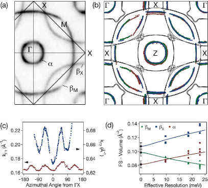
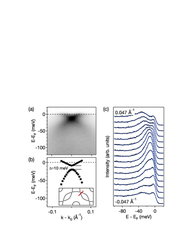
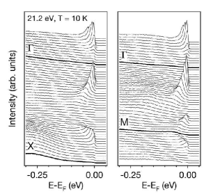
Representative spectra along X and M of the orthorhombic Brillouin zone (BZ) are shown in Fig. 1. The data clearly show the spectroscopic hall marks of a Landau Fermi liquid: well defined, dispersive quasiparticle bands with peaks that sharpen up progressively as the they approach the Fermi level (), reflecting the diminishing phase space for electron–electron scattering Claessen et al. (1992). Fig. 2(a) shows the experimental FS map, obtained from high–resolution spectra, taken on a uniform k–space grid and integrated over the energy window meV. Note that the resulting map has not been symmetrized. Only the measured momentum–space region is shown. Similar data sets were measured on several samples with photon energies of 21.2 eV (He I) and 40.8 eV (He II) and showed good reproducibility and no excitation–energy dependence, consistent with a highly two–dimensional (2D) electronic structure. Two, nearly isotropic bands, which are both centered at the origin and back–folded by the orthorhombicity are observed, a large electron like band with an average Fermi wave vector of 0.66 Å-1 and a smaller hole–pocket with Å-1. A quantitative determination of the unfolded Fermi wave vectors (Fig. 2(c)) shows a slight 4–fold anisotropy, analogous to the shape of the sheets in Sr2RuO4, and indicative of a dominant character of the FS. The LDA calculation (Fig. 2(b)) confirms this experimental assignment and shows that the level is pushed below by level mixing and repulsion between the and the orbitals, as discussed in Ref. B.J. Kim et al. . At the orthorhombic zone boundary, a small non–crossing gap opens. This is investigated in more detail in Fig. 3 showing the band dispersion across the zone boundary. The gap is not directly resolved in the raw data, but the flattened peak shape of the energy distribution curves (EDCs) shown in Fig. 3 (c) hints at the presence of a non-crossing gap slightly smaller than the line width of about 20 meV at the energy where the two branches intersect. For a more quantitative analysis, we fit the data with the spectral function for a 2D Fermi liquid superimposed on a smooth background. To increase the reliablility of this analysis, we choose to fit all EDCs simultaneously with a common self–energy and a momentum independent intensity and convolved the fit–function in energy and momentum with the independently determined respective resolutions. The resulting band positions are shown in Fig. 3(b) to display a gap of meV. This is significantly larger than the Landau level splitting even in high magnetic fields. Thus, in a 2D approximation, the FS contains three closed contours, a central hole pocket (), the lens–shaped electron pockets at M (), and the square–shaped hole pockets at X ().
| FS–volume (% BZ) | 6.1(4) | 7.4(4) | 8.1(5) |
|---|---|---|---|
| occupation () | 3.878(8) | 0.296(16) | 1.838(10) |
| Fermi velocity (eVÅ) | 0.41(4) | 0.61(6) | 0.55(6) |
| cyclotron mass () | 3.0(3) | 2.2(2) | 2.6(3) |
| dHvA FS–vol. (% BZ) | 6.6 – 9.2 | ||
| LDA FS–vol. (% BZ) |
We have determined the volume of these pockets from extensive fits to multiple data sets to be 6.1(4), 7.4(4), 8.1(5)% BZ for , and , respectively, as summarized in Tab. 1. Each of these values represents the average of 8–10 equivalent pockets, measured on different samples and with different photon energies and energy resolutions, and contains a correction for the systematic shifts in the zero frequency MDC peak positions (see Fig. 2(d)) caused by the finite energy resolution of the experiment 111Details of the data-analysis will be published elsewhere.. The such derived values are in excellent agreement with the frequency range of 6.6 to 9.2% BZ, in which dHvA oscillations have been observed very recently R. Perry et al. . In order to estimate the Luttinger volume, we assume two–dimensionality. After backfolding the fundamental bands to the orthorhombic Brillouin zone, which contains two Rh atoms per plane, we count electrons for the hole–pocket at , in the lens–shaped electron pocket, and for the X–point hole pocket. The three pockets thus contain 3.006(10) electrons per Rh, consistent with a stoichiometric material and a fully occupied –band.
Carrier masses have been determined using Fermi velocities evaluated in typically 100 intensity distributions along –space lines normal to the FS contour of each pocket. The averaged values are given in Tab. 1. The cyclotron masses are then calculated for average Fermi wave vectors to be , , and . Again assuming two dimensionality, the specific heat is given by , where is the Boltzmann constant, Avogadro’s number, and the in–plane lattice constant. Accounting for the twofold degeneracy of the –pocket, we find , and mJ/molK2, in fair agreement with the preliminary experimental report of 17.7(7) mJ/molK2 R. Perry et al. .
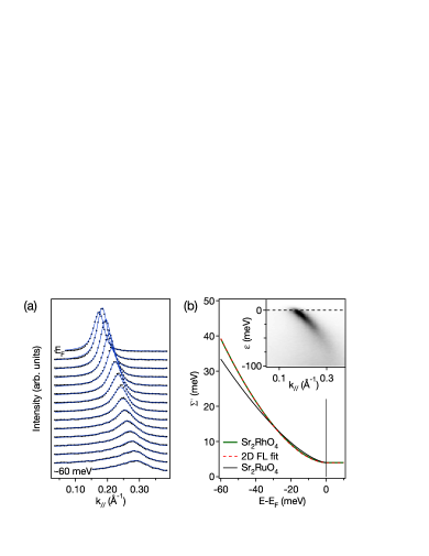
The QP self–energy has been derived from a detailed line–shape analysis summarized in Fig. 4. We first note that there exists no simple relation between the width of a single MDC (or EDC) and the self–energy for sharp and non–linear QP–bands as observed in Sr2RhO4, even for high–resolution data as those presented here. Therefore, we chose to perform 2D fits with a parametrized self–energy , as well as simultaneous 1D fits of all MDCs. Both of these methods are fully self–consistent and allow one to include a convolution with both, the energy and momentum resolution functions. The results of a typical 2D fit are shown in Fig. 4(c) and demonstrate that widths, asymmetries and intensities of the MDCs are well reproduced with a minimal parameter set. The imaginary part of the self–energy deduced in this way is , in close agreement with the analytical form for a 2D Fermi liquid with realistic parameter values and eV (see Fig. 4(b)) Hodges et al. (1971). Moreover, it is nearly identical with the recent result for Sr2RuO4 N.J.C. Ingle et al. (2005), hinting at comparable many–body interactions in both materials, dominated by electron–electron interactions. The strength of electron–phonon interactions cannot be determined reliably from the present data, since the coherent QP peaks can only be separated over a limited energy range barely larger than typical phonon frequencies. However, the quality of the above fit with a smooth QP–dispersion up to meV and a form of expected for electron–electron interactions only seems to indicate a minor importance of other degrees of freedom.
The correct volume counting of the expected number of electrons, the agreement with dHvA data, and the sheer observation of single, sharp QP peaks show that the 2-3 topmost unit cells of Sr2RhO4 which are probed by ARPES have a uniform and basically converged bulk electronic structure (for an example of ARPES data from a reconstructed surface, see e.g. Ref. K.M. Shen et al. (2001)). We therefore use the zero frequency line width of meV (corresponding to Å-1, % BZ) as an upper bound for possible systematic errors in the ARPES values caused by surface structural relaxations. This uncertainty is far smaller than the difference between ARPES and LDA Fermi wave vectors. The presented results thus establish for the first time a quantitative discrepancy between the experimental quasiparticle FS and the DFT–LDA FS in a Fermi–liquid–like correlated material. The two main discrepancies between calculation and experiment are the shape of the pocket and the volume ratios between the three main pockets, with LDA finding values of % BZ, % BZ, and % BZ, clearly incompatible with both, ARPES and dHvA. The disagreement could perhaps be explained by a deviation of the crystal structure from that assumed in the calculations or by an O deficiency. However, there is no experimental evidence for either. The Luttinger volume of 3.006(10) indicates good stoichiometry, and calculations performed with the experimental lattice structure from Ref. T. Vogt and D.J. Buttrey (1996), artificially distorted structures, and the fully relaxed LDA crystal structure all revealed rather similar LDA–FS that differ significantly from the experiment. We also searched for symmetry reducing rotations of the RhO6 octahedra, but found that only the reported symmetry T. Shimura et al. (1992); T. Vogt and D.J. Buttrey (1996) without out of plane tilt–distortions is stable within LDA.
It is thus compelling to attribute the observed discrepancy between LDA and ARPES to many–body interactions, not fully described within the LDA. The functional form of shown in Fig. 4. indicates that these interactions are dominated by electron–electron scattering, although it cannot fully exclude a more complex interplay of correlations and electron–phonon coupling, as it is e.g. observed in the metallic 4 compound Ca3Ru2O7 F. Baumberger et al. . The structural distortions in Sr2RhO4 may well be crucial for the marked difference in experimental and LDA FS since they reduce the band width and Fermi velocity by nearly a factor of two, compared to Sr2RuO4. Consequently, for Sr2RhO4, a band dependence of the real part of the self–energy at of meV as it was calculated by Liebsch and Lichtenstein for Sr2RuO4 A. Liebsch and A. Lichtenstein (2000) would be sufficient to explain the discrepancy between ARPES and LDA FS reported here.
Acknowledgements.
We thank B.J. Kim and C.Y. Kim for discussion and for provision of Ref. B.J. Kim et al. prior to publication. This work has been supported by the ONR grant N00014-01-1-0048. Additional support from SSRL is provided by the DOE’s office of Basic Energy Science, Division of Material Science with Contract DE-FG03-OIER45929-A001.References
- E. Pavarini et al. (2001) E. Pavarini et al., Phys. Rev. Lett. 87, 047003 (2001).
- S.A. Grigera et al. (2004) S.A. Grigera et al., Science 306, 1154 (2004).
- P. Aebi et al. (1994) P. Aebi et al., Phys. Rev. Lett. 72, 2757 (1994).
- A. Damascelli et al. (2003) A. Damascelli, Z. Hussain, and Z.-X. Shen, Rev. Mod. Phys. 75, 473 (2003).
- R.O. Jones and O. Gunnarson (1989) R.O. Jones and O. Gunnarson, Rev. Mod. Phys. 61, 689 (1989).
- T. Oguchi (1995) T. Oguchi, Phys. Rev. B 51, 1385(R) (1995).
- D.J. Singh (1995) D.J. Singh, Phys. Rev. B 52, 1358 (1995).
- A.P. Mackenzie et al. (1996) A.P. Mackenzie et al., Phys. Rev. Lett. 76, 3786 (1996).
- C. Bergemann et al. (2000) C. Bergemann et al., Phys. Rev. Lett. 84, 2662 (2000).
- A. Damascelli et al. (2000) A. Damascelli et al., Phys. Rev. Lett. 85, 5194 (2000).
- H. Ishida et al. (2005) H. Ishida, M.D. Johannes, and A. Liebsch, Phys. Rev. Lett. 94, 196401 (2005).
- A. Liebsch and A. Lichtenstein (2000) A. Liebsch and A. Lichtenstein, Phys. Rev. Lett. 84, 1591 (2000).
- M.Z. Hasan et al. (2004) M.Z. Hasan et al., Phys. Rev. Lett. 92, 246402 (2004).
- H.-B. Yang et al. (2005) H.-B. Yang et al., Phys. Rev. Lett. 95, 146401 (2005).
- M.D. Johannes et al. (2004) M.D. Johannes, I.I. Mazin, D.J. Singh, and D.A. Papaconstantopoulos, Phys. Rev. Lett. 93, 097005 (2004).
- S. Zhou et al. (2005) S. Zhou et al., Phys. Rev. Lett. 94, 206401 (2005).
- (17) R. Perry et al., in preparation, 2006.
- T. Shimura et al. (1992) T. Shimura, M. Itoh, and T. Nakamura, J. Solid State Chem. 98, 198 (1992).
- T. Vogt and D.J. Buttrey (1996) T. Vogt and D.J. Buttrey, J. Solid State Chem. 123, 186 (1996).
- D.J. Singh and L. Nordstrom (2006) D.J. Singh and L. Nordstrom, Planewaves, Pseudopotentials and the LAPW Method, 2nd Edition (Springer, Berlin, 2006).
- Claessen et al. (1992) R. Claessen et al., Phys. Rev. Lett. 69, 808 (1992).
- (22) B.J. Kim et al., submitted, 2006.
- N.J.C. Ingle et al. (2005) N.J.C. Ingle et al., Phys. Rev. B 72, 205114 (2005).
- Hodges et al. (1971) C. Hodges, H. Smith, and J.W. Wilkins, Phys. Rev. B 4, 302 (1971).
- K.M. Shen et al. (2001) K.M. Shen et al., Phys. Rev. B 64, R180502 (2001).
- (26) F. Baumberger et al., submitted, 2006.