Effect of post-growth annealing on the optical properties of InAs/GaAs quantum dots: A tight-binding study
Abstract
We present an atomistic study of the strain field, the one-particle electronic spectrum and the oscillator strength of the fundamental optical transition in chemically disordered InxGa1-xAs pyramidal quantum dots (QDs). Interdiffusion across the interfaces of an originally “pure” InAs dot buried in a GaAs matrix is simulated through a simple model, leading to atomic configurations where the abrupt hetero-interfaces are replaced by a spatially inhomogeneous composition profile . Structural relaxation and the strain field calculations are performed through the Keating valence force field (VFF) model, while the electronic and optical properties are determined within the empirical tight-binding (ETB) approach. We analyze the relative impact of two different aspects of the chemical disorder, namely: (i) the effect of the strain relief inside the QD, and (ii) the purely chemical effect due to the group-III atomic species interdiffusion. We find that these effects may be quantitatively comparable, significantly affecting the electronic and optical properties of the dot. Our results are discussed in comparison with recent luminescence studies of intermixed QDs.
pacs:
73.22.-f, 78.67.Hc, 71.15.ApI Introduction
Nanometer-size semiconductor quantum dots (QDs) have been the subject of many studies in the past years, due to their potential applications in optoelectronic devices and to their peculiar physical properties.Bimberg et al. (1999) As one particularly attractive feature, they offer the possibility to tailor the character of the QD electron (or hole) energy levels and of the energy of the fundamental optical transition by controlling the size, shape and composition of the QD through the growth process. Experimentally, InAs QDs in GaAs have been grown both by molecular beam epitaxy and metal-organic chemical vapor deposition. In most growth processes, nonuniform Ga incorporation in nominally InAs QDs has been reported.Scheerschmidt and Werner (2002); Joyce et al. (1998); Kegel et al. (2000); Xu et al. (1998); Fafard and Allen (1999); Lita et al. (1999); Garcia et al. (1997); Rosenauer et al. (1997); Bruls et al. (2002); Chu et al. (1999); Fry et al. (2000); Jayavel et al. (2004); Joyce et al. (2001); Lipinski et al. (2000); Zhi et al. (2004) Photoluminescence studies of annealed QDs have shown a blue-shift of their emission line, Leon et al. (1996); Malik et al. (1997); Lobo et al. (1998); Xu et al. (1998); Fafard and Allen (1999) which was suggested to reflect diffusion of Ga atoms from the matrix material into the QD during annealing. However, it is not clear to which extent the blue-shift is a consequence of chemical substitution (bulk GaAs has a wider band gap than InAs), and to which extent it is due to reduced strain in the QD after Ga interdiffusion, which also causes a band-gap widening. The recently observed change in the photoluminescence polarization anisotropy upon annealing Ochoa et al. (2003) represents a further interesting but not yet fully understood QD property.
From a theoretical point of view, a realistic treatment of elastic, electronic and optical properties of such heterostructures must consider a non-uniform InxGa1-xAs composition profile inside the QD, which we refer here as chemical disorder. Several theoretical works deal with chemical disorder, either from a macroscopic continuum approach, or within a microscopic model. Microscopic models provide an atomistic treatment, as required for a more reliable description of disordered heterostructures, taking into account the underlying zinc-blende structure and thus the correct symmetry of pyramidal QDs.Pryor et al. (1998) For the elastic properties, previously adopted macroscopic approaches involve a finite element analysisStoleru et al. (2002); Liao et al. (1999) or a Green’s function method,Califano and Harrison (2002) both in the framework of the continuum elasticity theory. Microscopic approaches rely on empiric interatomic potentials, such as the Tersoff type,Tersoff (1989) adopted for truncated pyramidal QDs,Migliorato et al. (2002) and the KeatingKeating (1966); Martin (1970) valence force field (VFF) model, used in the study of truncated conical QDs.Shumway et al. (2001)
A physical aspect indissociable from atomic interdiffusion is the strain relief mechanism due to the presence of chemical disorder, an effect that has not been highlighted by the previous theoretical studies. We study here square-based pyramidal InxGa1-xAs QDs within a combination of VFF and empirical tight-binding (ETB) models, where we distinguish between two different aspects of the chemical disorder on the electronic and optical properties, namely the effect of the strain relief inside and around the QD and the purely chemical effect due to the presence of new atomic species (Ga atoms) penetrating inside the QD.
From the structural point of view, we calculate the strain field inside and around the dot and directly compare these results with those from a pure InAs/GaAs QD of the same size and geometry. This allows a quantitative analysis of the strain relief mechanism due to alloying. To simulate the chemical disorder, we employ an atomistic diffusion model, where the degree of interdiffusion (and thus the degree of chemical disorder) can be controlled, so that a direct comparison between a chemically pure InAs/GaAs QD and chemically disordered InxGa1-xAs dots can be made.
Regarding the electronic properties, previous studies relied on macroscopic approaches such as the single band effective mass approximationBarker and O’Reilly (2000); Fry et al. (2000); Roy and Maksym (2003); Vasanelli et al. (2002) or the multiband model,Heinrichsdorff et al. (1998); Park et al. (2003); Sheng and Leburton (2003a, b); Stoleru et al. (2002) or on microscopic approaches as the empirical pseudopotential modelBester and Zunger (2003); Bester et al. (2003, 2004); Shumway et al. (2001) or the empirical tight-binding (ETB) model.Klimeck et al. (2002) The macroscopic models, working with envelope wavefunctions, are applicable to smooth composition gradings only Sheng and Leburton (2003a); Gunawan et al. (2005) and cannot properly address the effect of microscopic composition fluctuations, which are characteristic of annealed samples. We show here that, within ETB, it is possible to examine separately how two different aspects of chemical disorder affect the QD electronic and optical properties, namely the effect of the strain relief inside the QD and the purely chemical effect due to In Ga interdiffusion. We decouple these effects by performing two independent calculations of the single particle electronic bound states and the fundamental optical transition: One in a “physical” (strained) QD, and the other in an artificially strain-unaffected QD, where only chemical disorder effects play a role. Piezoelectric effects were not included here, since they become important only for larger QDs.Bester and Zunger (2005)
This paper is organized as follows: In Sec. II we present the diffusion model employed to simulate the chemical disorder, and we outline the procedure for the calculation of the electronic and optical properties within the ETB model. In Sec. III we present our results, and in Sec. IV a summary and conclusions.
II Formalism
II.1 Structural properties
We start with a square-based pyramidal InAs QD with {101} facets and a one-monolayer thick InAs wetting layer, all embedded in a GaAs matrix. We restrict ourselves for the present purpose to this simple QD shape since the relation between the blue-shift and degree of interdiffusion was found to be only weakly shape-dependent.Gunawan et al. (2005) The pyramid base is 6.8 nm, the height is 3.4 nm, and the external dimensions of the GaAs matrix are , where Å is the lattice constant of bulk GaAs. The system contains 85000 atoms, and periodic boundary conditions are applied. Chemical disorder is introduced in the system by allowing the interdiffusion of Ga and In atoms across the QD boundaries. Since the anion sublattice occupation is not affected by disorder, we discuss the model in terms of the group-III species fcc sublattice. Interdiffusion is modeled atomistically, i.e., each In atom may exchange its position with one of its Ga nearest neighbors according to a probability proportional to the concentration of Ga atoms around the In atom (, where is the number of Ga atoms among its 12 fcc nearest neighbors). If an exchange takes place, the affected Ga atom is picked randomly among the Ga nearest neighbors. We stress that the microscopic rules employed to model diffusion are compatible with Fick’s law of chemical diffusion on the macroscopic level. In our diffusion model, one era of duration is completed after all cations in the system have been attempted to move. The interdiffusion process is iterated for a discrete number of eras, and the resulting final atomic configuration at defines the QD to be analyzed. The parameter quantifies the extent of alloying in the system, and simulates the anneal temperature in controlled intermixing experiments.Fafard and Allen (1999) In order to give some insight about the overall behavior to be expected from our assumptions, we present initially a description for the evolution of the average occupation probabilities at each site. More explicitly, we call the probability of having an In atom in a cation lattice site at the position at a given time step (). This probability defines the average local concentration of In atoms. Obviously, the probability of having a Ga atom at the same position and at the same time step is . The average spatial and temporal evolution of is described by the equation
where is the -th nearest neighbor position-vector in the fcc sublattice. The following points should be mentioned:
-
1.
In and Ga atoms are treated symmetrically, thus the evolution of is given by an equation analogous to (II.1), where the roles of In and Ga are interchanged. It follows that the diffusion of In atoms into a GaAs-rich region proceeds identically to the diffusion of Ga atoms into an InAs-rich region.
-
2.
Ga (In) atoms can penetrate at most lattice constants into the QD (into the matrix), and corresponds to no interdiffusion taking place.
-
3.
The global concentration does not vary, i.e., the total number of cations of each species (In or Ga) in the system remains constant.
A VFF model, parameterized as described in Refs. Pryor et al., 1998; Santoprete et al., 2003, is then applied to determine the atomic relaxations that minimize the total elastic energy for the given distribution of species. In the minimization process, each atom is moved along the direction of the force acting on it, and the procedure is iterated until the force in each atom is smaller than eV/Å.
II.2 Electronic and optical properties
The electronic and optical properties are studied within an ETB method, adopting a parametrization with interactions up to second nearest neighbors and spin-orbit coupling.Boykin (1997) Strain effects are included by considering both bond length and bond angle deviations from ideal bulk InAs and GaAs.Santoprete et al. (2003) Bond length deviations with respect to the bulk equilibrium distances affect the ETB Hamiltonian off-diagonal elements as
| (2) |
where is the actual bond-length and is the bulk matrix element as given in Ref. Boykin, 1997 ( and label the different matrix elements). The exponent is a parameter determined to reproduce the volume deformation potentials of InAs and GaAs, whose value was previously determinedSantoprete et al. (2003) as for all and . Strain effects may be easily removed from the ETB Hamiltonian. The effects of the bond length deformations are completely removed from the Hamiltonian by taking in Eq. (2). An equivalent transformation causes the effect of bond angle deviations from the ideal tetrahedral angles to be eliminated from the ETB Hamiltonian. Single bound hole states and electron bound bound states are calculated as eigenvectors of the ETB Hamiltonian, using the folded spectrum method.Capaz et al. (1993); Wang and Zunger (1994) The optical transitions in the QD, treated within the electric dipole approximation, are quantified in terms of the dimensionless oscillator strength
| (3) |
where is the initial QD hole bound state, is the final QD electron bound state, is the transition energy, is the free electron mass, and is the polarization unit-vector. Within ETB, the electron and hole states are given by
| (4) |
and the electric dipole transition matrix element can be approximately written as Koiller et al. (1991)
| (5) | |||||
where represents a general ETB basis vector ( runs over the and ETB orbitals, labels the spins, the atomic sites), and and are the expansion coefficients of the hole and electron QD bound states in the ETB basis.
Expression (5) can be easily evaluated, since it involves all known quantities. Similarly to the electronic properties, for the optical properties the strain effects may also be completely removed from the calculation. This is easily done by using in Eq. (5) the strain-unaffected ETB Hamiltonian matrix elements for , the strain-unaffected wave function expansion coefficients for and , and the ideal (bulk) zinc-blende interatomic vectors ().
III Results
III.1 Strain field
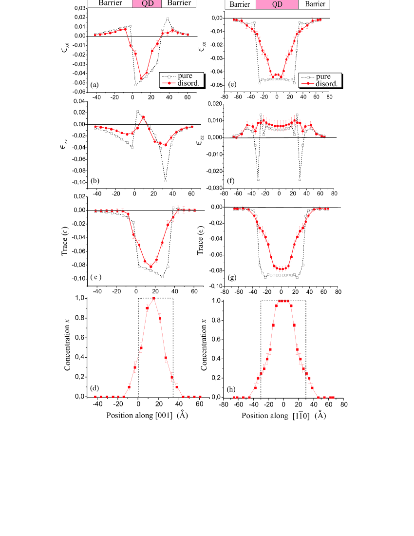
Fig. 1 shows a comparison of the average strain field and the local In average concentration between the chemically pure QD (corresponding to ), given by the dotted lines, and the chemically disordered QD (chosen here with ), given by the solid lines. For the size of QD in this study, allows for all but the innermost In atoms of the QD to diffuse out. For disordered QD’s, the results in Fig. 1 for each property were obtained by averaging over those calculated for an ensemble of 10 different simulation supercells, all corresponding to , but generated from different sequences of random numbers at each interdiffusion step. In this way, the effect of composition fluctuations around the average values given in Eq. (II.1) is reduced.
The panels on the left side show the component [panel (a)], the component [panel (b)] and the trace [panel (c)] of the local strain tensor, as well as the concentration of In atoms [panel (d)], along a line oriented along the [001] direction and passing through the tip of the pyramid. The panels on the right side [(e) - (h)] show the corresponding quantities calculated along a line in the [10] direction and intersecting the [001]-oriented pyramid axis at height from the base of the pyramid, where is the pyramid height. We observe from frames (d) and (h) that, according to our interdiffusion model, corresponds to a penetration of the Ga atoms inside the QD (and consequently of the In atoms inside the GaAs matrix) of about 6 monolayers, i.e. about 17 Å. The error bars shown in the figure indicate standard deviations , where =10. From the figure we may conclude that
-
1.
Chemical disorder significantly reduces the absolute value of the strain field in the regions directly affected by the diffusion process, in agreement with experimental results.Leon et al. (1996) On the other hand, very small changes in the strain field occur in the regions not affected by interdiffusion, i.e. in the core of the pyramid and in the GaAs matrix, at large distances from the dot.
-
2.
If interdiffusion takes place, the strain field varies more smoothly than in the case of a chemically pure QD. This is a direct consequence of the smooth variation of the concentration of In atoms across the heterointerfaces of the disordered dots.
III.2 Electronic and optical properties
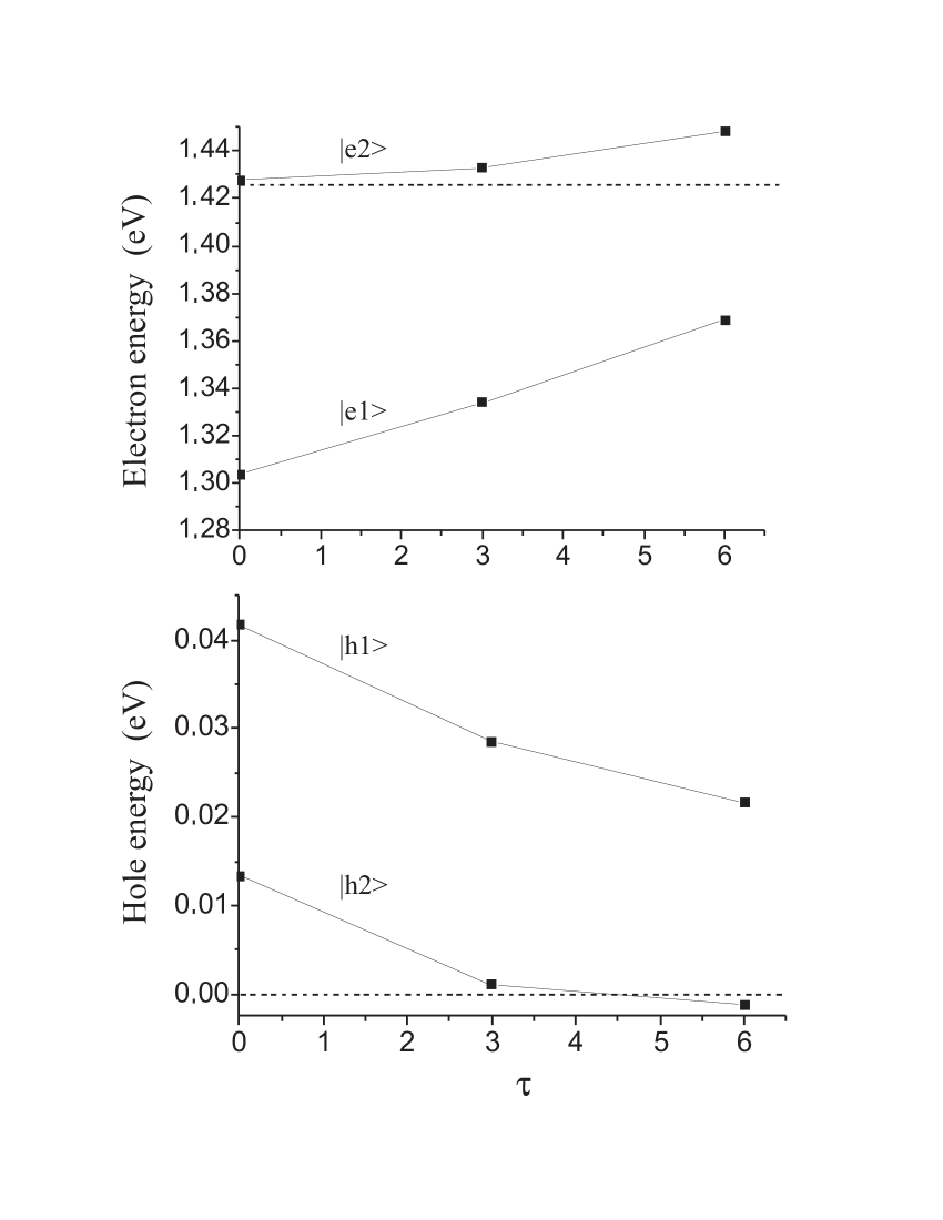
Fig. 2 shows the calculated eigenenergies of the QD bound states as a function of the degree of chemical disorder (characterized by the parameter ). The first two electron states ( and ) are represented in the upper panel, while the first two hole states are shown in the lower panel. A chemically pure QD corresponds to . The dashed horizontal lines represent the energies of the GaAs bulk conduction (upper panel) and valence (lower panel) band edges, delimiting approximately the energy range where a QD state is bound. The figure shows that the electron state energies increase with increasing chemical disorder, while the hole state energies decrease, in agreement with previous empirical pseudopotential calculationsShumway et al. (2001). This behavior results in an increase of the frequency of the optical emission (blueshift), a phenomenon which has been experimentally observed.Leon et al. (1996); Lobo et al. (1998); Malik et al. (1997); Ochoa et al. (2003) The figure shows that, for , the QD gap is about 7% larger than for .
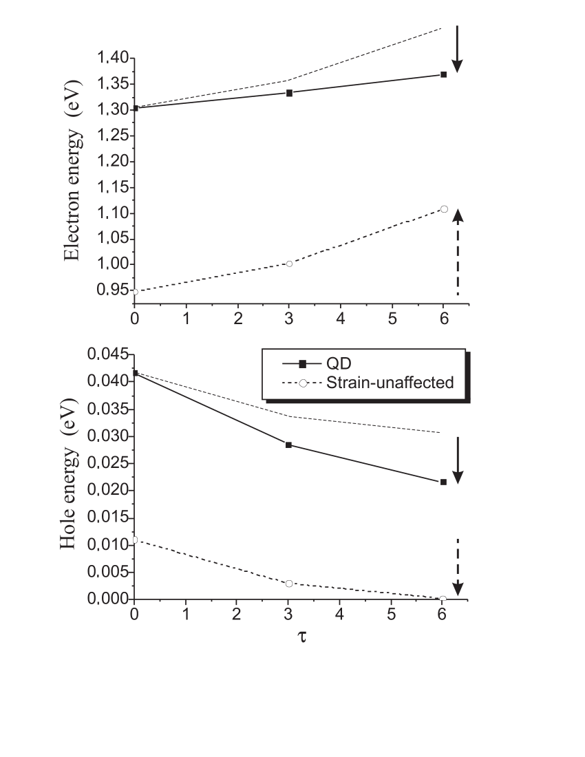
Chemical disorder contributes to the results of Fig. 2 in two ways, namely by the strain relief around the QD interfaces (see Fig. 1), and by the chemical effect due to the presence of Ga atoms inside the QD. These two effects can be decoupled by comparing the bound state energies of a strained QD with those of an artificially strain-unaffected QD, as a function of the degree of disorder. Fig. 3 shows such comparison for the energy of the electron ground state (upper panel) and of the hole ground state (lower panel). On each panel, the uppermost dashed line is a guide for the eye, parallel to the QD strain-unaffected energy curve and starting from the result for the physical QD. The strain relief contribution (represented by the solid arrow) can be directly compared with the purely chemical effect of the disorder, represented by the dashed arrow. We see that these two effects are comparable, contributing in opposite directions for the electron state, and in the same direction for the hole state. The purely chemical effect can be easily understood: As the interdiffusion increases, the concentration of In atoms in the inhomogeneous alloy InxGa1-xAs inside the QD decreases. The increase (decrease) of the electron (hole) bound state energy as decreases is an alloying effect, so that the electron (hole) state energy tends (for ) to the bulk GaAs conduction band minimum (valence band maximum). Results in Fig. 3 show that the chemical effects of disorder are partially canceled (enhanced) by the strain relief contribution for the electron (hole) state.
We now address the optical properties, focusing on the fundamental transition . In Table 1 we compare the results for a chemically pure QD (interdiffusion = off) with those for a chemically disordered QD (interdiffusion = on) with . For both cases, an additional comparison is made between a strained QD (strain = on) and an artificially strain-unaffected QD (strain = off). On the first two lines, we show the charge fraction inside the QD for both the ground electron state and the hole ground state . For the calculation of in the chemically disordered case, the QD border is taken the same as in the chemically pure case. The third line shows the oscillator strength of the transition in the QD for unpolarized light, normalized to the oscillator strength of the fundamental transition in bulk InAs for unpolarized light. The fourth line gives the degree of anisotropy of the QD fundamental transition with respect to light polarization within the pyramid basal plane, defined as
| (6) |
where and are unitary vectors along the inequivalent basal plane directions [110] and [10] respectively. The fifth line shows the oscillator strength of the QD fundamental transition for light linearly polarized along the [001] direction, normalized with respect to . Finally, the last line of Table 1 gives the relative change of the optical QD gap with respect to the gap corresponding to the case “strain off” and “interdiffusion off”. As for the case of the electronic properties, the direct comparison of the “physical” results with those of the disordered case and of the strain-unaffected case allows us to distinguish between the strain relief effect and the chemical effect, both of which are due to chemical disorder.
| Strain | off | on | off | on |
| Interdiffusion | off | off | on | on |
| 75% | 64% | 74% | 34% | |
| 11% | 54% | 11% | 31% | |
| 12% | 19% | 10% | 26% | |
| 111within our numerical precision | 2.5% | 11footnotemark: 1 | % | |
| 11footnotemark: 1 | 8% | 11footnotemark: 1 | % | |
| 0 | 35% | 18% | 44% |
From the results in Table 1, we arrive at the following conclusions:
-
1.
The first two lines show that chemical disorder reduces the confinement of the QD bound states through the partial relief of the strain field, while the chemical effect does not directly contribute. In fact, chemical disorder reduces the charge fractions and , while no changes are observed for the strain-unaffected calculation. The smaller confinement of the QD bound state wave functions in the chemically disordered case is consistent with the results of Fig. 2, where all electron and hole bound states become shallower when chemical disorder increases.
-
2.
The third line indicates that chemical disorder significantly enhances (by about , in the case considered here) the oscillator strength of the fundamental optical transition, in qualitative agreement with experimental results.Malik et al. (1997); Leon et al. (1996) This effect is primarily due to the modification of the strain field due to chemical disorder, because in the strain-unaffected case does not significantly vary.
-
3.
From the fourth line, we observe that chemical disorder strongly reduces the in-plane asymmetry , in accordance with previous experimental results.Ochoa et al. (2003) This is a direct consequence of the partial relief of strain due to disorder. In fact,Santoprete et al. in a pyramidal QD the asymmetry of the oscillator strength of the fundamental optical transition between the directions [110] and [10] is a direct consequence of asymmetry of the strain field between these directions, which is in turn a consequence of the symmetry. This can be deduced observing that in the strain-unaffected case vanishes. This result could be experimentally exploited to detect, among different samples containing QDs of similar geometry, those characterized by the higher chemical purity. In fact, these samples will be those with the higher asymmetry of the absorption coefficient of the fundamental optical transition (which is proportional to ), for in-plane polarized light.
-
4.
The fifth line of Table 1 implies that chemical disorder weakens the fundamental optical transition for perpendicularly polarized light. This is a consequence of the strain relief inside the QD: In the limit of complete relief (QD strain-unaffected) this transition is strictly forbidden.Santoprete et al.
-
5.
The last line summarizes the different effects contributing to the blue shift in the fundamental optical transition with respect to a hypothetical transition energy where both effects are removed. We see that strain and chemical disorder increase the QD gap by the same order of magnitude. We note that calculations for the relative blue shift presented in Ref. Gunawan et al., 2005 systematically underestimate this quantity as compared to the experimental results in Ref. Fafard and Allen, 1999 (see Fig. 7 in Ref. Gunawan et al., 2005). This discrepancy is probably due to the simplified theoretical description adopted there, where strain effects were not taken into account.
Finally, we analyzed the -component of the built-in dipole moment of the electron-hole pair, and how disorder affects it. Such dipole moment experimentally shows up as a Stark shift of the emitted light from a QD-LED under applied electrical field.Fry et al. (2000) For pure pyramidal InAs/GaAs QDs, this dipole moment points towards the base of the pyramid, i.e. the center of mass of the electron ground state lies above that of the hole ground state.Stier et al. (1999) However, in the case of truncated pyramidal InxGa1-xAs QDs, with increasing from the base to the tip of the pyramid, the dipole moment may have an opposite orientation, i.e. the center of mass of the hole state can sit above that of the electron state.Fry et al. (2000) Some authors have argued that such inversion occurs also for QDs having an In-rich core with an inverted-cone shape. This inverted-cone shape has been observed in truncated-cone nominal In0.8Ga0.2As QDsLenz et al. (2002) and In0.5Ga0.5As QDs.Liu et al. (2000) In our case, the dipole moment is always directed towards the base of the pyramid, i.e. the electron ground state sits always above the hole ground state, both for the pure and the disordered QD. This is because we have neither a truncated pyramidal shape nor an In-concentration increasing from the base to the tip of the pyramid (see Fig. 1). However, we observe that the disorder decreases the dipole moment of the dot. In fact, in the strained disordered case, the center of mass of the electron state lies 2.8 Å above that of the hole state, while in the strained pure case this separation is 3.5 Å.
IV Summary and Conclusions
We presented an atomistic interdiffusion model to simulate the composition profile of chemically disordered pyramidal InxGa1-xAs QDs buried in GaAs matrices. Calculations for the strain field inside and around the disordered QDs were compared to the strain field of chemically pure InAs QDs, showing that chemical disorder significantly reduces the absolute value of the strain field inside the QD, giving rise to smoother variations of this field across the heterointerfaces. Furthermore, we analyzed the consequences of chemical disorder for the electronic and optical properties within an ETB model. Our treatment allowed us to distinguish between two effects of the chemical disorder, namely the relief of the strain inside the QD, and the purely chemical effect due to the presence of new atomic species (Ga atoms) penetrating inside the QD. We showed that these two components of disorder have comparable effects on the QD electronic spectrum, while for the optical properties the strain relief effects are more relevant. In particular, we showed that strain relief (i) reduces the charge confinement (inside the QD) of the electron and hole bound state wave functions, (ii) significantly enhances the oscillator strength of the fundamental optical transition, (iii) strongly reduces the asymmetry of the oscillator strength of the fundamental optical transition between the directions [110] and [10] for in-plane polarized light, and (iv) strongly reduces the oscillator strength of the fundamental optical transition for perpendicularly polarized light.
Our results help to explain experimental findings for the optical properties of intermixed InAs/GaAs QDs.
Acknowledgements.
This work was partially supported by the Brazilian agencies CNPq, FAPERJ and Instituto do Milênio de Nanociências-MCT, and by Deutsche Forschungsgemeinschaft within Sfb 296. BK thanks the hospitality of the CMTC at the University of Maryland.References
- Bimberg et al. (1999) D. Bimberg, M. Grundmann, and N. N. Ledentsov, Quantum Dot Heterostructures (Wiley, New York, 1999).
- Scheerschmidt and Werner (2002) K. Scheerschmidt and P. Werner, Nano-Optoelectronics (Springer, Berlin, 2002), p. 67.
- Joyce et al. (1998) P. B. Joyce, T. J. Krzyzewski, C. G. Bell, B. A. Joyce, and T. S. Jones, Phys. Rev. B 58, R15981 (1998).
- Kegel et al. (2000) L. Kegel, T. H. Metzger, A. Lorke, J. Peisl, J. Stangl, G. Bauer, J. M. Garcia, and P. M. Petroff, Phys. Rev. Lett. 85, 1694 (2000).
- Xu et al. (1998) S. J. Xu, X. C. Wang, S. J. Chua, C. H. Wang, W. J. Fan, J. Jiang, and X. G. Xie, Appl. Phys. Lett. 72, 3335 (1998).
- Fafard and Allen (1999) S. Fafard and C. N. Allen, Appl. Phys. Lett. 75, 2374 (1999).
- Lita et al. (1999) B. Lita, R. S. Goldman, J. D. Phillips, and P. K. Bhattacharya, Appl. Phys. Lett. 75, 2797 (1999).
- Garcia et al. (1997) J. M. Garcia, G. Medeiros-Ribeiro, K. Schmidt, T. Ngo, J. L. Feng, A. Lorke, J. Kotthaus, and P. M. Petroff, Appl. Phys. Lett. 71, 2014 (1997).
- Rosenauer et al. (1997) A. Rosenauer, U. Fischer, and D. Gerthsen, Appl. Phys. Lett. 71, 3868 (1997).
- Bruls et al. (2002) B. M. Bruls, J. W. A. M. Vugs, P. M. Koenraad, H. W. M. Salernink, J. H. Wolter, M. Hopkinson, M. S. Skolnick, F. Long, and S. P. A. Gill, Appl. Phys. Lett. 81, 1708 (2002).
- Chu et al. (1999) L. Chu, M. Arzberger, G. Böhm, and G. Abstreiter, J. Appl. Phys. 85, 2355 (1999).
- Fry et al. (2000) P. W. Fry, I. E. Itskevich, D. J. Mowbray, M. S. Skolnick, J. J. Finley, J. A. Barker, E. P. O’Reilly, L. R. Wilson, I. A. Larkin, P. A. Maksym, et al., Phys. Rev. Lett. 84, 733 (2000).
- Jayavel et al. (2004) P. Jayavel, H. Tanaka, T. Kita, O. Wada, H. Ebe, M. Sugawara, J. Tatebayashi, Y. Arakawa, Y. Nakata, and T. Akiyama, Appl. Phys. Lett. 84, 1820 (2004).
- Joyce et al. (2001) P. B. Joyce, T. J. Krzyzewski, G. R. Bell, T. S. Jones, S. Malik, D. Childs, and R. Murray, J. Cryst. Growth 227-228, 1000 (2001).
- Lipinski et al. (2000) M. O. Lipinski, H. Schuler, O. G. Schmidt, K. Eberl, and N. Y. Jin-Phillipp, Appl. Phys. Lett. 77, 1789 (2000).
- Zhi et al. (2004) D. Zhi, M. Wei, R. E. Dunin-Borkowski, P. A. Midgley, D. W. Pashley, T. S. Jones, B. A. Joyce, P. F. Fewster, and P. J. Goodhew, Microelectronic Engineering 73-74, 604 (2004).
- Leon et al. (1996) R. Leon, Y. Kim, C. Jagadish, M. Gal, J. Zou, and D. J. H. Cockayne, Appl. Phys. Lett. 69, 1888 (1996).
- Malik et al. (1997) S. Malik, C. Roberts, R. Murray, and M. Pate, Appl. Phys. Lett. 71, 1987 (1997).
- Lobo et al. (1998) C. Lobo, R. Leon, S. Fafard, and P. G. Piva, Appl. Phys. Lett. 72, 2850 (1998).
- Ochoa et al. (2003) D. Ochoa, A. Polimeni, M. Capizzi, A. Patané, M. Henini, L. Eaves, and P. C. Main, J. Cryst. Growth 251, 192 (2003).
- Pryor et al. (1998) C. Pryor, J. Kim, L. W. Wang, A. J. Williamson, and A. Zunger, J. Appl. Phys. 83, 2548 (1998).
- Stoleru et al. (2002) V. G. Stoleru, D. Pal, and E. Towe, Physica E 15, 131 (2002).
- Liao et al. (1999) X. Z. Liao, J. Zou, D. J. H. Cockayne, R. Leon, and C. Lobo, Phys. Rev. Lett. 82, 5148 (1999).
- Califano and Harrison (2002) M. Califano and P. Harrison, J. Appl. Phys. 91, 389 (2002).
- Tersoff (1989) J. Tersoff, Phys. Rev. B 39, 5566 (1989).
- Migliorato et al. (2002) M. A. Migliorato, A. G. Cullis, M. Fearn, and J. H. Jefferson, Phys. Rev. B 65, 115316 (2002).
- Keating (1966) P. Keating, Phys. Rev. 145, 637 (1966).
- Martin (1970) R. Martin, Phys. Rev. B 1, 4005 (1970).
- Shumway et al. (2001) J. Shumway, A. J. Williamson, A. Zunger, A. Passeo, M. DeGiorgi, R. Cingolani, M. Catalano, and P. Crozier, Phys. Rev. B 64, 125302 (2001).
- Barker and O’Reilly (2000) J. A. Barker and E. P. O’Reilly, Phys. Rev. B 61, 13840 (2000).
- Roy and Maksym (2003) M. Roy and P. A. Maksym, Phys. Rev. B 68, 235308 (2003).
- Vasanelli et al. (2002) A. Vasanelli, R. Ferreira, H. Sasaki, G. Bastard, and R. Congolani, Phys. Stat. Sol. A 190, 551 (2002).
- Park et al. (2003) S. H. Park, D. Ahn, Y. T. Lee, and S. L. Chuang, Jpn. J. Appl. Phys. 42, 144 (2003).
- Sheng and Leburton (2003a) W. Sheng and J. P. Leburton, Phys. Rev. B 67, 125308 (2003a).
- Sheng and Leburton (2003b) W. Sheng and J. P. Leburton, Phys. Status Solidi B 237, 394 (2003b).
- Heinrichsdorff et al. (1998) F. Heinrichsdorff, M. Grundmann, O. Stier, A. Krost, and D. Bimberg, J. Cryst. Growth 195, 540 (1998).
- Bester et al. (2003) G. Bester, S. Nair, and A. Zunger, Phys. Rev. B 67, 161306 (2003).
- Bester et al. (2004) G. Bester, J. Shumway, and A. Zunger, Phys. Rev. Lett. 93, 47401 (2004).
- Bester and Zunger (2003) G. Bester and A. Zunger, Phys. Rev. B 68, 73309 (2003).
- Klimeck et al. (2002) G. Klimeck, F. Oyafuso, T. B. Boykin, R. C. Bowen, and P. von Allmen, CMES - Computer Modeling in Engineering & Sciences 3, 601 (2002).
- Gunawan et al. (2005) O. Gunawan, H. S. Djie, and B. S. Ooi, Phys. Rev. B 71, 205319 (2005).
- Bester and Zunger (2005) G. Bester and A. Zunger, Phys. Rev. B p. 45318 (2005).
- Santoprete et al. (2003) R. Santoprete, B. Koiller, R. B. Capaz, P. Kratzer, Q. K. K. Liu, and M. Scheffler, Phys. Rev. B 68, 235311 (2003).
- Boykin (1997) T. B. Boykin, Phys. Rev. B 56, 9613 (1997).
- Wang and Zunger (1994) L.-W. Wang and A. Zunger, J. Chem. Phys. 100, 2395 (1994).
- Capaz et al. (1993) R. B. Capaz, G. C. de Araujo, B. Koiller, and J. P. von der Weid, J. Appl. Phys. 74, 5531 (1993).
- Koiller et al. (1991) B. Koiller, R. Osório, and L. M. Falicov, Phys. Rev. B 43, 4170 (1991). In the present calculations the Hamiltonian and the orbitals are spin-dependent, the distance vectors extend up to second neighbors and incorporate distortions in the “strain on” cases.
- (48) R. Santoprete, B. Koiller, R. B. Capaz, P. Kratzer, and M. Scheffler, 27th International Conference on the Physics of Semiconductor. Editors J. Menendez and C. Van de Walle, AIP Conference Proceedings Volume 772, American Institute of Physics, Melville, NY, 2005, pp. 745-746.
- Stier et al. (1999) O. Stier, M. Grundmann, and D. Bimberg, Phys. Rev. B 59, 5688 (1999).
- Lenz et al. (2002) A. Lenz, R. Timm, H. Eisele, C. Henning, S. K. Becker, R. L. Sellin, U. W. Pohl, D. Bimberg, and M. Dähne, Appl. Phys. Lett. 81, 5150 (2002).
- Liu et al. (2000) N. Liu, J. Tersoff, O. Backlenov, J. A. L. Holmes, and C. K. Shih, Phys. Rev. Lett. 84, 334 (2000).