The healing mechanism for excited molecules near metallic surfaces
Abstract
Radiation damage prevents the ability to obtain images from individual molecules. We suggest that this problem can be avoided for organic molecules by placing them in close proximity with a metallic surface. The molecules will then quickly dissipate any electronic excitation via their coupling to the metal surface. They may therefore be observed for a number of elastic scattering events that is sufficient to determine their structure.
pacs:
68.43.-h 73.40.Gk 87.64.-tExamining individual molecules at an atomic length scale is one of the most challenging modern problems of science. An analysis of elastic and inelastic cross sections provides a basis for understanding why molecules can become fragmented beyond recognition long before the time necessary to accumulate sufficient counts for image reconstruction. In the case of organic molecules, most structural knowledge has been gained using crystalline samples, where damaging exposure is distributed over many identical molecules, and the image is almost the same as that of an undamaged molecule. For molecules which cannot be crystallized, there is an urgent need for methods that can give information on their structures.
In single molecule diffraction techniques proposed using either femtosecond x-ray pulses nat_fel or high energy electron beams prl_smd , short wavelengths are used to image the atomic structure of individual molecules, but the radiation doses destroy the molecule after providing a diffraction pattern. Imaging with coherent electrons from Low Energy Electron Point Sources (LEEPS) is a less destructive method to characterize single molecules leeps . A holographic interference pattern can be generated by LEEPS from which one can reconstruct the object’s structure. For example, one can reconstruct the structure of single polymer strands like DNA fink_nat . Recently, another method using interference patterns with standing x-ray waves has been used to map the perylene-tetracarboxylic-dianhydride (PTCDA) molecule on a silver surface nixsw05 . The use of positrons for microscopy is not as well known, but it has tremendous potential. Positrons, owing to their positive charge, have negative work functions in several materials, and they can be reemitted spontaneously. This feature allows the control of slow positron beams having energies in the epithermal region. A positron microscope for imaging single molecules has been recently proposed by Mills and Platzman mills01 . In order to get proper structural information with sufficient resolution, Mills and Platzman have estimated that each atom in a molecule of atoms should suffer about annihilations in the course of the positron diffraction. After the annihilation, the molecule is in an ionic excited state, and under normal circumstances, such a large number of ionizations would destroy the molecule.
Clearly, while exploring the opportunities of imaging single molecules with either x-rays, electrons or positrons, it is vital to reduce radiation damage at the level of individual molecules. In this letter, we utilize the neutralization process when a molecule is placed near a metallic surface - here called the healing mechanism - and propose that it may be applied to heal the radiation damage. The main idea is to fill the vacant molecular orbital by an electron from the metal in a time short compared to some characteristic vibration time of the molecule. Therefore, charge neutrality is restored before the onset of the vibrational modes which would lead to the destruction of the molecule. This healing mechanism can be decomposed in two main steps. In the first step, when the positive ion is sufficiently close to the surface, one conduction electron of the metal will tunnel under the influence of the strong electric field produced by the ion. In the second step, the tunneling electron will fill the hole via an Auger process involving an electron-hole pair of the metal. The physics of the healing mechanism is closely related to various neutralization processes relax0 ; relax1 ; relax2 ; relax3 ; relax4 ; relax5 ; relax6 ; relax7 and to the intermolecular Coulombic decay icd . Most of this work describes collisions of slow ions with solids. Our study involves the neutralization of physisorbed molecules on metallic surfaces. Given the wide range of estimates of the neutralization time in previous work relax1 , we must perform a conservative estimate to demonstrate the feasibility of the healing mechanism in our case.
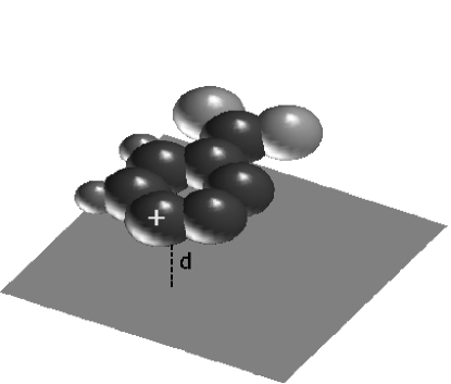
A schematic of the healing mechanism set-up is shown in Fig. 1. The molecule, in this example PTCDA, is on a metallic substrate, which should be chemically inert to preserve the integrity of the molecule. Gold, silver or other noble metals are possible substrates. In reality, the PTCDA geometry might slightly bend near the metallic substrate because of the readiness of some PTCDA atoms to form weak bonds with the surface nixsw05 .
When a positive charge is suddenly created on the molecule, the resulting electric field attracts the electrons of the metal. The probability that one electron escapes from the metal depends on the work function and on the distance of the positive charge from the surface. An accurate determination of the tunneling probability would require solving the Schödinger equation, for example using the matching wave function method fowler . However, the result is mostly dominated by an exponential factor given by the WBK approximation probst
| (1) |
where is the width of the potential barrier and is the Fermi energy of the metal.
The actual shape of the potential barrier is complicated, but a linear approximation of the electrostatic potential at the metal-molecule interface yields a simple triangular barrier, and the penetration probability becomes footnote1
| (2) |
where atomic units have been used.
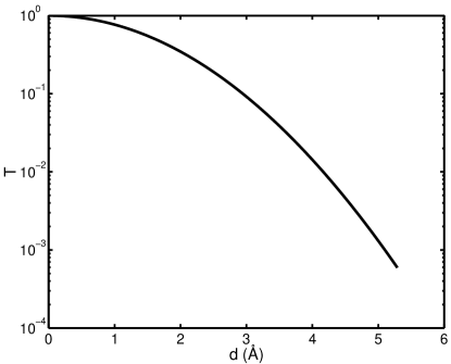
Fig. 2 shows that under a barrier characterized by a work function eV is still about for a distance . Actually, our major assumption - the triangular barrier - underestimates since the electric field gets stronger as an electron approaches the molecule. Our assumption, as we shall see, therefore leads to an overestimate of the healing time.
An electron of the metal is expected to fall into the vacant orbital of the molecule. Indeed, a photoemission study kahn96 of the PTCDA organic molecule on a metal surface reveals the energy-level alignment shown in the schematic diagram of Fig. 3, where the highest occupied molecular orbital (HOMO) level is about eV below . A similar diagram of energy levels has been obtained via first-principles calculations picozzi .
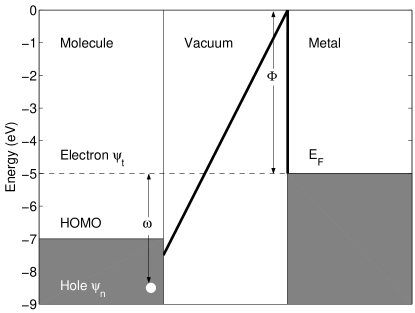
The energy separation (see Fig. 3) between the state of the electron spilling out from the metal footnote_ef and the molecular orbital of the hole must be dissipated during the recombination. An Auger process involving an electron-hole excitation in the metal can mediate the capture of the electron in the state into the vacant molecular state . There are several possible electron transfer processes between molecules and surfaces surfsci . For chemisorbed systems, a hole lifetime is much shorter than a typical vibration time gunnarsson , so that normally the hole would be filled before the molecule can fly apart. For N2 molecules physisorbed on graphite, experiments suggest that the neutralization occurs with a characteristic time s nilsson . In general, for a molecule far from the surface, the corresponding hole may not be filled for a while. In this case, the relevant process is the Auger neutralization as described by Propst probst . The actual calculation of the Auger neutralization rate is a difficult problem including subtle screening effects at the metal-vacuum interface relax1 . Nevertheless, it has been suggested by numerical calculations that, as a good approximation, the dependence on the distance and the energy factorize relax2 as
| (3) |
where is obtained using the formula for the molecule immersed in the bulk and is an unknown function, usually approximated by an exponential relax2 , which decays away from the metal surface. Propst probst , however, has shown that the distance of the molecule from the surface comes into play mostly through the WKB transmission probability. Therefore, we can assume . The matrix element of the Auger process in the bulk involves the ground state of the metal, the excited state of the metal and the density operator for excitations with momentum transfer . The actual form of bock reads
| (4) |
where is a form factor given by
| (5) |
is the Coulomb interaction Fourier transform
| (6) |
and is the volume of the metallic sample. The rate of the Auger recombination can be calculated by using Fermi’s golden rule
| (7) |
where is the energy difference between the metal ground-state and the excited state . Eq. 7 can be rewritten in the form
| (8) |
where is the dynamical structure factor pines defined by
| (9) |
The fluctuation-dissipation theorem relates the dynamical structure factor to the dielectric function by the formula
| (10) |
Substituting Eq. 10 in Eq. 8, we obtain
| (11) |
To extract an Auger rate estimate, one can use the Lindhard RPA formula pines for the dielectric function . Density inhomogeneity effects, due to the lattice and the surface, and correlation corrections beyond the RPA are not considered in this study. For , one can use a Slater type of orbital slater (STO) given by
| (12) |
where provides the size of the molecular orbital. Therefore, assuming that is slowly varying, the main form factor contribution to the integral in Eq. 11 is given by
| (13) |
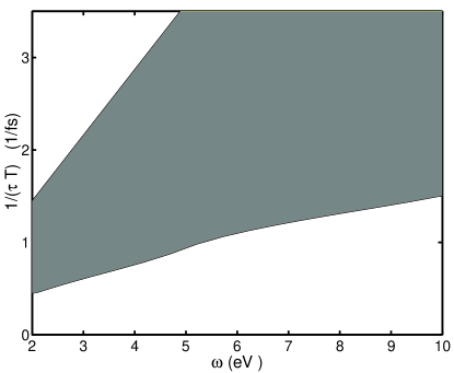
Fig. 4 shows typical values in the energy range eV for a gold surface (corresponding to a electron density parameter ). Since this rate is proportional to the penetration probability , we show in the ordinate axis of the plot in Fig 4. The grey region in Fig. 4 corresponds to a STO exponent ranging from to . For a distance of , since is about the Auger rate becomes of the order of 1/s in the lower part of the grey region in Fig. 4. This value is comparable to the highest vibration modes in molecules. All this implies that, within distances of less than , the time to fill the hole is estimated to be fast enough to keep the molecule from being damaged by molecular vibrations. As mentioned previously, the value of the penetration probability given by Eq. 2 is a lower bound. This fact adds to the robustness of the present model.
In conclusion, the effects of radiation damage on molecules are of concern, because they can be a major obstacle to the emergence of microscopy as a tool to determine the structure of delicate biological molecules. This is true whether x-rays, electrons or positrons are used as the illumination. We have shown that in favorable conditions, ionized molecules on a metal surface can quickly dissipate any electronic excitation via their coupling to the metal. The relaxation is achieved via an electron tunneling from the metal to the molecule and falling into the vacant orbital. The falling electron gives up its energy to an excitation of the metal produced by an Auger process. If the molecule is within distances of about from the metal surface, short lifetimes of the hole on the molecule (of the order of fs) prevent the destruction of the molecule. Our model may in fact overstimate the healing time. This healing mechanism is particularly suitable for the positron microscope newly proposed by Mills and Platzman mills01 , but it can be applied also to other microscopes imaging single molecules. In all these cases, the molecule on the metallic surface may be observed without being damaged for a number of scattering events sufficient to determine its structure by speckle diffraction. These measurements would be ideal for studying delicate biological molecules which cannot be crystallized.
We acknowledge Rolando Saniz, Olle Gunnarsson and Dan Nissenbaum for useful discussions. This work is supported by the US Department of Energy contract DE-AC03-76SF00098 and benefited from the allocation of computer time at NERSC and Northeastern University’s Advanced Scientific Computation Center (ASCC).
References
- (1) R. Neutze et al., Nature 406, 752 (2000).
- (2) J. C. H. Spence and R. B. Doak, Phys. Rev. Lett. 92, 198102 (2004).
- (3) H. W. Fink, W. Stocker, and H. Schmid, Phys. Rev. Lett. 65, 1204 (1990).
- (4) H. W. Fink and C. Schönenberger, Nature 398, 407 (1999).
- (5) A. Hauschild et al., Phys. Rev. Lett. 94, 036106 (2005).
- (6) A. P. Mills and P. M. Platzman in New Directions in Antimatter Chemistry and Physics, C. M. Surko and F. A. Gianturco, eds., Kluwer Academic Publishers, The Netherlands, (2001).
- (7) R.A. Baragiola and C. A. Dukes, Phys. Rev. Lett. 76, 2547 (1996).
- (8) M.A. Cazalilla et al., Phys. Rev. B 58, 13991 (1998).
- (9) Y. Bandurin, V. A. Esaulov, L. Guillemot, and R. C. Monreal, Phys. Rev. Lett. 92, 017601 (2004).
- (10) J. Burgdorfer et al., Nucl. Instr. and Meth. B 205, 690 (2003).
- (11) L. Wirtz et al., Phys. Rev. A 67, 012903 (2003).
- (12) J. Stöckl et al., Phys. Rev. Lett. 93, 263201 (2004).
- (13) T. Fonden and A. Zwartkruis, Phys. Rev. B 48, 15 603 (1993).
- (14) S. Wethekam, A. Mertens and H. Winter, Phys. Rev. Lett. 90, 037602 (2003).
- (15) R. Santra and L.S. Cederbaum, Phys. Rep. 368, 1 (2002).
- (16) See e.g. J. W. Gadzuk and E. W. Plummer, Rev. Mod. Phys. 45, 487 (1973).
- (17) F.M. Propst, Phys. Rev. 129, 7 (1963).
- (18) We have assumed that the charge distribution on the molecule is spherical and we have used the method of images to determine the electric field. Near the metal-vacuum interface, along the direction from the metal surface to the positive charge, the electric field intensity is ; see e.g. L. Landau and E. M. Lifchitz, Electrodynamics of Continuous Media, Pergamon, New York, (1960).
- (19) Y. Hirose et al., Phys. Rev. B 54, 13748 (1996).
- (20) S. Picozzi et al., Phys. Rev. B 68, 195309 (2003).
- (21) We have assumed that is at the Fermi level .
- (22) See e.g. W. Sesselmann et al., Surf. Sci. 146, 17 (1984) and D. Lovric et al., Surf. Sci. 189/190, 59 (1987).
- (23) O. Gunnarsson, private communication.
- (24) O. Björneholm et al., Phys. Rev. Lett. 68, 1892 (1991).
- (25) Related lifetimes have been used in the context of electron relaxation in quantum dots, see e.g. U. Bockelmann and T. Egeler, Phys. Rev. B 46, R15574 (1992).
- (26) D. Pines, Elementary Excitations in Solids, W.A. Benjamin Inc, New York, Amsterdam (1963).
- (27) J.C. Slater, Phys. Rev. 36, 57 (1930).