Charged exciton emission at 1.3 m from single InAs quantum dots grown by metalorganic chemical vapor deposition
Abstract
We have studied the emission properties of self-organized InAs quantum dots (QDs) grown in an InGaAs quantum well by metalorganic chemical vapor deposition. Low-temperature photoluminescence spectroscopy shows emission from single QDs around 1300 nm; we clearly observe the formation of neutral and charged exciton and biexciton states, and we obtain a biexciton binding energy of 3.1 meV. The dots exhibit an s-p shell splitting of approximately 100 meV, indicating strong confinement.
pacs:
81.07.Ta, 78.67.Hc, 73.21.La, 81.15.Gh, 78.55.CrSemiconductor self-assembled quantum dots (QDs) are of considerable interest for future telecommunication applications, such as low-threshold lasers and non-classical light sources for quantum key distribution systems. Efficient single-photon emission has recently been demonstrated at visible wavelengths using semiconductor QD structures,michler00 ; santori02 ; zwiller03 and there have been many detailed investigations into the low-temperature optical characteristics of QDs emitting at 1150 nm or less.lomascolo02 ; moskalenko02 ; kaiser02 However, to date there have been only a small number of spectroscopic experiments on single QDs emitting in the important telecommunications window around 1300 nm:ward04 biexcitonic features have been identified in low-temperature photoluminescence (PL) from QDs grown by molecular beam epitaxy (MBE),alloing05 whereas similar investigations for QDs fabricated by metalorganic chemical vapor deposition (MOCVD) show an unclear power dependence in the emission.song05
Quantum dot structures grown by MOCVD have potentially a large commercial value due to the high growth rates achievable; however, for applications at telecommunication wavelengths the growth is complicated by large strain effects and complex surface dynamics within the dot layers.passaseo04 Therefore, there is a strong motivation for studying the optical characteristics of these structures in relation to other fabrication techniques. Here, we report on the emission properties of single QDs in a novel dots-in-well (DWELL) heterostructure grown by MOCVD. We present low-temperature PL spectra from individual QDs with an emission wavelength of 1300 nm; power-dependent measurements clearly reveal the formation of an exciton-biexciton system, with a biexciton binding energy of more than 3 meV. We also identify recombination from charged exciton and biexciton complexes, and we observe a large energy difference between s- and p-shell states.
The QDs were fabricated using conventional low-pressure MOCVD on a (100) GaAs substrate: an InAs(:Bi) dot layer was deposited in a 5 nm In0.12Ga0.88As(:Bi) quantum well (QW), and the DWELL heterostructure grown between GaAs barrier layers and InGaP cladding layers. Bismuth doping was found to significantly improve the PL intensity and emission wavelength of the dots. The DWELL structure results in a pronounced red-shift relative to similar InAs/GaAs systems due to effects such as strain relaxationnishi99 and alloy decomposition.guffarth01 Atomic force microscopy (AFM) measurements on similar samples suggest a dot size of 15 nm with elongation along the axis; the QD sheet density is estimated as cm-2. A more detailed description of the growth will be published elsewhere. In order to obtain single dot spectroscopy, mesa structures were fabricated by electron-beam lithography and dry etching, with sizes between 22 m2 and 200200 nm2. Micro-PL measurements were taken using an Ar+ laser (488 nm) focused to a 2 m spot; the luminescence was dispersed in a 0.5 m spectrometer and detected with a nitrogen cooled InGaAs photodiode array (). Unless otherwise stated, the sample temperature was maintained at 5 K in a continuous-flow He cryostat.
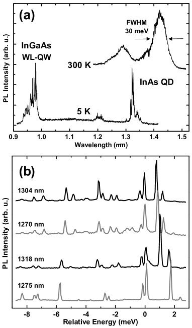
Figure 1(a) shows PL spectra from an unetched region of the sample at 300 K, and from a 200 nm mesa at 5 K. In the former case the full width at half maximum (FWHM) of the QD peak is 30 meV, indicating a good growth uniformity. The shorter wavelength peak is from p-shell states, as discussed later. At 5 K, emission is observed below 1.0 m (1.27 eV) from the hybrid wetting layer – quantum well (WL–QW) that forms in DWELL structures.chang05
In Fig. 1(b) we show PL spectra from a selection of 200 nm mesas, obtained with a power density of 20 W cm-2. Each spectrum has been shifted in energy to align the main emission peak observed at low power. The other emission lines exhibit very close similarities in energy and intensity between the different spectra; this is strong evidence that each spectrum originates from a single QD, and that the different lines arise from various exciton complexes within the dot.
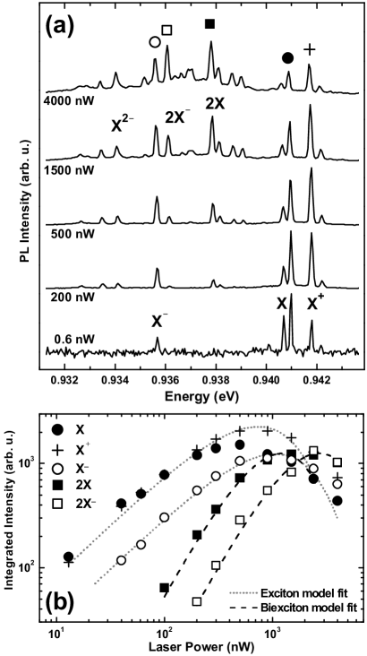
To investigate the origin of these lines, PL spectra were taken for a range of excitation powers as shown in Fig. 2(a). At low powers the spectra are composed of four narrow lines ( 100 eV, resolution limited). All of these lines are present at the lowest powers measured ( 30 mW cm-2) for many different mesas [see Fig. 1(b)] and exhibit an almost identical linear increase in intensity over low excitation powers before saturating at 500 nW [Fig. 2(b)]. These lines are assigned to recombination from neutral (X) and charged (X-/X+) exciton states. Emission from the neutral exciton state exhibits a polarization-dependent fine-structure splitting of approximately 300 eV. This is attributed to splitting of the bright exciton angular momentum states due to dot asymmetry;bayer02b the origin and nature of this fine-structure will be discussed in detail elsewhere.
With increasing power, we observe the appearance of additional lines below the exciton energy. In particular, the intensities of the lines labeled 2X and 2X- are found to increase superlinearly with excitation power [Fig. 2(b)], which is consistent with emission from biexciton states. The intensity dependence of these lines can be fit very well over low powers by rate-equation models based on random capture of excitons into a dot.bacher99 The other lower energy lines are attributed to charged exciton complexes consisting of two or more electrons bound to a hole; this is consistent with theoretical predictions,zunger04 and other experimental observations.warburton00 The positive trion X+ appears at a higher energy than the exciton as the hole wavefunction has a smaller lateral extent than the electron wavefunction .lelong96 The lack of any observable exchange energy splitting in polarization dependent PL is further evidence of the charged nature of these complexes.bayer02b
The assignment of the 2X- (X2-) state has been corroborated by measuring the energy difference between this line and the 2X (X-) line in a study of 9 dots; this was found to be 1.77 meV (1.51 meV), with a standard deviation of 100 eV. From this survey the mean binding energies of the X- (X+) and 2X states are 5.6 meV (-1.1 meV) and 3.1 meV respectively; the first value is consistent with the shifts calculated by Finley et al. finley01b for dots with nm. The biexciton binding energy is also in agreement with the values obtained by Kaiser et al. kaiser02 for a similar strongly confined DWELL system.
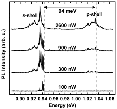
Finally, Fig. 3 shows broad-spectrum power dependence of the same dot shown in Fig. 2. At 300 nW (10 W cm-2) we see the appearance of a new set of lines approximately 100 meV above the ground-state s-shell multiplet. These appear concomitantly with the 2X lines and therefore can be attributed to recombination of electron-hole pairs in p-shell states. The magnitude of this s-p shell splitting is consistent with the large confinement potential expected for structures with strain-reducing layers.liu05
In summary, we have observed 1.3 m emission at 5 K from individual InAs QDs grown in an InGaAs QW by MOCVD. This suggests that this structure can be utilized as a single-photon source at telecommunication wavelengths. Recombination from neutral and charged biexciton states was clearly observed, the former having a binding energy of 3.1 meV. The negative (positive) trion is found to have a binding energy of 5.6 meV (-1.1 meV). These values, and the large energy separation between s- and p-shell recombination, indicate strong confinement and small dot size.
This work was partly supported by the National Institute of Information and Communications Technology (NICT).
References
- (1) P. Michler, A. Kiraz, C. Becher, W. V. Schoenfeld, P. M. Petroff, L. Zhang, E. Hu, and A. Imamolu, Science 290, 2282 (2000).
- (2) C. Santori, D. Fattal, J. Vučković, G. S. Solomon, and Y. Yamamoto, Nature (London) 419, 594 (2002).
- (3) V. Zwiller, T. Aichele, W. Seifert, J. Persson, and O. Benson, Appl. Phys. Lett. 82, 1509 (2003).
- (4) M. Lomascolo, A. Vergine, T. K. Johal, R. Rinaldi, A. Passaseo, R. Cingolani, S. Patan, M. Labardi, M. Allegrini, F. Troiani, and E. Molinari, Phys. Rev. B 66, 041302 (2002).
- (5) E. S. Moskalenko, K. F. Karlsson, P. O. Holtz, B. Monemar, W. V. Schoenfeld, J. M. Garcia, and P. M. Petroff, J. Appl. Phys. 92, 6787 (2002).
- (6) S. Kaiser, T. Mensing, L. Worschech, F. Klopf, J. P. Reithmaier, and A. Forchel, Appl. Phys. Lett. 81, 4898 (2002).
- (7) M. B. Ward, D. C. Unitt, Z. Yuan, P. See, R. M. Stevenson, K. Cooper, P. Atkinson, I. Farrer, D. A. Ritchie, and A. J. Shields, Physica E 21, 390 (2004).
- (8) B. Alloing, C. Zinoni, V. Zwiller, L. H. Li, C. Monat, M. Gobet, G. Buchs, A. Fiore, E. Pelucchi, and E. Kapon, Appl. Phys. Lett. 86, 101908 (2005).
- (9) H. Z. Song, T. Usuki, S. Hirose, K. Takemoto, Y. Nakata, N. Yokoyama, and Y. Sakuma, Appl. Phys. Lett. 86, 113118 (2005).
- (10) A. Passaseo, V. Tasco, M. De Giorgi, M. T. Todaro, M. De Vittorio, and R. Cingolani, Appl. Phys. Lett. 84, 1868 (2004).
- (11) K. Nishi, H. Saito, S. Sugou, and J.-S. Lee, Appl. Phys. Lett. 74, 1111 (1999).
- (12) F. Guffarth, R. Heitz, A. Schliwa, O. Stier, N. N. Ledentsov, A. R. Kovsh, V. M. Ustinov, and D. Bimberg, Phys. Rev. B 64, 085305 (2001).
- (13) W.-H. Chang, H.-Y. Chen, H.-S. Chang, W.-Y. Chen, T. M. Hsu, T.-P. Hsieh, J.-I. Chyi, and N.-T. Yeh, Appl. Phys. Lett. 86, 131917 (2005).
- (14) M. Bayer, G. Ortner, O. Stern, A. Kuther, A. A. Gorbunov, A. Forchel, P. Hawrylak, S. Fafard, K. Hinzer, T. L. Reinecke, S. N. Walck, J. P. Reithmaier, F. Klopf, and F. Schafer, Phys. Rev. B 65, 195315 (2002).
- (15) G. Bacher, R. Weigand, J. Seufert, V. D. Kulakovskii, N. A. Gippius, A. Forchel, K. Leonardi, and D. Hommel, Phys. Rev. Lett. 83, 4417 (1999).
- (16) A. Zunger and G. Bester, Physica E 21, 204 (2004).
- (17) R. J. Warburton, C. Schaflein, F. Haft, F. Bickel, A. Lorke, K. Karrai, J. M. Garcia, W. Schoenfeld, and P. M. Petroff, Nature (London) 405, 926 (2000).
- (18) Ph. Lelong and G. Bastard, Solid State Commun. 98, 819 (1996).
- (19) J. J. Finley, P. W. Fry, A. D. Ashmore, A. Lemaitre, A. I. Tartakovskii, R. Oulton, D. J. Mowbray, M. S. Skolnick, M. Hopkinson, P. D. Buckle, and P. A. Maksym, Phys. Rev. B 63, 161305 (2001).
- (20) W.-S. Liu and J.-I. Chyi, J. Appl. Phys. 97, 024312 (2005).