Lifetimes of Stark-shifted image states
Abstract
The inelastic lifetimes of electrons in image-potential states at Cu(100) that are Stark-shifted by the electrostatic tip-sample interaction in the scanning tunneling microscope are calculated using the many-body GW approximation. The results demonstrate that in typical tunneling conditions the image state lifetimes are significantly reduced from their field-free values. The Stark-shift to higher energies increases the number of inelastic scattering channels that are available for decay, with field-induced changes in the image state wave function increasing the efficiency of the inelastic scattering through greater overlap with final state wave functions.
pacs:
73.20.At,68.37.Ef,72.15.LhI Introduction
The scanning tunneling microscope (STM) is a versatile and powerful probe of surface electronic structure; but it is not ideal. The electric field between the probe tip and the surface of the sample affects the surface. This influence can be exploited to positive effect, most dramatically through the controlled modification of surface atomic structure eig90_ ; cro93_ . More prosaically, the influence of the tip must be allowed for when interpreting STM measurements, especially at semiconductor surfaces where tip-induced band bending occurs mce93_ .
Recently a significant Stark-effect – the shift in energy due to the electric
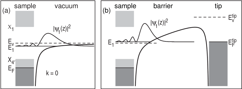
field – has been identified in scanning tunneling spectroscopy (STS) of Shockley surface-state electrons at a metal surface lim03_ . Surface states, in which the electron is caught between the barrier potential outside the surface and a band gap in the crystal, have been extensively studied by STM and STS, with a particular focus on their dynamics jli98_ ; kli00a ; bur99_ ; bra02_ and interactions rep00_ , and the recognition of a Stark-shift in the case of Shockley states reconciles a discrepancy that has existed between STS-derived binding energies and those from photoelectron spectroscopy rei01_ . A more pronounced Stark-effect has been known for some time bec85_ ; bin85_ in the case of a second class of surface electron state, namely the image-potential states that arise when an electron outside a conductor polarises the surface and is attracted to the resulting “image charge”, shown schematically in Fig. 1. Image-potential states are more weakly bound than Shockley surface states (which lie close to the Fermi energy ), forming a hydrogenic-like series with energies
| (1) |
converging on the vacuum level of the surface . In (1) is a quantum defect that depends upon the surface. Tunneling via image states requires significantly greater bias voltages than Shockley states, and the image state electrons are Stark-shifted to higher energies by several tenths of an eV bec85_ ; bin85_ ; wah03_ .
The presence of a measurable Stark-shift in the surface state energies raises the important question as to whether there are also changes in the inelastic interactions of the surface state electrons in the presence of the STM tip. Electronic excitations in the surface state bands decay on a femtosecond timescale through interactions with the electrons and phonons of the surface and bulk, and there has been considerable activity in recent years directed at an understanding of these interactions ech04_ . A significant electric-field induced lifetime change would have important consequences for the interpretation of STM and STS experiments investigating the dynamical properties of image-potential states, for example in nanostructures where the lateral resolution of the the STM is paramount.
To investigate this issue we have performed many-body calculations of the lifetimes of image-potential states at Cu(100) in the presence of an electric-field due to the tip of an STM. Our calculations are based upon the approach introduced by Chulkov et al. chu98_ and used subsequently in numerous surface state lifetime studies with considerable success ech04_ . The damping rate or inverse lifetime of the image state is calculated from the expectation value of the imaginary part of the non-local self energy operator
| (2) |
The energy of the image state is , and the wave function. The imaginary part of the self energy is calculated in the GW approximation of many-body theory, which uses the first term only in the series expansion of in terms of the screened Coulomb interaction :
| (3) |
We use the zero’th order approximation to the Green function; in the spectral representation
| (4) |
where the are one-electron eigenfunctions with eigenenergies and a positive infinitesimal. The screened interaction is evaluated in the random phase approximation (RPA)
| (5) | |||||
where is the bare Coulomb interaction and is the density-density response function of the non-interacting electron system:
| (6) | |||||
This GW-RPA approach has been shown to give decay rates for image states at Cu surfaces that are within 1 meV of those found using the more complete GW-TDLDA approximation sar99_ , in which exchange-correlation effects that are omitted in GW-RPA are included in both (5) and (6). Note that the phonon contribution to the decay rate of image-potential states at Cu(100) is meV and so safely ignored here eig03_ .
We calculate the Green function using a one-dimensional pseudopotential which by construction reproduces the Cu(100) bulk band edge energies and , and the energies of the unoccupied surface resonance and first image state at the field-free surface, and which also accurately predicts the energies of the higher image states chu99_ . In the direction parallel to the surface we assume parabolic dispersion with effective masses () fitted to ab-initio band structures. To model the influence of the STM we follow Limot et al. lim03_ and include a linear potential due to the bias voltage between the STM tip and the sample, and modify the image potential to include the multiple images present in the tunnel junction geometry. Using this potential we are able to reproduce the sequence of Stark-shifted image state energies and increments in tip-sample distance observed at Cu(100) in spectroscopy by Wahl et al., wah03_ who were also able to describe them using a model that omitted the effect of multiple-images.
In Fig. 2 we show the calculated damping rates (inverse lifetimes) of the Stark-shifted image potential state at Cu(100).

Calculation parameters have been systematically varied to ensure that decay rates are converged to within 1 meV. At the field-free Cu(100) surface the image state lies at an energy of eV relative to the vacuum energy, and we find meV for this state, corresponding to a lifetime of fs. This compares well with the lifetime fs measured using time-resolved two-photon photoemission (2PPE) hof97_ ; ber02_ , and fs found in previous GW calculations sar99_ . In the presence of the electric field due to the STM tip, the image state electrons are Stark-shifted to higher energies. At currents of 0.1-1 nA the level is observed in spectroscopy at bias voltages near V, corresponding to an energy eV wah03_ . For the results in Fig. 2 the tip-sample separation has been varied so that the bias voltage coincides with the image state energy in the presence of the electric field. This situation corresponds to the onset of tunneling into the level (Fig. 1(b)). In these conditions the resulting image state energy increases linearly with the applied electric field, and we find that the decay rates also increase linearly. The rate of change is , so that when the image state is Stark-shifted to eV the lifetime is only 15 fs, a reduction of 60% from the field-free value.
Recently the phase relaxation time of electrons in the image

state at Cu(100) has been studied using the STM by Wahl et al. wah03_ , who measured the spatial decay of quantum interference patterns near steps. This technique measures the lifetime of electrons with non-vanishing momentum parallel to the surface, corresponding to energies above the image state band minimum: . In Fig. 3 we compare calculated decay rates as a function of lateral energy with those reported in Ref. wah03_ . There it was concluded that the STM tip did not substantially alter the dynamical properties of the image-potential states, but subsequently an error has been recognised in the identification of the phase relaxation length that was used cra05_ so that the values displayed in Fig. 3 have been multiplied by 2 to correct for this. The calculations are performed for fields which give eV. Also shown are calculated results for the field-free case, along with values from 2PPE measurements ber02_ which correspond to this case. The overall agreement between theoretical and experimental lifetimes shown in Fig. 3 is very good, and confirms the existance of a significant field-induced change in the inelastic lifetimes of the image state electrons.
We now consider the origin of this effect. In Fig. 4 we compare

the probability density and the imaginary part of the self-energy at the Cu(100) surface calculated both with and without the electric field caused by the tip of the STM. The changes in show that accompanying the Stark-shift to higher energies is a redistribution of the weight of the surface state towards the metal surface, which increases the spatial overlap with the non-local self-energy. By displacing the image state electron towards the surface, the inelastic scattering channels are rendered more efficient. Calculating using the wave function of the Stark-shifted state but the self energy of the field-free surface at the unperturbed image state energy accounts for approximately three-quarters of the full increase in , as shown by the dashed line in Fig. 2.
The remaining change in the decay rate originates in the increase in the magnitude of that can also be seen in Fig. 4. We find that calculating the self-energy using in (3) either the screened interaction of the field-free surface or of the surface in the presence of the electric field gives comparable results, i.e., the changes in the electron wave functions caused by the electric field of the STM tip do not have a significant effect on the screening of the Coulomb interactions that dominate the inelastic scattering of the image state. Instead, the change in is due to the increase in the number of final states into which the image state can decay, i.e. the number of states between and the image state energy, which is Stark-shifted to higher energies by the tip-surface interaction of the STM. Thus the decreased lifetime of the Stark-shifted image-potential states results from an increase in the number of final states available for inelastic scattering along with increased efficiency of inelastic channels due to the greater spatial overlap of initial and final state wave functions.
Given the magnitude of the tip-induced change in the lifetimes of image-potential states it is worthwhile to consider whether similar changes affect STM-derived lifetimes of Shockley surface states jli98_ ; kli00a ; bur99_ ; bra02_ . Our calculations for Cu(111) indicate that the effect is minor. In this case at the Shockley state lies at eV which means that under typical tunneling conditions the electrostatic tip-surface interaction gives rise to fields that are 5–10 times smaller than those present when tunneling into image-potential states, and the resulting Stark-shift is much smaller: 10-15 meV kro04_ . This causes only a minor change in the number of final states that are available for decay. Furthermore, unlike image potential states which lie predominantly outside the surface, much of the Shockley state lies inside the metal, and is screened from the tip-induced electric field; there is a negligible change in the wave function penetration in the presence of the fields. Overall the electron-electron scattering decay rate which contributes two thirds of the total decay rate of meV ech04_ changes by less than 5% in the electric field. Although it is safe to dismiss the effect for this particular case, it is clear that the tip-induced field will have an increasingly important effect on the lifetimes of Shockley states that lie further from , especially at positive energies.
To conclude, we have used the many-body GW-RPA method to calculate the inelastic lifetimes of electrons in image states at Cu(100) including the electric field due to the tip-surface interaction in the STM. We find that under typical tunneling conditions the lifetime of electrons in the image state band is reduced by some 60% compared to in the absence of the STM tip. The Stark-shift to higher energies increases the number of inelastic decay channels that are available, whilst the the electric field moves the image state electrons closer to the metal surface, which significantly increases the efficiency of the scattering channels due to increased spatial overlap with final state wave functions. This tip-induced change in electron lifetimes must be taken into account when using the STM to study the dynamical properties of higher-lying surface electron states.
References
- (1) D. M. Eigler and E. K. Schweizer, Nature 344, 524 (1990).
- (2) M. F. Crommie, C. P. Lutz, and D. M. Eigler, Science 262, 218 (1993).
- (3) M. McEllistrem, G. Haase, D. Chen, and R. J. Hamers, Phys. Rev. Lett. 70, 2471 (1993).
- (4) L. Limot, T. Maroutian, P. Johansson, and R. Berndt, Phys. Rev. Lett. 91, 196801 (2003).
- (5) J. Li, W. -D. Schneider, R. Berndt, O. R. Bryant, and S. Crampin, Phys. Rev. Lett. 81, 4464 (1998).
- (6) J. Kliewer, R. Berndt, E. V. Chulkov, V. M. Silkin, P. M. Echenique and S. Crampin, Science 288, 1399 (2000).
- (7) L. Bürgi, O. Jeandupeux, H. Brune, and K. Kern, Phys. Rev. Lett. 82, 4516 (1999).
- (8) K. -F. Braun and K. -H. Rieder, Phys. Rev. Lett. 88, 096801 (2002).
- (9) J. Repp, F. Moresco, G. Meyer, K. -H. Rieder, P. Hyldgaard, and M. Persson, Phys. Rev. Lett. 85, 2981 (2000).
- (10) F. Reinert, G. Nicolay, S. Schmidt, D. Ehm, and S. Hüfner, Phys. Rev. B 63, 115415 (2001).
- (11) R. S. Becker, J. A. Golovchenko, and B. S. Swartzentruber, Phys. Rev. Lett. 55, 987 (1985).
- (12) G. Binnig, K. H. Frank, H. Fuchs, N. Garcia, B. Reihl, H. Rohrer, F. Salvan, and A. R. Williams, Phys. Rev. Lett. 55, 991 (1985).
- (13) P. Wähl, M.A. Schneider, L. Diekhöner, R. Vogelgesang, and K. Kern, Phys. Rev. Lett. 91, 106802 (2003).
- (14) For a recent review, see P. M. Echenique, R. Berndt, E. V. Chulkov, Th. Fauster, A. Goldmann, and U. Höfer, Surf. Sci. Rep. 52, 219 (2004).
- (15) E. V. Chulkov, I. Sarría, V. M. Silkin, J. M. Pitarke, and P. M. Echenique, Phys. Rev. Lett. 80, 4947 (1998).
- (16) I. Sarría, J. Osma, E. V. Chulkov, J. M. Pitarke, and P. M. Echenique, Phys. Rev. B 60, 11795 (1999).
- (17) A. Eiguren, B. Hellsing, E. V. Chulkov, P. M. Echenique, J. Electron Spectrosc. Relat. Phenom. 129, 111 (2003).
- (18) E. V. Chulkov, V. M. Silkin, and P. M. Echenique, Surf. Sci. 437 330 (1999).
- (19) U. Höfer, I. L. Shumay, Ch. Reuss, U. Thomann, W. Wallauer, and Th. Fauster, Science 277, 1480 (1997).
- (20) W. Berthold, U. Höfer, P. Feulner, E. V. Chulkov, V. M. Silkin, and P. M. Echenique, Phys. Rev. Lett. 88, 056805 (2002).
- (21) S. Crampin, J. Kröger, H. Jensen, and R. Berndt, cond-mat/0410542; P. Wahl, private communication.
- (22) J. Kröger, L. Limot, H. Jensen, R. Berndt, and P. Johansson, Phys. Rev. B. 70, 033401 (2004).