Sub-unit cell layer-by-layer growth of Fe3O4, MgO, and Sr2RuO4 thin films
Abstract
The use of oxide materials in oxide electronics requires their controlled epitaxial growth. Recently, it was shown that Reflection High Energy Electron Diffraction (RHEED) allows to monitor the growth of oxide thin films even at high oxygen pressure. Here, we report the sub-unit cell molecular or block layer growth of the oxide materials Sr2RuO4, MgO, and magnetite using Pulsed Laser Deposition (PLD) from stoichiometric targets. Whereas for perovskites such as SrTiO3 or doped LaMnO3 a single RHEED intensity oscillation is found to correspond to the growth of a single unit cell, in materials where the unit cell is composed of several molecular layers or blocks with identical stoichiometry, a sub-unit cell molecular or block layer growth is established resulting in several RHEED intensity oscillations during the growth of a single unit-cell.
pacs:
61.14.Hg, 74.76.Db, 75.70.-i, 81.15.FgThe physical properties of thin films are strongly influenced by their microstructure and morphology which, in turn, are determined by the deposition conditions and the growth mode. RHEED has proven to be a useful surface sensitive tool for monitoring in situ the growth of semiconductor thin films Lagally:93 . RHEED has also been successfully used for studying the growth of the complex copper oxide superconductors under high vacuum conditions Terashima:90 ; Karl:92 ; Bozovic:95 . Recently, high pressure RHEED systems have been developed allowing for the analysis of oxide thin films grown by Pulsed Laser Deposition (PLD) at high oxygen partial pressure of up to several 10 Pa Rijnders:97 ; Klein:99 . A common way of using RHEED in the analysis of thin film growth is the observation of the electron diffraction pattern during epitaxial growth. In this case intensity oscillations of the diffraction spots are associated with a layer-by-layer or Frank-van der Merwe growth mode Terashima:90 . The deposited material nucleates on the substrate surface forming two dimensional islands which are coalescing with increasing coverage. This process results in a periodic roughening and flattening of the film surface translating into RHEED intensity oscillations.
In this Letter, we report on the study of RHEED intensity oscillations during the PLD growth of the oxides materials Sr2RuO4, MgO, and magnetite (Fe3O4) from stoichiometric targets. We have used an ultra high vacuum Laser Molecular Beam Epitaxy (L-MBE) system with in-situ high pressure RHEED and a 248 nm KrF excimer laser Gross:2000a . It is well known that due to the ionic bond character in oxides the different atomic layers in general will not be charge neutral and therefore the energetically most favorable growth unit often is a molecular layer which is composed of one or several atomic layers to obtain charge neutrality. Therefore, molecular layer or block layer epitaxyGross:2000a is established for most oxides, whereas atomic layer epitaxy is common for semiconductor superlattice growth. Our results clearly show that under suitable deposition conditions a molecular or block layer growth is indeed established for MgO, Sr2RuO4 and Fe3O4. However, in the present study the observed thickness of the charge neutral molecular layers is exceeding that of the minimum charge neutral building blocks of the respective material, since stoichiometric targets have been used in the PLD process. In this case, the minimum observed building blocks are those satisfying both the requirement of charge neutrality and the chemical composition supplied from the stoichiometric target material. Let us illustrate this for the example of SrTiO3. This material can be grown both in alternating charge neutral SrO and TiO2 molecular layers supplied from SrO and TiO2 targets, and in SrTiO3 layers provided from a stoichiometric target. Note that in the first case every molecular layer will produce a RHEED intensity oscillation resulting in two RHEED oscillations per unit cell. In contrast, in the latter case only a single oscillation per unit cell is observed, since SrO and TiO2 are supplied simultaneously and the minimum charge neutral block compatible with the supplied stoichiometry consists of the whole unit cell. We observed an equivalent behavior for the growth of MgO, Sr2RuO4, and Fe3O4 using stoichiometric targets. Due to the complex unit cells of MgO and Sr2RuO4 consisting of identical stoichiometric layers shifted by lattice translations two RHEED oscillations per unit cell are observed. For Fe3O4, the unit cells consists of even 4 stoichiometric sub-units resulting in 4 RHEED oscillations per unit cell.
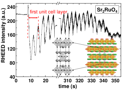
We first discuss the growth of the unconventional superconductor Sr2RuO4. This material is isostructural to La2CuO4, which already has been studied by RHEED Terashima:90 . For structural reasons, a similar growth is expected for both materials. Fig. 1 shows the RHEED intensity oscillations of the diffraction spot recorded during the PLD growth of a -axis oriented Sr2RuO4 film on a NdGaO3 substrate. The layer-by-layer growth mode is achieved for a substrate temperature C, an oxygen pressure of mTorr, a laser repetition rate of Hz, and a laser energy density on the target of J/cm2. RHEED was performed with 15 keV electrons at an incident angle of . To obtain the number of RHEED oscillations per unit cell, the film thickness has been determined precisely by X-ray reflectometry and then divided by the number of observed RHEED oscillations. The derived block thickness corresponds to half a unit cell (Å). Considering the crystal structure shown in the inset of Fig. 1, this result is intuitive: The unit cell consists of two identical layers which are displaced within the -plane by , where and are the in-plane lattice vectors. Note that after the third oscillation the intensity of the maxima remains about constant. The growth can be stopped and continued with almost unchanged modulation depth. After a certain film thickness the oscillations start to flatten out with an overall decrease of reflected intensity due to surface roughening because of a transition to island growth. The layer-by-layer growth mode can be re-established by thermal annealing. An interesting feature is that the RHEED intensity starts to increase immediately after starting the PLD process. However, this effect is only present for starting the growth on the substrate, but is absent for starting on the surface of an already grown Sr2RuO4 film. Therefore, we believe that this effect is not related to the diffraction conditions, but more likely to a change of the surface structure and morphology of the substrate due to the partial coverage with the thin film material. A similar effect has been observed for isostructural La2CuO4 Terashima:90 as well as for GaAs Dobson:87 . We finally note, that our high quality Sr2RuO4 films have lower resistivity values (cm at 300 mK) than those reported so far in literature Schlom:98 . However, these values are still about ten times larger than those required for superconductivity (cm).
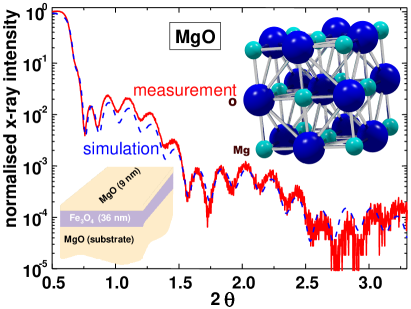
As the second example we discuss the growth of a MgO cap layer on a magnetite thin film, which will be discussed below. Both films were grown under the same growth conditions (C, mbar, Hz, and J/cm2) leading for both materials to a layer-by-layer growth mode. First the magnetite thin film was grown on a MgO substrate with atomically flat surface (3 Å rms roughness) followed by the MgO cap layer. Fig. 2 shows X-ray reflectometry data of a Fe3O4 (36 nm)/MgO (9 nm) bilayer. Fitting the datarefsim gives the thickness of the MgO and Fe3O4 layers with an error of less than %. As already described above, from the MgO layer thickness and the observed number of RHEED oscillations we obtain the result that two RHEED oscillations correspond to the growth of a single MgO unit cell shown in Fig. 2. Again, the intuitive explanation for this observation is the fact that the unit cell of MgO consists of two identical stoichiometric layers displaced along the -plane by . After the deposition of the MgO cap layer, the surface morphology was examined in-situ by Atomic Force Microscopy. The rms surface roughness averaged over an area of 1 m2 was below 2 Å, i.e. even below that of the MgO substrate. This result is consistent with a dominant layer-by-layer growth mode observed for the whole bilayer.
As the last example we discuss the growth of Fe3O4 thin films on MgO substrates. Fig. 3 shows the RHEED intensity vs. time which has several interesting features. First, the modulation depth decreases continuously with increasing film thickness, if the ablation is not interrupted. Since the intensity of the maxima decreases while the intensity of the minima increases, we ascribe this to a gradual transition to the step-flow-growth mode which leaves the reflected intensity unchanged. The layer-by-layer growth mode can be re-established by annealing the sample for about 100 s in the deposition atmosphere. Second, there is a huge increase of the RHEED intensity directly after starting the PLD process. In contrast to the case of Sr2RuO4, this effect is observed reproducibly after each growth stop and not only if the deposition is started on the bare substrate. Note that the RHEED intensity vs. time curves in Fig. 3 are obtained from the diffraction spot. For this spot the electrons reflected from different growth planes interfere constructively (in-Bragg condition). In this case for purely coherent scattering no RHEED oscillations are expected. The fact that we do observe RHEED oscillations is caused by multiple and diffuse, incoherent scattering which results in an increasing (decreasing) intensity with increasing (decreasing) step density Korte:97 . Therefore, starting from a smooth surface the RHEED intensity first increases due to the increasing step density. That is, a -phase shift of the intensity oscillation relative to those recorded for off-Bragg condition is obtained. A similar behavior has also been found for semiconductor growth Dobson:87 ; Joyce:88 ; Braun:99 . This interpretation is consistent with a third observation: after stopping the growth of Fe3O4 the intensity decreases. Since it is natural to assume that annealing leads to a smoother surface, i.e. smaller step density, it is evident that the measured intensity of the spot is decreasing with time. The relevance of multiple scattering processes is also manifested by the presence of Kikuchi lines. The RHEED pattern in the inset of Fig. 3 indicates that the resonant enhancement is due to both, coupling to surface bound states, and three-dimensional diffraction Braun:99 .
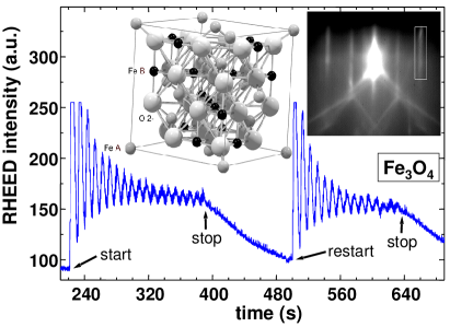
From the measured film thickness and the number of RHEED oscillations, for Fe3O4 we obtain four oscillations per unit cell. Considering the unit cell of Fe3O4 with the inverse spinel Fdm structure ( nm), one can identify the corresponding molecular layers each consisting of the composition Fe(A)Fe(B)Fe(B)O. The letters A and B refer to the tetrahedral A-site and the octahedral B-site, where the B-site is equally occupied by Fe3+ and Fe2+ ions. These building blocks cannot be mapped onto each other by lattice translations. We note that our high quality, 30-50 nm thick magnetite films have a saturation magnetization at room temperature close to the theoretically expected value of 4.0 /f.u., that is, the saturation magnetization is comparable to the best results reported in literature so far Kale:01 . We further note, that recently four RHEED intensity oscillations per unit cell have been observed for the layered manganites. Interestingly, there the basic building block corresponds to only half the layer thickness expected according the chemical composition (La,Sr)3Mn2O7 Philipp:02 .
In summary, we have grown Sr2RuO4, MgO, and Fe3O4 epitaxial thin films by PLD from stoichiometric targets. Our results show that high pressure RHEED allows to monitor the epitaxial growth of the complex oxide materials on a sub-unit cell level. We have observed a molecular or block layer growth mode where the basic building blocks are determined by the chemical composition provided by the stoichiometric target material. For materials with units cells consisting of several block layers of identical stoichiometry, several RHEED oscillations per unit cell are observed.
This work was supported in part by the Deutsche Forschungsgemeinschaft (project Al/560) and the BMBF (project 13N8279). We thank S. Schymon for the preparation and characterization of Sr2RuO4 thin films.
References
- (1) M. G. Lagally and D. E. Savage, MRS Bull. XVIII, 24 (1993).
- (2) T. Terashima, Y. Bando, K. Iijima, K. Yamamoto, K. Hirata, K. Hayashi, K. Kamigaki, and H. Terauchi, Phys. Rev. Lett. 65, 2684 (1990).
- (3) H. Karl and B. Stritzker, Phys. Rev. Lett. 69, 2939 (1992).
- (4) I. Bozovic and J. N. Eckstein, MRS Bull. XX, 32 (1995).
- (5) G. J. H. M. Rijnders, G. Koster, D. H. A. Blank, H. Rogalla, Appl. Phys. Lett. 70, 1888 (1997).
- (6) J. Klein, C. Höfener, L. Alff, and R. Gross, Supercond. Sci. Technol. 12, 1023 (1999), see also J. Magn. Magn. Mater. 211, 9 (2000).
- (7) R. Gross, J. Klein, B. Wiedenhorst, C. Höfener, U. Schoop, J. B. Philipp, M. Schonecke, F. Herbstritt, L. Alff, Yafeng Lu, A. Marx, S. Schymon, S. Thienhaus, W. Mader, SPIE Conf. Proc. 4058 (2000), pp. 278-294.
- (8) P. J. Dobson, B. A. Joyce, J. H. Neave, J. Cryst. Growth 81, 1 (1987).
- (9) D. G. Schlom, Y. Jia, L.-N. Zou, J. H. Haeni, S. Briczinski, M. A. Zurbuchen, C. W. Leitz, S. Madhavan, S. Wozniak, Y. Liu, M. E. Hawley, G. W. Brown, A. Dabkowski, H. A. Dabkowska, R. Uecker, and P. Reiche, SPIE Conf. Proc. 3481 (1998), pp. 226-240.
- (10) Bruker AXS Windows RefSim simulation software, based on dynamical scattering theory.
- (11) U. Korte and P. A. Maksym, Phys. Rev. Lett. 78, 2381 (1997).
- (12) B. A. Joyce, J. H. Neave, J. Zhang, P. J. Dobson, in Reflection High Energy Electron Diffraction and Reflection Electron Imaging of Surfaces, P. K. Larsen and P. J. Dobson eds. (Plenum, London 1988) pp. 397-417.
- (13) W. Braun, Applied RHEED, Springer Tracts in Modern Physics 154, Springer-Verlag Berlin Heidelberg (1999).
- (14) S. Kale, S. M. Bhagat, S. E. Lofland, T. Scabarozi, S. B. Ogale, A. Orozco, S. R. Shinde, T. Venkatesan, B. Hannoyer, B. Mercey, and W. Prellier, Phys. Rev. B 64, 205413 (2001).
- (15) J. B. Philipp, J. Klein, C. Recher, T. Walther, W. Mader, M. Schmid, R. Suryanarayanan, L. Alff, and R. Gross, Phys. Rev. B 65, 184411 (2002).